Saket * Call Girls in Delhi - Phone 9711199012 Escorts Service at 6k to 50k a...
BACK OF THIGH AND POPLITEAL FOSSA.pptx
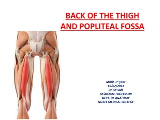
1. BACK OF THE THIGH
AND POPLITEAL FOSSA
MBBS 1st year
15/02/2023
Dr. SK SAH
ASSOCIATE PROFESSOR
DEPT. OF ANATOMY
NOBEL MEDICAL COLLEGE
2. BACK OF THE THIGH
• The muscles of the posterior
compartment of the thigh are called
the hamstrings.
• To be called a hamstring, the muscle
must arise from the ischial tuberosity.
• The hamstrings are:
1. Biceps femoris
2. Semimembranosus
3. Semitendinosus
4. Hamstring part of the adductor
magnus.
• These muscles extend the thigh at the
hip and flex the leg at the knee.
3. • The hamstrings are a group of muscles and
their tendons at the rear of the upper leg.
They include the biceps femoris,
semitendinosus, and semimembranosus.
• The hamstrings flex the knee joint and extend
the thigh to the backside of the body.
• They are used in walking, running, and many
other physical activities.
4. • The common criteria of any hamstring muscles
are:
Muscles should originate from ischial tuberosity.
Muscles should be inserted over the knee joint,
in the tibia or in the fibula.
Muscles will be innervated by the tibial branch
of the sciatic nerve.
Muscle will participate in flexion of the knee
joint and extension of the hip joint.
• Those muscles which fulfill all of the four criteria
are called true hamstrings.
5.
6.
7.
8.
9. BICEPS FEMORIS
• ORIGIN:
• the long head originates with
the semitendinosus muscle
from the inferomedial part of
the upper area of the ischial
tuberosity;
• the short head arises from the
lateral lip of the linea aspera
on the shaft of the femur.
• Insertion: The main part of
the tendon inserts into the
lateral surface of the head of
the fibula.
10. SEMITENDINOSUS
• It originates with the long head of the
biceps femoris muscle from the
inferomedial part of the upper area of
the ischial tuberosity.
• The spindle-shaped muscle belly ends
in the lower half of the thigh and
forms a long cord-like tendon, which
lies on the semimembranosus muscle
and descends to the knee.
• The tendon curves around the medial
condyle of the tibia and inserts into
the medial surface of the tibia just
posterior to the tendons of the gracilis
and sartorius muscles.
11. SEMIMEMBRANOSUS
• It is attached above to the
superolateral impression on the
ischial tuberosity.
• and below mainly to the groove
and adjacent bone on the
medial and posterior surfaces of
the medial tibial condyle.
• Expansions from the tendon
also insert into and contribute
to the formation of ligaments
and fascia around the knee
joint.
12. Arteries of Posterior Thigh
• The arteries of the
posterior compartment
of the thigh arise from
two major arteries:
• inferior gluteal artery.
• perforating branches of
the profunda femoris .
13. Nerves of Posterior Thigh
• The muscles of the posterior
compartment of the thigh are
innervated by the tibial (medial)
part of the SCIATIC NERVE.
14. POPLITEAL FOSSA
• Diamond shaped dipression behind
the knee joint.
• Homologous with the cubital fossa
of upper limb.
• BOUNDARIES:
Superolaterally– Biceps femoris
Superomedially– semitendinosus
and semimembranosus.
Inferolaterally– lateral head of
gastrocnemius supplimented by
plantaris.
Inferomedially– medial head of
gatrocnemius.
15. • Roof: is formed by deep fascia or popliteal fascia.
• Floor: is formed from above downward by ;
-- popliteal surface of the femur.
-- capsule of the knee joint.
-- the strong popliteal fascia covering the popliteus
muscle.
16. CONTENTS OF POPLITEAL FOSSA
I. The Popliteal artery and its branches.
II. The Popliteal vein and its tributaries.
III. The Tibial nerve and its branches.
IV. The Common peroneal nerve and its branches.
• The fossa also contains;
I. Posterior cut. N of thigh
II. Genicular branch of obturator N.
III. The popliteal lymph node.
IV. Fat.
19. APPLIED ANATOMY
• Blood pressure in the lower limb is recorded from
the popliteal artery.
• When the popliteal artery is affected by
atherosclerosis, the lower part of the artery usually
remains patent where grafts can be tried.
• The popliteal artery is more prone to aneurysm than
many other arteries of the body.
20. • Pulsations of the femoral artery can be felt at the
midingunal point.
• The femoral vein is most commonly used for the
intravenous infusions in infants and in patients with
peripheral circulatory failure.
Femoral artery is used for embalming procedure.
The femoral and obturator nerve which supply the hip
joint, also supply the knee joint. Therefore, diseases of
the hip may produce reffered pain in the knee and also
in the cutaneous area innervated by these nerves.
21. Wrist drop– paralysis of Radial Nerve
Foot drop– paralysis of Common
peroneal nerve
Sleeping foot– compression of Sciatic
nerve