SlideShare ist ein Scribd-Unternehmen logo
1 von 35
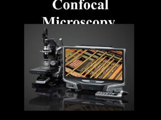
Confocal
Microscopy
INTRODUCTION
• An optical imaging technique for increasing optical
resolution and contrast of a micrograph.
• Radiations emitted from laser cause sample to
fluoresce.
• Uses pinhole screen to produce high resolution
images.
• Eliminates out of focus.
• So images have better contrast and are less hazy.
• A series of thin slices of the specimen are assembled
to generate a 3dimensinal image.
• Is an updated version of fluorescence microscopy.
HISTORY
• Two investigators at Cambridge, Brad Amos and John White
attempted to look at the mitotic divisions in the first few
divisions in embryos of C. elegans.
• They were doing antitubulin immunofluorescence and were
trying to determine the cleavage planes of the cells,
• But were frustrated in their attempt in that the majority of
the fluorescence they observed was out of focus.
• They looked at the technique called confocal imaging which
was first proposed by Nipkow and pioneered by a postdoc at
Harvard named Minsky.
• He made the first stage scanning confocal microscope in 1957.
• By illuminating single point at a time, Minsky avoided most of
the unwanted scattered light.
• For buiiding the image, Minsky scanned the specimen by
moving the stage rather than light rays.
COMPONENTS OF CONFOCAL
MICROSCOPY
• Laser
• Photomultiplier
• Filters
LASER
LASER
• Light Amplification by Stimulated Emission of
Radiation (Laser).
• Lasers are used because they are an intense
coherent monochromatic source of light, capable
of being expanded to fill an aperture or focused
to a spot.
• The laser beam is usually linearly polarized. The
main drawback in using lasers is that to cover a
large excitation range you will need several lasers.
• However a single laser line may not optimally
excite your molecule.
PHOTOMULIPLIER
PHOTOMULTIPLIER
• A photomultiplier tube, useful for light detection of very weak
signals,it is a photo emissive device in which the absorption of
a photon results in the emission of an electron.
• These detectors work by amplifying the electrons generated
by a photocathode exposed to a photon flux.
FILTERS
•
• Microscopy Filters are used in a variety of microscopy applications for
increasing contrast, blocking ambient light, removing harmful ultraviolet
or infrared light, or for selectively transmitting only wanted wavelengths.
• Most of the microscopes, are based on optical filters.
• A typical system has three basic filters: an excitation filter (or exciter), a
dichroic beamsplitter (or dichromatic mirror), and an emission filter (or
barrier filter).
• An excitation filter is a high quality optical-glass filter commonly used in
fluorescence microscopy and spectroscopic applications for selection of the
excitation wavelength of light from a light source.
• Barrier filters are filters which are designed to suppress or block (absorb) the
excitation wavelengths and permit only selected emission wavelengths to
pass toward the eye or other detector.
• A third version of the beam splitter is a dichroic mirrored prism assembly
which uses dichroic optical coatings to divide an incoming light beam into a
number of spectrally distinct output beams. The dichroic beam splitter
controls which wavelengths of light go to their respective filter.
DICHORIC MIRROR
INSTRUMENTAL DESIGN
Ray diagram for confocal microscope
CONFOCAL MICROSCOPE
Schematic diagram
confocal microscope
PRINCIPLE
In confocal microscopy two pinholes are typically used:
• A pinhole is placed in front of the illumination source to
allow transmission only through a small area
• This illumination pinhole is imaged onto the focal plane
of the specimen, i.e. only a point of the specimen is
illuminated at one time.
• Fluorescence excited in this manner at the focal plane is
imaged onto a confocal pinhole placed right in front of
the detector.
• Only fluorescence excited within the focal plane of the
specimen will go through the detector pinhole.
• Scanning of small sections is done and joined them
together for better view.
WORKING MECHANISM
Confocal microscope incorporates 2 ideas:
1. Point-by-point illumination of the specimen.
2. Rejection of out of focus of light.
Light source of very high intensity is used—Zirconium arc lamp in
Minsky’s design & laser light source in modern design.
a)Laser provides intense blue excitation light.
b)The light reflects off a dichoric mirror, which directs it to an
assembly of vertically and horizontally scanning mirrors.
c)These motor driven mirrors scan the laser beam across the
specimen.
d) The specimen is scanned by moving the stage back & forth in
the vertical & horizontal directions and optics are kept
stationary.
Contd….
• Dye in the specimen is excited by the laser light &
fluoresces.
• The fluorescent (green) light is descanned by the same
mirrors that are used to scan the excitation (blue) light
from the laser beam
• Then it passes through the dichoric mirror
• Then it is focused on to pinhole.
• The light passing through the pinhole is measured by the
detector such as photomultiplier tube.
• For visualization, detector is attached to the computer,
which builds up the image at the rate of 0.1-1 second for
single image
LIGHT PATHWAY
• Light from
excitation filter
passes through
objective lens and
light absorbed by
specimen.
• Light emitted goes
back through
objective lens,
barrier filter, then
detector.
x y
z
OPTICAL SECTIONING AND 3-D RECONSTRUCTION
x y
z
OPTICAL SECTIONING AND 3-D RECONSTRUCTION
x y
z
=
OPTICAL SECTIONING AND 3-D RECONSTRUCTION
3-D Imaging with Confocal Microscopy
ADVANTAGES
• The specimen is everywhere illuminated axially, rather than
at different angles, thereby avoiding optical aberrations.
• Entire field of view is illuminated uniformly.
• The field of view can be made larger than that of the static
objective by controlling the amplitude of the stage
movements.
• Image formed are of better resolution.
• Cells can be live or fixed. Serial optical sections can be
collected.
• Taking a series of optical slices from different focus levels in
the specimen generates a 3D data set.
DRAWBACKS
• Resolution : It has inherent resolution limitation due to
diffraction. Maximum best resolution of confocal microscopy is
typically about 200nm.
• Pin hole size : Strength of optical sectioning depends on the
size of the pinhole.
• Intensity of the incident light
• Fluorophores :
• a)The fluorophore should tag the correct part of the specimen.
• b)Fluorophore should be sensitive enough for the given
excitation wave length.
• c)It should not significantly alter the dynamics of the organism
in the living specimen.
• Photobleaching: photochemical alteration of a dye or a
fluorophore molecule such that it permanently is unable to
fluorescence.
APPLICATIONS
1.Confocal
microscopy allows
analysis of
fluorescent
labelled thick
specimens without
physical
sectioning.
Zebra fish embryo wholemount Neurons (green) and
Cell adhesion molecule (red)
APPLICATIONS
2. Three-Dimensional
Reconstruction of
Specimen .
3D shadow projection
3. Improved Resolution
APPLICATIONS
An Intestine SectionKidney cells (fluorescence vs Confocal
microscope
4. More Colour Possibilities
Because the images are
detected by a computer rather
than by eye, it is possible to
detect more color differences.
APPLICATIONS
IMAGE TAKEN BY CONFOCAL MICROSCOPE
Colour coded image of actin filaments in a cancer cell
IMAGE TAKEN BY CONFOCAL MICROSCOPE
Pancreatic islets of mouse
IMAGE TAKEN BY CONFOCAL MICROSCOPE
Drosophila brain
with triple
antibodies
IMAGE TAKEN BY CONFOCAL MICROSCOPE
Hepatocyte
sphenoid
Confocal vs. Widefield
Confocal Widefield
Tissue culture cell with 60x / 1.4NA objective
CONFOCAL VS. WIDEFIELD
Tissue culture cell with 60X /1.4NA Objective
CONFOCAL VS. WIDEFIELD
20 micrometer rat intestine section recorded with 60 x/1.4NA Objective
EXAMPLES
Drosophila S2 cell expressing GFP-H2B
and mChery-tubulin [ Nico suurname
and Ron vale ].
S.Cerevisae expressing mitochondrially
targeted RFP [Sussane Rafeilski
,Marshall lab ].
THANK YOU

Weitere ähnliche Inhalte

Was ist angesagt?

Fluorescence Microscopy
Fluorescence MicroscopyFluorescence Microscopy
Fluorescence MicroscopyAalap Tripathy
 
Confocal laser scanning microscopy (clsm)
Confocal laser scanning microscopy (clsm)Confocal laser scanning microscopy (clsm)
Confocal laser scanning microscopy (clsm)Nethravathi Siri
 
Fluorescence microscope by Subhankar Das
Fluorescence microscope by Subhankar DasFluorescence microscope by Subhankar Das
Fluorescence microscope by Subhankar DasSubhankar Das
 
Fluorescence microscopy presentation
Fluorescence microscopy presentationFluorescence microscopy presentation
Fluorescence microscopy presentationMandira bhosale
 
Phase contrast microscope
Phase contrast microscopePhase contrast microscope
Phase contrast microscopeSalman Khan
 
FLUORESCENCE MICROSCOPY
FLUORESCENCE MICROSCOPYFLUORESCENCE MICROSCOPY
FLUORESCENCE MICROSCOPYprachann
 
Phase contrast microscope
Phase contrast microscopePhase contrast microscope
Phase contrast microscopeSuganyaPaulraj
 
Fluorescence microscopy
Fluorescence microscopyFluorescence microscopy
Fluorescence microscopyJyotsna Verma
 
Principles and application of light, phase constrast and fluorescence microscope
Principles and application of light, phase constrast and fluorescence microscopePrinciples and application of light, phase constrast and fluorescence microscope
Principles and application of light, phase constrast and fluorescence microscopeMaitriThakor
 
Phase contrast and Fluorescent microscope
Phase contrast and Fluorescent microscopePhase contrast and Fluorescent microscope
Phase contrast and Fluorescent microscopeNithiya Pappuraj
 

Was ist angesagt? (20)

Confocal microscopy
Confocal microscopyConfocal microscopy
Confocal microscopy
 
Fluorescence microscope
Fluorescence microscopeFluorescence microscope
Fluorescence microscope
 
Fluorescence Microscopy
Fluorescence MicroscopyFluorescence Microscopy
Fluorescence Microscopy
 
Confocal laser scanning microscopy (clsm)
Confocal laser scanning microscopy (clsm)Confocal laser scanning microscopy (clsm)
Confocal laser scanning microscopy (clsm)
 
Fluorescence microscope by Subhankar Das
Fluorescence microscope by Subhankar DasFluorescence microscope by Subhankar Das
Fluorescence microscope by Subhankar Das
 
Confocal microscopy
Confocal microscopyConfocal microscopy
Confocal microscopy
 
Fluorescence microscopy presentation
Fluorescence microscopy presentationFluorescence microscopy presentation
Fluorescence microscopy presentation
 
Phase contrast microscope
Phase contrast microscopePhase contrast microscope
Phase contrast microscope
 
FLUORESCENCE MICROSCOPY
FLUORESCENCE MICROSCOPYFLUORESCENCE MICROSCOPY
FLUORESCENCE MICROSCOPY
 
Light microscope
Light microscopeLight microscope
Light microscope
 
Bright field microscope
Bright field microscopeBright field microscope
Bright field microscope
 
Phase contrast microscope
Phase contrast microscopePhase contrast microscope
Phase contrast microscope
 
Fluorescence microscopy
Fluorescence microscopyFluorescence microscopy
Fluorescence microscopy
 
Principles and application of light, phase constrast and fluorescence microscope
Principles and application of light, phase constrast and fluorescence microscopePrinciples and application of light, phase constrast and fluorescence microscope
Principles and application of light, phase constrast and fluorescence microscope
 
phase contrast microscope
phase contrast microscopephase contrast microscope
phase contrast microscope
 
microscopy
 microscopy microscopy
microscopy
 
Confocal microscopy
Confocal microscopyConfocal microscopy
Confocal microscopy
 
Light Microscopy
Light MicroscopyLight Microscopy
Light Microscopy
 
Phase contrast and Fluorescent microscope
Phase contrast and Fluorescent microscopePhase contrast and Fluorescent microscope
Phase contrast and Fluorescent microscope
 
Fluorescence Microscopy
Fluorescence MicroscopyFluorescence Microscopy
Fluorescence Microscopy
 

Ähnlich wie Confocal microscope presentation pt

Various types of microscopes and microscopy
Various types of microscopes and microscopy   Various types of microscopes and microscopy
Various types of microscopes and microscopy Dr.Dinesh Jain
 
MICROSCOPY.pdf..........................
MICROSCOPY.pdf..........................MICROSCOPY.pdf..........................
MICROSCOPY.pdf..........................YolywinSanico
 
microscope- lecture 2.pptx
microscope- lecture 2.pptxmicroscope- lecture 2.pptx
microscope- lecture 2.pptxOsmanAli92
 
confocalmicroscopy-121115061847-phpapp02.pdf
confocalmicroscopy-121115061847-phpapp02.pdfconfocalmicroscopy-121115061847-phpapp02.pdf
confocalmicroscopy-121115061847-phpapp02.pdfvadivel415
 
Microscopy presentation
Microscopy presentationMicroscopy presentation
Microscopy presentationmoavia Atiq
 
Confocal Microscopy.pptx
Confocal Microscopy.pptxConfocal Microscopy.pptx
Confocal Microscopy.pptxRajadr
 
Microscopy 121125060708-phpapp01
Microscopy 121125060708-phpapp01Microscopy 121125060708-phpapp01
Microscopy 121125060708-phpapp01Ghassan Hadi
 
2. Microscopy (Biochemistry)
2. Microscopy (Biochemistry)2. Microscopy (Biochemistry)
2. Microscopy (Biochemistry)Jay Khaniya
 
Microscopy 4 anna
Microscopy 4 annaMicroscopy 4 anna
Microscopy 4 annaAnnakurian9
 
2 Microscopy and staining.pptx
2 Microscopy and staining.pptx2 Microscopy and staining.pptx
2 Microscopy and staining.pptxNeelGamers
 
Microscope lecture notes for laboratory).ppt
Microscope lecture notes for laboratory).pptMicroscope lecture notes for laboratory).ppt
Microscope lecture notes for laboratory).pptSheikhAlshoteri2
 
Light microscopy.pptx bright dark phase contrast microscope
Light microscopy.pptx bright dark phase contrast microscopeLight microscopy.pptx bright dark phase contrast microscope
Light microscopy.pptx bright dark phase contrast microscopeDarshanN65
 
ELECTRON MICROSCOPE-1.pptx
ELECTRON MICROSCOPE-1.pptxELECTRON MICROSCOPE-1.pptx
ELECTRON MICROSCOPE-1.pptxOpenPhone
 
Microscope anwesha.pptx
Microscope anwesha.pptxMicroscope anwesha.pptx
Microscope anwesha.pptxAnwesha Mandal
 

Ähnlich wie Confocal microscope presentation pt (20)

Compound microscope
Compound microscopeCompound microscope
Compound microscope
 
Various types of microscopes and microscopy
Various types of microscopes and microscopy   Various types of microscopes and microscopy
Various types of microscopes and microscopy
 
MICROSCOPY.pdf..........................
MICROSCOPY.pdf..........................MICROSCOPY.pdf..........................
MICROSCOPY.pdf..........................
 
Microscopy
MicroscopyMicroscopy
Microscopy
 
microscope- lecture 2.pptx
microscope- lecture 2.pptxmicroscope- lecture 2.pptx
microscope- lecture 2.pptx
 
MICROSCOPY.pptx
MICROSCOPY.pptxMICROSCOPY.pptx
MICROSCOPY.pptx
 
Microscope
MicroscopeMicroscope
Microscope
 
confocalmicroscopy-121115061847-phpapp02.pdf
confocalmicroscopy-121115061847-phpapp02.pdfconfocalmicroscopy-121115061847-phpapp02.pdf
confocalmicroscopy-121115061847-phpapp02.pdf
 
Microscopy presentation
Microscopy presentationMicroscopy presentation
Microscopy presentation
 
Confocal Microscopy.pptx
Confocal Microscopy.pptxConfocal Microscopy.pptx
Confocal Microscopy.pptx
 
Microscopy 121125060708-phpapp01
Microscopy 121125060708-phpapp01Microscopy 121125060708-phpapp01
Microscopy 121125060708-phpapp01
 
MICROSCOPY.pptx
MICROSCOPY.pptxMICROSCOPY.pptx
MICROSCOPY.pptx
 
2. Microscopy (Biochemistry)
2. Microscopy (Biochemistry)2. Microscopy (Biochemistry)
2. Microscopy (Biochemistry)
 
Microscopy 4 anna
Microscopy 4 annaMicroscopy 4 anna
Microscopy 4 anna
 
2 Microscopy and staining.pptx
2 Microscopy and staining.pptx2 Microscopy and staining.pptx
2 Microscopy and staining.pptx
 
Microscope lecture notes for laboratory).ppt
Microscope lecture notes for laboratory).pptMicroscope lecture notes for laboratory).ppt
Microscope lecture notes for laboratory).ppt
 
Light microscopy.pptx bright dark phase contrast microscope
Light microscopy.pptx bright dark phase contrast microscopeLight microscopy.pptx bright dark phase contrast microscope
Light microscopy.pptx bright dark phase contrast microscope
 
Microscope 120109025117-phpapp02
Microscope 120109025117-phpapp02Microscope 120109025117-phpapp02
Microscope 120109025117-phpapp02
 
ELECTRON MICROSCOPE-1.pptx
ELECTRON MICROSCOPE-1.pptxELECTRON MICROSCOPE-1.pptx
ELECTRON MICROSCOPE-1.pptx
 
Microscope anwesha.pptx
Microscope anwesha.pptxMicroscope anwesha.pptx
Microscope anwesha.pptx
 

Kürzlich hochgeladen

POINT- BIOCHEMISTRY SEM 2 ENZYMES UNIT 5.pptx
POINT- BIOCHEMISTRY SEM 2 ENZYMES UNIT 5.pptxPOINT- BIOCHEMISTRY SEM 2 ENZYMES UNIT 5.pptx
POINT- BIOCHEMISTRY SEM 2 ENZYMES UNIT 5.pptxSayali Powar
 
1029-Danh muc Sach Giao Khoa khoi 6.pdf
1029-Danh muc Sach Giao Khoa khoi  6.pdf1029-Danh muc Sach Giao Khoa khoi  6.pdf
1029-Danh muc Sach Giao Khoa khoi 6.pdfQucHHunhnh
 
Kisan Call Centre - To harness potential of ICT in Agriculture by answer farm...
Kisan Call Centre - To harness potential of ICT in Agriculture by answer farm...Kisan Call Centre - To harness potential of ICT in Agriculture by answer farm...
Kisan Call Centre - To harness potential of ICT in Agriculture by answer farm...Krashi Coaching
 
A Critique of the Proposed National Education Policy Reform
A Critique of the Proposed National Education Policy ReformA Critique of the Proposed National Education Policy Reform
A Critique of the Proposed National Education Policy ReformChameera Dedduwage
 
BASLIQ CURRENT LOOKBOOK LOOKBOOK(1) (1).pdf
BASLIQ CURRENT LOOKBOOK  LOOKBOOK(1) (1).pdfBASLIQ CURRENT LOOKBOOK  LOOKBOOK(1) (1).pdf
BASLIQ CURRENT LOOKBOOK LOOKBOOK(1) (1).pdfSoniaTolstoy
 
Ecosystem Interactions Class Discussion Presentation in Blue Green Lined Styl...
Ecosystem Interactions Class Discussion Presentation in Blue Green Lined Styl...Ecosystem Interactions Class Discussion Presentation in Blue Green Lined Styl...
Ecosystem Interactions Class Discussion Presentation in Blue Green Lined Styl...fonyou31
 
Measures of Central Tendency: Mean, Median and Mode
Measures of Central Tendency: Mean, Median and ModeMeasures of Central Tendency: Mean, Median and Mode
Measures of Central Tendency: Mean, Median and ModeThiyagu K
 
APM Welcome, APM North West Network Conference, Synergies Across Sectors
APM Welcome, APM North West Network Conference, Synergies Across SectorsAPM Welcome, APM North West Network Conference, Synergies Across Sectors
APM Welcome, APM North West Network Conference, Synergies Across SectorsAssociation for Project Management
 
Sanyam Choudhary Chemistry practical.pdf
Sanyam Choudhary Chemistry practical.pdfSanyam Choudhary Chemistry practical.pdf
Sanyam Choudhary Chemistry practical.pdfsanyamsingh5019
 
Web & Social Media Analytics Previous Year Question Paper.pdf
Web & Social Media Analytics Previous Year Question Paper.pdfWeb & Social Media Analytics Previous Year Question Paper.pdf
Web & Social Media Analytics Previous Year Question Paper.pdfJayanti Pande
 
The basics of sentences session 2pptx copy.pptx
The basics of sentences session 2pptx copy.pptxThe basics of sentences session 2pptx copy.pptx
The basics of sentences session 2pptx copy.pptxheathfieldcps1
 
The Most Excellent Way | 1 Corinthians 13
The Most Excellent Way | 1 Corinthians 13The Most Excellent Way | 1 Corinthians 13
The Most Excellent Way | 1 Corinthians 13Steve Thomason
 
Paris 2024 Olympic Geographies - an activity
Paris 2024 Olympic Geographies - an activityParis 2024 Olympic Geographies - an activity
Paris 2024 Olympic Geographies - an activityGeoBlogs
 
SOCIAL AND HISTORICAL CONTEXT - LFTVD.pptx
SOCIAL AND HISTORICAL CONTEXT - LFTVD.pptxSOCIAL AND HISTORICAL CONTEXT - LFTVD.pptx
SOCIAL AND HISTORICAL CONTEXT - LFTVD.pptxiammrhaywood
 
Grant Readiness 101 TechSoup and Remy Consulting
Grant Readiness 101 TechSoup and Remy ConsultingGrant Readiness 101 TechSoup and Remy Consulting
Grant Readiness 101 TechSoup and Remy ConsultingTechSoup
 
microwave assisted reaction. General introduction
microwave assisted reaction. General introductionmicrowave assisted reaction. General introduction
microwave assisted reaction. General introductionMaksud Ahmed
 
Beyond the EU: DORA and NIS 2 Directive's Global Impact
Beyond the EU: DORA and NIS 2 Directive's Global ImpactBeyond the EU: DORA and NIS 2 Directive's Global Impact
Beyond the EU: DORA and NIS 2 Directive's Global ImpactPECB
 

Kürzlich hochgeladen (20)

POINT- BIOCHEMISTRY SEM 2 ENZYMES UNIT 5.pptx
POINT- BIOCHEMISTRY SEM 2 ENZYMES UNIT 5.pptxPOINT- BIOCHEMISTRY SEM 2 ENZYMES UNIT 5.pptx
POINT- BIOCHEMISTRY SEM 2 ENZYMES UNIT 5.pptx
 
1029-Danh muc Sach Giao Khoa khoi 6.pdf
1029-Danh muc Sach Giao Khoa khoi  6.pdf1029-Danh muc Sach Giao Khoa khoi  6.pdf
1029-Danh muc Sach Giao Khoa khoi 6.pdf
 
Advance Mobile Application Development class 07
Advance Mobile Application Development class 07Advance Mobile Application Development class 07
Advance Mobile Application Development class 07
 
Kisan Call Centre - To harness potential of ICT in Agriculture by answer farm...
Kisan Call Centre - To harness potential of ICT in Agriculture by answer farm...Kisan Call Centre - To harness potential of ICT in Agriculture by answer farm...
Kisan Call Centre - To harness potential of ICT in Agriculture by answer farm...
 
A Critique of the Proposed National Education Policy Reform
A Critique of the Proposed National Education Policy ReformA Critique of the Proposed National Education Policy Reform
A Critique of the Proposed National Education Policy Reform
 
BASLIQ CURRENT LOOKBOOK LOOKBOOK(1) (1).pdf
BASLIQ CURRENT LOOKBOOK  LOOKBOOK(1) (1).pdfBASLIQ CURRENT LOOKBOOK  LOOKBOOK(1) (1).pdf
BASLIQ CURRENT LOOKBOOK LOOKBOOK(1) (1).pdf
 
Ecosystem Interactions Class Discussion Presentation in Blue Green Lined Styl...
Ecosystem Interactions Class Discussion Presentation in Blue Green Lined Styl...Ecosystem Interactions Class Discussion Presentation in Blue Green Lined Styl...
Ecosystem Interactions Class Discussion Presentation in Blue Green Lined Styl...
 
Measures of Central Tendency: Mean, Median and Mode
Measures of Central Tendency: Mean, Median and ModeMeasures of Central Tendency: Mean, Median and Mode
Measures of Central Tendency: Mean, Median and Mode
 
APM Welcome, APM North West Network Conference, Synergies Across Sectors
APM Welcome, APM North West Network Conference, Synergies Across SectorsAPM Welcome, APM North West Network Conference, Synergies Across Sectors
APM Welcome, APM North West Network Conference, Synergies Across Sectors
 
Sanyam Choudhary Chemistry practical.pdf
Sanyam Choudhary Chemistry practical.pdfSanyam Choudhary Chemistry practical.pdf
Sanyam Choudhary Chemistry practical.pdf
 
Web & Social Media Analytics Previous Year Question Paper.pdf
Web & Social Media Analytics Previous Year Question Paper.pdfWeb & Social Media Analytics Previous Year Question Paper.pdf
Web & Social Media Analytics Previous Year Question Paper.pdf
 
The basics of sentences session 2pptx copy.pptx
The basics of sentences session 2pptx copy.pptxThe basics of sentences session 2pptx copy.pptx
The basics of sentences session 2pptx copy.pptx
 
Código Creativo y Arte de Software | Unidad 1
Código Creativo y Arte de Software | Unidad 1Código Creativo y Arte de Software | Unidad 1
Código Creativo y Arte de Software | Unidad 1
 
The Most Excellent Way | 1 Corinthians 13
The Most Excellent Way | 1 Corinthians 13The Most Excellent Way | 1 Corinthians 13
The Most Excellent Way | 1 Corinthians 13
 
Paris 2024 Olympic Geographies - an activity
Paris 2024 Olympic Geographies - an activityParis 2024 Olympic Geographies - an activity
Paris 2024 Olympic Geographies - an activity
 
SOCIAL AND HISTORICAL CONTEXT - LFTVD.pptx
SOCIAL AND HISTORICAL CONTEXT - LFTVD.pptxSOCIAL AND HISTORICAL CONTEXT - LFTVD.pptx
SOCIAL AND HISTORICAL CONTEXT - LFTVD.pptx
 
Grant Readiness 101 TechSoup and Remy Consulting
Grant Readiness 101 TechSoup and Remy ConsultingGrant Readiness 101 TechSoup and Remy Consulting
Grant Readiness 101 TechSoup and Remy Consulting
 
microwave assisted reaction. General introduction
microwave assisted reaction. General introductionmicrowave assisted reaction. General introduction
microwave assisted reaction. General introduction
 
Beyond the EU: DORA and NIS 2 Directive's Global Impact
Beyond the EU: DORA and NIS 2 Directive's Global ImpactBeyond the EU: DORA and NIS 2 Directive's Global Impact
Beyond the EU: DORA and NIS 2 Directive's Global Impact
 
Mattingly "AI & Prompt Design: Structured Data, Assistants, & RAG"
Mattingly "AI & Prompt Design: Structured Data, Assistants, & RAG"Mattingly "AI & Prompt Design: Structured Data, Assistants, & RAG"
Mattingly "AI & Prompt Design: Structured Data, Assistants, & RAG"
 

Confocal microscope presentation pt

  • 2. INTRODUCTION • An optical imaging technique for increasing optical resolution and contrast of a micrograph. • Radiations emitted from laser cause sample to fluoresce. • Uses pinhole screen to produce high resolution images. • Eliminates out of focus. • So images have better contrast and are less hazy. • A series of thin slices of the specimen are assembled to generate a 3dimensinal image. • Is an updated version of fluorescence microscopy.
  • 3. HISTORY • Two investigators at Cambridge, Brad Amos and John White attempted to look at the mitotic divisions in the first few divisions in embryos of C. elegans. • They were doing antitubulin immunofluorescence and were trying to determine the cleavage planes of the cells, • But were frustrated in their attempt in that the majority of the fluorescence they observed was out of focus. • They looked at the technique called confocal imaging which was first proposed by Nipkow and pioneered by a postdoc at Harvard named Minsky. • He made the first stage scanning confocal microscope in 1957. • By illuminating single point at a time, Minsky avoided most of the unwanted scattered light. • For buiiding the image, Minsky scanned the specimen by moving the stage rather than light rays.
  • 4. COMPONENTS OF CONFOCAL MICROSCOPY • Laser • Photomultiplier • Filters
  • 6. LASER • Light Amplification by Stimulated Emission of Radiation (Laser). • Lasers are used because they are an intense coherent monochromatic source of light, capable of being expanded to fill an aperture or focused to a spot. • The laser beam is usually linearly polarized. The main drawback in using lasers is that to cover a large excitation range you will need several lasers. • However a single laser line may not optimally excite your molecule.
  • 8. PHOTOMULTIPLIER • A photomultiplier tube, useful for light detection of very weak signals,it is a photo emissive device in which the absorption of a photon results in the emission of an electron. • These detectors work by amplifying the electrons generated by a photocathode exposed to a photon flux.
  • 9. FILTERS • • Microscopy Filters are used in a variety of microscopy applications for increasing contrast, blocking ambient light, removing harmful ultraviolet or infrared light, or for selectively transmitting only wanted wavelengths. • Most of the microscopes, are based on optical filters. • A typical system has three basic filters: an excitation filter (or exciter), a dichroic beamsplitter (or dichromatic mirror), and an emission filter (or barrier filter). • An excitation filter is a high quality optical-glass filter commonly used in fluorescence microscopy and spectroscopic applications for selection of the excitation wavelength of light from a light source. • Barrier filters are filters which are designed to suppress or block (absorb) the excitation wavelengths and permit only selected emission wavelengths to pass toward the eye or other detector. • A third version of the beam splitter is a dichroic mirrored prism assembly which uses dichroic optical coatings to divide an incoming light beam into a number of spectrally distinct output beams. The dichroic beam splitter controls which wavelengths of light go to their respective filter.
  • 11. INSTRUMENTAL DESIGN Ray diagram for confocal microscope
  • 13.
  • 14. PRINCIPLE In confocal microscopy two pinholes are typically used: • A pinhole is placed in front of the illumination source to allow transmission only through a small area • This illumination pinhole is imaged onto the focal plane of the specimen, i.e. only a point of the specimen is illuminated at one time. • Fluorescence excited in this manner at the focal plane is imaged onto a confocal pinhole placed right in front of the detector. • Only fluorescence excited within the focal plane of the specimen will go through the detector pinhole. • Scanning of small sections is done and joined them together for better view.
  • 15. WORKING MECHANISM Confocal microscope incorporates 2 ideas: 1. Point-by-point illumination of the specimen. 2. Rejection of out of focus of light. Light source of very high intensity is used—Zirconium arc lamp in Minsky’s design & laser light source in modern design. a)Laser provides intense blue excitation light. b)The light reflects off a dichoric mirror, which directs it to an assembly of vertically and horizontally scanning mirrors. c)These motor driven mirrors scan the laser beam across the specimen. d) The specimen is scanned by moving the stage back & forth in the vertical & horizontal directions and optics are kept stationary.
  • 16. Contd…. • Dye in the specimen is excited by the laser light & fluoresces. • The fluorescent (green) light is descanned by the same mirrors that are used to scan the excitation (blue) light from the laser beam • Then it passes through the dichoric mirror • Then it is focused on to pinhole. • The light passing through the pinhole is measured by the detector such as photomultiplier tube. • For visualization, detector is attached to the computer, which builds up the image at the rate of 0.1-1 second for single image
  • 17. LIGHT PATHWAY • Light from excitation filter passes through objective lens and light absorbed by specimen. • Light emitted goes back through objective lens, barrier filter, then detector.
  • 18. x y z OPTICAL SECTIONING AND 3-D RECONSTRUCTION
  • 19. x y z OPTICAL SECTIONING AND 3-D RECONSTRUCTION
  • 20. x y z = OPTICAL SECTIONING AND 3-D RECONSTRUCTION
  • 21. 3-D Imaging with Confocal Microscopy
  • 22. ADVANTAGES • The specimen is everywhere illuminated axially, rather than at different angles, thereby avoiding optical aberrations. • Entire field of view is illuminated uniformly. • The field of view can be made larger than that of the static objective by controlling the amplitude of the stage movements. • Image formed are of better resolution. • Cells can be live or fixed. Serial optical sections can be collected. • Taking a series of optical slices from different focus levels in the specimen generates a 3D data set.
  • 23. DRAWBACKS • Resolution : It has inherent resolution limitation due to diffraction. Maximum best resolution of confocal microscopy is typically about 200nm. • Pin hole size : Strength of optical sectioning depends on the size of the pinhole. • Intensity of the incident light • Fluorophores : • a)The fluorophore should tag the correct part of the specimen. • b)Fluorophore should be sensitive enough for the given excitation wave length. • c)It should not significantly alter the dynamics of the organism in the living specimen. • Photobleaching: photochemical alteration of a dye or a fluorophore molecule such that it permanently is unable to fluorescence.
  • 24. APPLICATIONS 1.Confocal microscopy allows analysis of fluorescent labelled thick specimens without physical sectioning. Zebra fish embryo wholemount Neurons (green) and Cell adhesion molecule (red)
  • 26. 3. Improved Resolution APPLICATIONS An Intestine SectionKidney cells (fluorescence vs Confocal microscope
  • 27. 4. More Colour Possibilities Because the images are detected by a computer rather than by eye, it is possible to detect more color differences. APPLICATIONS
  • 28. IMAGE TAKEN BY CONFOCAL MICROSCOPE Colour coded image of actin filaments in a cancer cell
  • 29. IMAGE TAKEN BY CONFOCAL MICROSCOPE Pancreatic islets of mouse
  • 30. IMAGE TAKEN BY CONFOCAL MICROSCOPE Drosophila brain with triple antibodies
  • 31. IMAGE TAKEN BY CONFOCAL MICROSCOPE Hepatocyte sphenoid
  • 32. Confocal vs. Widefield Confocal Widefield Tissue culture cell with 60x / 1.4NA objective CONFOCAL VS. WIDEFIELD Tissue culture cell with 60X /1.4NA Objective
  • 33. CONFOCAL VS. WIDEFIELD 20 micrometer rat intestine section recorded with 60 x/1.4NA Objective
  • 34. EXAMPLES Drosophila S2 cell expressing GFP-H2B and mChery-tubulin [ Nico suurname and Ron vale ]. S.Cerevisae expressing mitochondrially targeted RFP [Sussane Rafeilski ,Marshall lab ].