
Empfohlen
Weitere ähnliche Inhalte
Was ist angesagt?
Was ist angesagt? (20)
Ähnlich wie Pitutary gland
Ähnlich wie Pitutary gland (20)
Kürzlich hochgeladen
Kürzlich hochgeladen (20)
Pitutary gland
- 1. PITUTARY GLANDMADE BY SHITIJ GUPTA
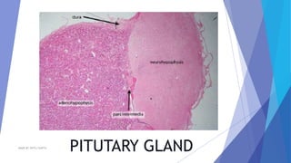
- 2. ANATOMY AND POSITION The pituitary gland is a pea-shaped structure that measures 1–1.5 cm (0.5 in.) in diameter It is suspended near to the 3rd ventricle lies in the hypophyseal fossa of the sella turcica of the sphenoid bone. It attaches to the hypothalamus by a stalk, the infundibulum has two anatomically and functionally separate portions: the anterior pituitary and the posterior pituitary. MADE BY SHITIJ GUPTA
- 3. ADENOHYPOPHYSIS The anterior pituitary (anterior lobe), also called the adenohypophysis, accounts for about 75% of the total weight of the gland and is composed of epithelial tissue. The anterior pituitary consists of two parts in an adult: 1. The pars distalis is the larger portion 2. the pars tuberalis forms a sheath around the infundibulum. secretes hormones that regulate a wide range of bodily activities, from growth to reproduction. Release of anterior pituitary hormones is stimulated by releasing hormones and suppressed by inhibiting hormones from the hypothalamus. Thus, the hypothalamic hormones are an important link between the nervous and endocrine systems MADE BY SHITIJ GUPTA
- 4. MADE BY SHITIJ GUPTA
- 5. Somatotrophs secrete human growth hormone (hGH), also known as somatotropin Thyrotrophs secrete thyroid-stimulating hormone (TSH), also known as thyrotropin. TSH controls the secretions and other activities of the thyroid gland. Gonadotrophs secrete two gonadotropins: follicle-stimulating hormone (FSH) and luteinizing hormone (LH). Lactotrophs secrete prolactin (PRL) Corticotrophs secrete adrenocorticotropic hormone (ACTH), also known as corticotropin , which stimulates the adrenal cortex to secrete glucocorticoids such as cortisol. Some corticotrophs, remnants of the pars intermedia, also secrete melanocyte- stimulating hormone (MSH). Types of cells in adenohypophysis MADE BY SHITIJ GUPTA
- 6. HYPOTHALAMIC RELATION WITH ADENOHYPOPHYSIS The function of adenohypophysis is not regulated by direct innervation but instead by vascular connection with hypothalamus. The arterial blood reaching the hypothalamus enters the specilaised region known as MEDIAN EMINENCE . Where vessel into network of primary arteries And enters the hypophyseal portal system MADE BY SHITIJ GUPTA
- 7. HYPOPHSEAL PORTAL SYSTEM Hypothalamic hormones that release or inhibit anterior pituitary hormones reach the anterior pituitary through a portal system. In the hypophyseal portal system ,blood flows from capillaries in the hypothalamus into portal veins that carry blood to capillaries of the anterior pituitary The superior hypophyseal arteries, branches of the internal carotid arteries, bring blood into the hypothalamus . At the junction of the median eminence of the hypothalamus and the infundibulum, these arteries divide into a capillary network called the primary plexus of the hypophyseal portal system. From the primary plexus, blood drains into the hypophyseal portal veins that pass down the outside of the infundibulum. In the anterior pituitary, the hypophyseal portal veins divide again and form another capillary network called the secondary plexus of the hypophyseal portal system. Its major function is the transport and exchange of hormones to allow a fast communication between both glands MADE BY SHITIJ GUPTA
- 8. MADE BY SHITIJ GUPTA
- 9. HYPOTHALAMIC RELATION WITH NEUROHYPOPHYSIS NEURAL CONNECTIONS BETWEEN THE HYPOTHALAMUS AND THE POSTERIOR LOBE OF PITUATARY ARE MAINTAINED DURING DEVELOPMENT. THE POSTERIOR PITUATARY CONTAINS AXONS FROM END TERMINALS OF NEURON THAT HAVE THEIR CELL BODIES WITHIN THE HYPOTHALAMUS. THESE NEURONS TERMINATE CLOSE TO NUMEROUS CAPILLARIES LOCATED THROUGHOUT THE POSTERIOR PITUATARY, THE CELL BODIES OF THESE NEURONS ARE FOUND WITHIN TWO DISTINCT AREAS OF THE HYPOTHALAMUS, THE SUPRAOPTIC NUCLEUS AND THE PARAVENTRICULAR NUCLEUS MADE BY SHITIJ GUPTA
- 10. NEUROHYPOPHYSIS The posterior pituitary (posterior lobe), also called the neurohypophysis, is composed of neural tissue. It also consists of two parts: 1. the pars nervosa 2. the larger bulbar portion 3. the infundibulum. Pituicytes are similar to astrocytes, another type of glial cell. Their main role is to assist in the storage and release of hormones of the posterior pituitary. Pituicytes surround axonal endings and regulate hormone secretion by releasing their processes from these endings Herring bodies or neurosecretory bodies are structures found in the posterior pituitary. They represent the terminal end of the axons from the hypothalamus, and hormones are temporarily stored in these locations. They are neurosecretory terminals. MADE BY SHITIJ GUPTA
- 11. MADE BY SHITIJ GUPTA
- 12. INTERMEDIATE LOBE A third region of the pituitary gland called the pars intermedia atrophies during human foetal development and ceases to exist as a separate lobe in adults However, some of its cells migrate into adjacent parts of the anterior pituitary, where they persist. MADE BY SHITIJ GUPTA
- 13. DEVELOPMENT OF HYPOPHYSIS MADE BY SHITIJ GUPTA
- 14. 0verview of functioning of pituitary gland HYPOPHYSEAL CEREBRI CONSIDERED AS MASTER OF ENDOCRINE ORCHESTRA BUT UNDER THE CONTROL OF HYPOTHALAMUS MADE BY SHITIJ GUPTA
- 15. HYPOTHALAMIC CONTROL OF PITUATRY GLAND MADE BY SHITIJ GUPTA
- 16. HORMONES OF ADENOHYPOPHYSIS MADE BY SHITIJ GUPTA
- 17. Growth hormone or Somatotropin It is a peptide hormone. The overall effect of growth hormone is to promote tissue growth .In this regard it is considered as ANABOLIC HORMONE. While many effects of GH is similar to insulin ,some are exactly opposite to insulin. GH is therefore said to have both insulin like effects and anti-insulin or “diabetogenic” effects. 1. ANTI INSULIN EFFECT- Growth hormone is often said to have anti-insulin activity, because it supresses the abilities of insulin to stimulate uptake of glucose in peripheral tissues and enhance glucose synthesis in the liver. Stimulates the breakdown of fat stores. 2. INSULIN –LIKE EFFECTS- It directly stimulates the uptake of amino acids from blood to muscle cells and also stimulates protein synthesis in muscle , GH stimulates liver protein synthesis. actions of the growth hormone it is necessary to divide its effects into two groups: 1. Direct effect: the growth hormone binds to receptors on target cells. Fat cells (adipocytes), for example, have growth hormone receptors. So, the growth hormone causes fat cells to break down into triglycerides and suppresses their ability to take up and accumulate circulating lipids. 2. Indirect effect: the growth hormone causes secretion of IGF-1, an insulin-like growth factor hormone. The liver and other tissues secrete IGF-1 in response to growth hormone. Growth effects of the growth hormone are mostly related to the action of IGF-I. MADE BY SHITIJ GUPTA
- 18. Growth hormone MADE BY SHITIJ GUPTA
- 19. MADE BY SHITIJ GUPTA
- 20. Chondrocytes of bone Other organ and tissues MUSCLE LIVER ADIPOSE Amino acid uptake Protein synthesis Glucose uptake Glucose uptake Gluconeogenesis Increased muscle mass Decreased adiposity Somatomedin production SOMATOMEDINS LipolysisProtein synthesis Collagen synthesis Protein synthesis Cell proliferation IGF-IIIGF-I Protein synthesis RNA synthesis DNA synthesis Cell size & number INCREASED LINEAR GROWTH INCREASED TISSUE GROWTH AND ORGAN SIZE MADE BY SHITIJ GUPTA
- 21. EFFECTS OF SOMATOMEDINS In response to somatotropin liver tissues secretes somatomedins(IGF-I AND IGF-II) EFFECTS OF IGF-1 EFFECTS OF IGF-II INVOLVES IN INCREASED LINAER GROWTH. INVOLVES IN INCREASED TISSUE GROWTH AND ORGAN SIZE. IT STIMULATES SKELETAL GROWTH BY INCREASING THE FORMATION OF CARTILAGE IN EPIPHYSEAL PLATES. THE CARTILAGE EVENTUALLY BE REPLACED BY BONE MINERAL AND HENCE BONE GROWS. IGF-I PROMOTES BONE GROWTH BY STIMULATING CHONDROCYTES (AS IT INCRESAES COLLAGEN SYNTHESIS). IT STIMULATES PROTEIN ,RNA AND DNA SYNTHESIS THEREFORE IT IS ABLE TO ELICIT A GENERAL ANABOLIC RESPONSE IN A VARIETY OF DIFFERENT ORGANS .THIS RESULTS IN GENERALISED TISSUE GROWTH. MADE BY SHITIJ GUPTA
- 22. FACTORS AFFECTING SECRETION OF GH STIMULATORS 1. GH-RH 2. DEEP SLEEP 3. HYPOGLYCEMIA 4. STRESS – PHYSICAL TRAUMA , INFECTION , PSYCOLOGICAL STRESS 5. AMINO ACIDS SUCH AS ARGININE INHIBITORS 1. R.E.M. SLEEP(RAPID EYE MOVEMENT SLEEP) 2. HYPERGLYCEMIA MADE BY SHITIJ GUPTA
- 23. TSH OR THYROTROPIN This hormone is synthesised by the anterior pituitary and its release is stimulated by TRH from the hypothalamus. It stimulates growth and activity of the thyroid gland, which secretes the hormones thyroxine (T4) and triiodothyronine (T3). Release is lowest in the early evening and highest during the night. Secretion is regulated by a negative feedback mechanism . When the blood level of thyroid hormones is high, secretion of TSH is reduced, and vice versa. MADE BY SHITIJ GUPTA
- 24. MADE BY SHITIJ GUPTA
- 25. GONADOTROPINS After puberty two gonadotrophins (sex hormones) are secreted by the anterior pituitary in response to luteinising hormone releasing hormone (LHRH), also known as gonadotrophin releasing hormone (GnRH). In both males and females these are: • follicle stimulating hormone (FSH) • luteinising hormone (LH). In both sexes. FSH stimulates production of gametes (ova or spermatozoa). In females. LH and FSH are involved in secretion of the hormones oestrogen and progesterone during the menstrual cycle . As the levels of oestrogen and progesterone rise secretion of LH and FSH is suppressed. In males. LH, also called interstitial cell stimulating hormone (ICSH) stimulates the interstitial cells of the testes to secrete the hormone testosterone MADE BY SHITIJ GUPTA
- 26. MADE BY SHITIJ GUPTA
- 27. MADE BY SHITIJ GUPTA
- 28. PROLACTIN MADE BY SHITIJ GUPTA
- 29. CORTICOTROPIN OR ACTH Corticotrophs secrete mainly adrenocorticotropic hormone(ACTH). ACTH controls the production and secretion of cortisol and other glucocorticoids by the cortex (outer portion) of the adrenal glands. Corticotrophin-releasing hormone (CRH) from the hypothalamus stimulates secretion of ACTH by corticotrophs. Stress-related stimuli, such as low blood glucose or physical trauma, and interleukin-1, a substance produced by macrophages, also stimulate release of ACTH. Glucocorticoids inhibit CRH and ACTH release via negative feedback. MADE BY SHITIJ GUPTA
- 30. MADE BY SHITIJ GUPTA
- 31. HORMONES OF NEUROHYPOPHYSIS MADE BY SHITIJ GUPTA
- 32. OXYTOCIN During and after delivery of a baby, oxytocin affects two target tissues: the mother’s 1. uterus 2. breasts. During delivery, stretching of the cervix of the uterus stimulates the release of oxytocin which, in turn, enhances contraction of smooth muscle cells in the wall of the uterus after delivery, it stimulates milk ejection (“let-down”) from the mammary glands in response to the mechanical stimulus provided by a suckling infant. The function of oxytocin in males and in non-pregnant females is not clear. Experiments with animals have suggested that it has actions within the brain that foster parental caretaking behaviour toward young offspring. It may also be responsible, in part, for the feelings of sexual pleasure during and after intercourse. MADE BY SHITIJ GUPTA
- 33. MADE BY SHITIJ GUPTA
- 34. CLINICAL RELATIONSHIP BETWEEN OXYTOCIN AND CHILDBIRTH Years before oxytocin was discovered, it was common practice in midwifery to let a first-born twin nurse at the mother’s breast to speed the birth of the second child. Now we know why this practice is helpful—it stimulates the release of oxytocin. Even after a single birth, nursing promotes expulsion of the placenta (afterbirth) and helps the uterus regain its smaller size. Synthetic oxytocin (Pitocin) often is given to induce labor or to increase uterine tone and control haemorrhage just after giving birth. MADE BY SHITIJ GUPTA
- 35. ANTI-DIURETIC HORMONE OR VASSOPRESSIN It is a hormone which decreases urine. ADH causes the kidneys to return more water to the blood, thus decreasing urine volume. In the absence of ADH, urine output increases more than tenfold, from the normal 1 to 2 litres to about 20 litres a day. Drinking alcohol often causes frequent and copious urination because alcohol inhibits secretion of ADH. ADH also decreases the water lost through sweating and causes constriction of arterioles, which increases blood pressure. This hormone’s other name, vasopressin, reflects this effect on blood pressure. The amount of ADH secreted varies with blood osmotic pressure and blood volume MADE BY SHITIJ GUPTA
- 36. MADE BY SHITIJ GUPTA
- 37. HORMONES OF PARS INTERMEDIA MADE BY SHITIJ GUPTA
- 38. MELANOCYTE STIMULATING HORMONE (MSH) Melanocyte-stimulating hormone (MSH) increases skin pigmentation in amphibians by stimulating the dispersion of melanin granules in melanocytes. Its exact role in humans is unknown, but the presence of MSH receptors in the brain suggests it may influence brain activity. There is little circulating MSH in humans. However, continued administration of MSH for several days does produce a darkening of the skin. Excessive levels of corticotropin-releasinghormone (CRH) can stimulate MSH release dopamine inhibits MSH release. MADE BY SHITIJ GUPTA