
Empfohlen
Weitere ähnliche Inhalte
Ähnlich wie Cellular Innate Immune Responses Triggered by PRRs
Ähnlich wie Cellular Innate Immune Responses Triggered by PRRs (20)
Mehr von ShafqatJaffer786
Mehr von ShafqatJaffer786 (20)
Kürzlich hochgeladen
Kürzlich hochgeladen (20)
Cellular Innate Immune Responses Triggered by PRRs
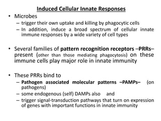
- 1. Induced Cellular Innate Responses • Microbes – trigger their own uptake and killing by phagocytic cells – In addition, induce a broad spectrum of cellular innate immune responses by a wide variety of cell types • Several families of pattern recognition receptors –PRRs– present (other than those mediating phagocytosis) on these immune cells play major role in innate immunity • These PRRs bind to – Pathogen associated molecular patterns –PAMPs– (on pathogens) – some endogenous (self) DAMPs also and – trigger signal-transduction pathways that turn on expression of genes with important functions in innate immunity
- 2. • Proteins encoded by these genes are antimicrobial molecules such as – antimicrobial peptides and interferons, – chemokines and cytokines • These proteins recruit and activate – other cells, – enzymes such as iNOS that generate antimicrobial molecules, and – proinflammatory mediators (i.e., components that promote inflammation).
- 3. • Some PRRs are expressed on the plasma membrane • Other PRRs are actually found inside our cells, (either in endosomes/ lysosomes or in the cytosol). • This ensures that the cell can recognize PAMPs on both extracellular and intracellular pathogens. • DAMPs released by cell and tissue damage also can be recognized by both cell surface and intracellular PRRs.
- 4. • Many cell types in the body express these PRRs: – all types of myeloid white blood cells (monocytes, macrophages, neutrophils, eosinophils, mast cells, basophils, dendritic cells) and – subsets of the three types of lymphocytes (B cells, T cells, and NK cells) – the skin, mucosal and glandular epithelial cells, – vascular endothelial cells that line the blood vessels, and – fibroblasts and stromal support cells in various tissues.
- 5. Cell wall components of Gram-negative and Gram-positive bacteria.
- 6. • There are four main families of mammalian PRRs that activate signaling pathways leading to protective responses. 1. Toll-like receptors (TLRs) • the first family of PRRs to be discovered; • best-characterized in terms of their structure, how they bind PAMPs and activate cells, and the extensive and varied set of innate immune responses that they induce. • recognize many types of pathogen molecules • As of 2011, 13 TLRs that function as PRRs have been identified in mice and humans • TLRs 1-10 are conserved between mice and humans; TLR10 is not functional in mice, TLRs 11-13 are expressed in mice but not in humans.
- 7. • These TLRs can detect a wide variety of PAMPs from bacteria, viruses, fungi, and parasites, as well as DAMPs from damaged cells and tissues • Each TLR has a distinct repertoire of specificities for conserved PAMPs; the TLRs and some of their known PAMP ligands are listed in Table 5-4. • TLRs do not promote phagocytosis
- 9. • TLRs are membrane spanning proteins that share a common structural element in their extracellular region called leucine-rich repeats (LRRs); • Multiple LRRs make up the horseshoe-shaped extracellular ligand-binding domain of the TLR polypeptide chain (Figure 5-11a).
- 10. FIGURE 5-11 Toll-like receptor (TLR) structure - Structure of a TLR polypeptide chain. Each TLR polypeptide chain is made up of a ligand-binding exterior domain that contains many leucine-rich repeats (LRRs, repeating segments of 24-29 amino acids containing the sequence LxxLxLxx, where L is leucine and x is any amino acid), a membrane-spanning domain (blue), and an interior Toll/IL-1R (TIR) domain, which interacts with the TIR domains of other members of the TLR signal-transduction pathway. Two such polypeptide chains pair to form Toll/IL-1R (TLR) dimers, the form that binds ligands.
- 11. Cellular location of TLRs. TLRs that interact with extracellular ligands reside in the plasma membrane; TLRs that bind ligands generated from endocytosed microbes are localized to endosomes and/or lysosomes. Upon ligand binding, the TLR4/4 dimer moves from the plasma membrane to the endosomal/lysosomal compartment, where it can activate different signaling components.
- 12. 2. C-Type Lectin Receptors – second family of cell surface PRRs – activate innate and inflammatory responses – CLRs are plasma membrane receptors expressed variably on monocytes, macrophages, dendritic cells, neutrophils, B cells, and T-cell subsets. – CLRs bind carbohydrates on the surfaces of extracellular pathogens; generally recognize carbohydrate components of fungi, mycobacteria, viruses, parasites, and some allergens (peanut and dust mite proteins). – Humans have at least 15 CLRs that function as PRRs – CLRs have a variety of functions: • some CLRs function as phagocytic receptors (see Table 5-3), and • all CLRs trigger signaling pathways that activate transcription factors that induce effector gene expression.
- 14. 3. Retinoic Acid-Inducible Gene-I-Like Receptors (RLRs) – are soluble PRRs that – reside in the cytosol of many cell types – play critical roles as sensors of viral infection – bind viral RNA in the cytosol of infected cells
- 15. 4. Nod-Like Receptors – Final family of PRRs is the NLRs. – large family of cytosolic proteins – activated by a variety of intracellular PAMPs and substances that alert cells to damage or danger (DAMPs and other harmful substances). – Play major roles in activating beneficial innate immune and inflammatory responses; – some NLRs also trigger inflammation that causes extensive tissue damage and disease.
- 16. • The PRR-activated signaling pathways – induce the transcription of genes that encode an arsenal of proteins that help us to mount protective responses. – Some of the induced proteins are antimicrobial and directly combat pathogens, – While others serve key roles in activating and enhancing innate and adaptive immune responses. – Some of the most common proteins and peptides that are secreted by cells following PAMP activation of PRRs and that contribute to innate and inflammatory responses are listed in Table 5-5. PRR signaling induce Expression of innate immunity proteins
- 17. Antimicrobial Peptides • Defensins and cathelicidins – are important in barrier protection, such as on the skin and the epithelial layers connected to the body’s openings (see Table 5-2). – Some cells and tissues constitutively (i.e., continually, without activation) express these peptides. – For example, α-defensins and some β-defensins are constitutively expressed in human intestinal Paneth epithelial cells – some defensins and the cathelicidin LL-37 are constitutively synthesized and packaged in the granules of neutrophils, ready to kill phagocytosed bacteria, fungi, viruses, and protozoan parasites.
- 18. • In histology, an intestinal gland (also crypt of Lieberkühn and intestinal crypt) is a gland found in the intestinal epithelium lining of the small intestine and large intestine (colon). The glands and intestinal villi are covered by epithelium, which contains multiple types of cells: enterocytes (absorbing water and electrolytes), goblet cells (secreting mucus), enteroendocrine cells (secreting hormones), cup cells, tuft cells, and at the base of the gland, Paneth cells (secreting anti-microbial peptides) and stem cells.
- 19. – In some other cell types, the expression of these antimicrobial peptides is induced or enhanced by signaling through PRRs, in particular TLRs and the NLRs. • such as mucosal and glandular epithelial cells, skin keratinocytes, and NK cells – Macrophages do not produce these antimicrobial peptides following PRR activation; there is an indirect pathway by which microbes induce cathelicidin in macrophages, which then can help the macrophages kill the pathogens.
- 21. Type I Interferons ( IFN-α,β) • Another major class of antimicrobial proteins transcriptionally induced directly by PRRs • Their production generally activated by those cell surface TLRs and intracellular TLRs, RLRs, and NLRs that recognize viral nucleic acids and other components and activate the transcription factors. • One particular type of dendritic cell, called the plasmacytoid dendritic cell (pDC) because of its shape, is a particularly effective producer of Type I IFNs. • key roles in controlling viral infections; other immune- related activities: activate NK cells and regulate activities of macrophages and T cells.
- 22. • Plasmacytoid dendritic cells (pDCs) are innate immune cells that circulate in the blood and are found in peripheral lymphoid organs. • As components of the innate immune system, these cells express intracellular Toll-like receptors 7 and 9 which detect ssRNA and unmethylated CpG DNA sequences, respectively. • Upon stimulation and subsequent activation, these cells produce large amounts (up to 1,000 times more than other cell type) of type I interferon (mainly IFN-α(alpha) and IFN- β (beta)), which are critical pleiotropic anti-viral compounds mediating a wide range of effects. • The number of circulating pDCs are found to be decreased during chronic HIV infection as well as HCV (Hepatitis C virus) infection.
- 23. Cytokines • Several key cytokines are among the proteins transcriptionally induced by PRR activation— not directly antimicrobial —which activate and regulate a wide variety of cells and tissues involved in innate, inflammatory, and adaptive responses. • Cytokines function as the protein hormones of the immune system, produced in response to stimuli and acting on a variety of cellular targets. • Three of the most important cytokines are IL-1, IL-6, and TNF- α, the major proinflammatory cytokines that act locally on blood vessels and other cells to increase vascular permeability and help recruit and activate cells at sites of infection; they also have systemic effects. • IL-1, IL-6, and GM-CSF also feed back on bone marrow hematopoiesis to enhance production of neutrophils and other myeloid cells that will contribute to pathogen clearance.
- 24. • Cytokines are a broad and loose category of small proteins (~5–20 kDa) that are important in cell signaling. • Their release has an effect on the behavior of cells around them. It can be said that cytokines are involved in autocrine signaling, paracrine signaling and endocrine signaling as immunomodulating agent • may include chemokines, interferons, interleukins, lymphokines, and tumour necrosis factors but generally not hormones or growth factors (despite some overlap in the terminology). • Cytokines are produced by a broad range of cells, including immune cells like macrophages, B lymphocytes, T lymphocytes and mast cells, as well as endothelial cells, fibroblasts, and various stromal cells; a given cytokine may be produced by more than one type of cell. • They act through receptors, and are especially important in the immune system; cytokines modulate the balance between humoral and cell- based immune responses, and they regulate the maturation, growth, and responsiveness of particular cell populations. • Some cytokines enhance or inhibit the action of other cytokines in complex ways.
- 25. • Interleukins are a group of cytokines (secreted proteins and signal molecules) that were first seen to be expressed by white blood cells (leukocytes). • The function of the immune system depends in a large part on interleukins, and rare deficiencies of a number of them have been described, all featuring autoimmune diseases or immune deficiency. • The majority of interleukins are synthesized by helper CD4 T lymphocytes, as well as through monocytes, macrophages, and endothelial cells. They promote the development and differentiation of T and B lymphocytes, and hematopoietic cells. • Tumor necrosis factor (TNF, tumor necrosis factor alpha, TNFα, cachexin, or cachectin) is a cell signaling protein (cytokine) involved in systemic inflammation and is one of the cytokines that make up the acute phase reaction. It is produced chiefly by activated macrophages, although it can be produced by many other cell types such as CD4+ lymphocytes, NK cells, neutrophils, mast cells, eosinophils, and neurons.[5] • The primary role of TNF is in the regulation of immune cells. • Granulocyte-macrophage colony-stimulating factor (GM-CSF), also known as colony-stimulating factor 2 (CSF2), is a monomeric glycoprotein secreted by macrophages, T cells, mast cells, natural killer cells, endothelial cells and fibroblasts that functions as a cytokine.
- 26. • some TLRs in monocytes, macrophages, and dendritic cells after activation also induce production of IL-12 and IL-18; – these cytokines play key roles in inducing naïve helper T cells to become TH1 cells, in particular by inducing production of IFN-γ. – The hallmark cytokine of TH1 cells is IFN-γ, which stimulates cell- mediated immunity and is an important macrophage-activating cytokine. Hence IL-12 and IL-18 are also considered proinflammatory. • IL-10 is another important cytokine specifically induced by some TLRs in macrophages, dendritic cells, other myeloid cells, and subsets of T, B, and NK cells. – IL-10 is anti-inflammatory, in that it inhibits macrophage activation and the production of proinflammatory cytokines by other myeloid cells. IL-10 levels increase over time and contribute to controlling the extent of inflammation-caused tissue damage.
- 27. Chemokines • These small protein chemoattractants (agents that induce cells to move toward higher concentrations of the agent) recruit cells into, within, and out of tissues. • Some chemokines are responsible for constitutive (homeostatic) migration of white blood cells throughout the body. • Other chemokines, produced in response to PRR activation, have key roles in the early stages of immune and inflammatory responses – they attract cells that contribute both to clearing the infection or damage and to amplifying the response.
- 28. – The first chemokine to be cloned, IL-8 (also called CXCL8), is produced in response to activation by PAMPs, DAMPs, or some cytokines – It leads to activation of a variety of cells at sites of infection or tissue damage, including macrophages, dendritic cells, epithelial cells, and vascular endothelial cells. – Its key roles occurs in the initial stages of infection or tissue damage; it serves as a chemoattractant for neutrophils, recruiting them to sites of infection.
- 29. – Other chemokines are specifically induced by PRR activation of epithelial cells in certain mucosal tissues and serve to recruit cells specifically to those sites, where they generate immune responses appropriate for clearing the invading pathogen. – For example, B cells are recruited to the lamina propria, the lymphocyte-rich tissue under the intestinal epithelium, by two chemokines, CCL28 and CCL20, produced by intestinal epithelial cells activated by PAMP binding to their TLRs. – These activated epithelial cells (as well as local dendritic cells activated by PAMPs) also produce cytokines that stimulate the B cells to produce IgA, the class of antibodies most effective in protecting against mucosal infections .
- 30. ……….continued
- 31. ……continued
- 32. Enzymes: iNOS and COX2 • Other genes activated in many cell types by PRR activated signaling pathways are those for two enzymes that contribute importantly to the generation of antimicrobial and proinflammatory mediators: inducible nitric oxide synthase (iNOS) and cyclooxygenase 2 (COX2). • The iNOS enzyme catalyzes an important step in the formation of nitric oxide, which kills phagocytosed microbes (see Figure 5- 8). • COX2, whose synthesis is induced by PRR activation in monocytes, macrophages, neutrophils, and mast cells, is key to converting the lipid intermediate arachidonic acid to prostaglandins, potent proinflammatory mediators.
- 33. Figure 5-8 Generation of antimicrobial reactive oxygen and nitrogen species. In the cytoplasm of neutrophils, macrophages, and dendritic cells, several enzymes, including phagosome NADPH oxidase, transform molecular oxygen into highly reactive oxygen species (ROS) that have antimicrobial activity. One of the products of this pathway, superoxide anion, can interact with a reactive nitrogen species (RNS), generated by inducible nitric oxide synthase (iNOS) to produce peroxynitrite, another RNS. NO can also undergo oxidation to generate the RNS nitrogen dioxide.