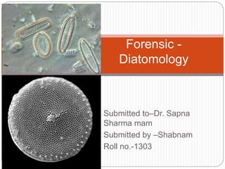
SlideShare ist ein Scribd-Unternehmen logo
1 von 46
Submitted to–Dr. Sapna
Sharma mam
Submitted by –Shabnam
Roll no.-1303
Forensic -
Diatomology
Content
1. Introduction
2. Nature
3. Location
4. Structure
5. Classification
6. Life-cycle of diatoms
6. Method of identification
7. Comparison
8. Applications
9. Reference
Introduction
 Diatoms are eukaryotic photosynthetic organisms
referred to as algae with a length/diameter of between 2
and 500 microns.
 They have a transparent cell wall (frustule) made of
silicon dioxide (SiO2), which is itself hydrated with a little
amount of water.
 Diatoms are the most diverse protists on earth and one
of the Heterokont algae.
 Estimates of the number of diatom species
approximately range from 20,000 - 2 million, out of which
92 genera and about 569 species are reported from
India.
 Scientists are discovering new species every year.
Introduction
Nature
 Diatoms belong to Bacillariophyceae which is mainly exist
in two forms, either bilateral or radial in symmetry.
 Depending on the mode of nutrition they may be
photosynthetic autotrophs or photosynthetic symbionts or
heterotrophs.
 The cells are surrounded by a rigid cell wall, called
frustule, consisting of upper epitheca and lower hypotheca;
arranged in the form of a box with its lid.
 The cell wall is composed of pectic substances
impregnated with high amount of siliceous substance.
 The wall may have secondary structures like spines,
bristles etc.
 The photosynthetic pigments are chlorophyll a, chlorophyll
c along with xanthophylls like fucoxanthin, diatoxanthin
and diadinoxanthin.
 Reserve food is oil, volutin and crysolaminarin.
Location
 Diatoms live in water, or even in moist habitats or soils.
Some diatoms live as free-floating cells in the plankton
of ponds, lakes and oceans.
 Such as diatoms are found in fresh water (Denticula
tenuis, Navicula pupula, Meridion circulare, Cymbella
ventricosa, Melosira variens, Amorpha ovalis etc.), sea
water (Corethron, Biddulphia, Sceletonema, Fragilaria,
Tropido- nensis etc.) and soil (Pinnularia, Navicula,
Frustulia etc.).
 The terrestrial species (Amorpha, Navicula, Pinnularia
etc.) are able to withstand desiccation for a long period.
Some diatoms (Gomphonima, Cymbella etc.) can grow
as epiphyte on other algae (Enteromorpha, Cladophora
etc.) and higher plant.
 Licmophora, a member of diatom, grows endozoically.
1.or 2. diatom cells attach to boulders, 3.growing on sediments
& pebbeles, 4.Growing around submerged tree branches, 5.Found
on submerged stem of phragmites, 6.Diatoms inhabiting the soil.
Location
 As they produce long-chain fatty acids and an
important source of these energy rich molecules that
are food for the entire food web, from zooplankton to
aquatic insects to fish to whales.
 Being autotrophic they are restricted to the photic
zone (water depths down to about 200m depending
on clarity) as they trap light to make food itself by
photosynthesis process. Both benthic and planktic
forms exist.
Morphology
 Diatoms are commonly unicellular and free-
living but some members form colonies of
various shapes like filaments, mucilaginous
colonies etc.
 Microscopic cells are of different shapes.
They may be oval, spherical, triangular,
boat- shaped etc.
 Plant bodies are posses either bilateral
(plane of symmetry) or round (radial in
symmetry).
 The wall may have secondary structures like
spines, bristles etc.
 Raphe is an elongated fissure in a valve
which is used by diatoms for movement and
used as diatom identification on the basis of
position and number of raphe.
Cell structure
 The cell consists of cell wall and protoplast. The cells are covered
by a siliceous wall, the frustule.The entire content present inside
the cell wall is the protoplast. The cell membrane encloses a large
central vacuole surrounded by cytoplasm. The cytoplasm contains
single nucleus, mitochondria, golgi bodies and chloroplasts. The
chloroplasts may be of different shapes like stellate, H-shaped,
discoid etc. In some species the chloroplasts contain pyrenoids.
 The photosynthetic pigments are chlorophyll a, c1 and c2, β-
carotene, fucoxanthin, diatoxanthin and diadinoxanthin.The latter
two are present in small quantity.The golden-brown colour of
diatom cells is due to the presence of xanthophylls like
fucoxanthin, diatoxanthin and diadinoxanthin.
 The term diatomin is used for the mixture of chlorophyll and
carotenoids, particularly carotene and several brown xanthophylls
pigments). The reserve food of diatoms is chrysolaminarin and oil
droplets (they do not store in the form of starch).
Frustule
 It consists of two overlapping halves, the theca. The upper
one is epitheca and lower one is hypotheca.
 Both the theca consist of two portions:
 (a) Valve — the upper flattened top and
 (b) Connecting band or cingulum (pl. cingula) — the
incurved region.
 The common region of the connecting bands, where both
the theca remain fitted together, is the girdle. [When the
diatoms are observed from the valve side i.e., valve side is
uppermost, called the valve view, but when viewed from
the connecting band, it is the girdle view].
 Depending on symmetry, the cells are divided into two
orders: Pennales (bilaterally symmetry) and Centrales
(radially symmetry).
CELL STRUCTURE OF DIATOMS
Movement of diatoms
 All diatoms with raphe are motile.
 Strand of mucilage are secreted through the raphe by
which diatom attach to the substratum and glide over
the substratum.
 Mucilage secretions can also be used to form
colonies of various patterns. The gliding movement is
caused by the circulation of cytoplasm within the
raphe by the release of mucilage.
 The rate of movement varies from 02-25 µm/sec. The
locomotion is affected by temperature, light etc.
Movement of diatoms
Locomotion by mucilage.
Classification
 Domain: Eukaryota
 Kingdom: Chromalveolata
 Phylum: Heterokontophyta
 Class: Bacillariophyceae
 Orders: Centrales ,Pennales
Classification
 The classification system developed by Simonsen
(1979) and further developed by Round et al.
(1990) is currently the most commonly accepted.
 The valve face of the diatom frustule is
ornamented with pores (areolae), processes,
spines, hyaline areas and other distinguishing
features.
 It is these skeletal features which are used to
classify and describe diatoms, which is an
advantage in terms of palaeontology since the
same features are used to define extant species
as extinct ones.
Classification
 The Centrales (now called the Biddulphiales) which have
valve striae arranged basically in relation to a point, an
annulus or a central areola and tend to appear radially
symmetrical.
 Diatoms commonly found in the marine plankton may
be divided into the centric diatoms including three sub-
orders based primarily on the shape of the cells, the
polarity and the arrangement of the processes.
 These are the Coscinodiscineae, with a marginal ring of
processes and no polarity to the symmetry, the
Rhizosoleniineae with no marginal ring of processes and
unipolar symmetry, and the Biddulphiineae with no
marginal ring of processes and bipolar symmetry.
Classification
 Pennales (now called Bacillariales) which have valve
striae arranged in relation to a line and tend to appear
bilaterally symmetrical.
 The pennate diatoms are divided into two sub-orders,
the Fragilariineae which do not posses a raphe
(araphid) and the Bacillariineae which posses a raphe.
Difference between diatoms
Pennate diatoms Centric diatoms
A. Bilateral symmetry.
B. Elongated chloroplast.
C. Raphe is present responsible
for movement.
D. Pennate striations are
present.
E. These are generally posses
boat shape.
A. Radial symmetry.
B. Discoid chloroplast, large
vacuole.
C. Raphe is absent.
D. Radiant striations are
present.
E. The shape and size is
generally varies, almost
immobile.
Life-cycle of diatoms
 When a cell divides each new cell takes as its
epitheca a valve of the parent frustule, and within ten
to twenty minutes builds its own hypotheca; this
process may occur between one and eight times per
day by means of vegitative reproduction and
continuous cell division some cells gradually become
reduced in size.
 During sexual reproduction auxospore is produce to
regain their normal size by the restorative process
instead of multiplication.
Vegetative reproduction
 Vegetative reproduction performs with the help of cell division. It takes
place usually at midnight or in the early morning.
 During cell division the protoplast of the cell enlarges slightly, thus the cell
increases in volume and slightly separates both the theca (epitheca and
hypotheca).
 Then the protoplast undergoes mitotic division and gets separated along
the longitudinal axis through the median line. Thus one half of protoplast
remains in epitheca and the other one in hypotheca. One side of the
protoplast thus remains naked. Now both the theca i.e., epitheca and
hypotheca of mother cell behave as epitheca of the daughter cells.
 Thus new silicious valves are deposited towards the naked sides of the
protoplast and always behave as hypotheca of the daughter cells.
Connecting bands are developed between the theca. Later on, the
daughter cells get separated.
 During cell division, both the theca i.e., epitheca and hypotheca of
the mother cell behave as epitheca of the daughter cells. So at the
side where the hypotheca behaves as epitheca, the cell becomes
reduced in size. Thus with continuous cell division some cells
gradually become reduced in size.
Sexual Reproduction
 The pattern of sexual reproduction differs in both orders — Pennales
and Centrales. During this process, auxospore is formed in both the
groups. During cell division, those cells become reduced in size, are
able to regain their normal size through the formation of auxospore, so
it is a “restorative process” rather than multiplication.
 Auxospore formation in Pennales: It takes place through gametic
union, autogamy and parthenogenesis.
 These are of the following types:
 1. Production of one auxospores by two conjugating cells- In this
process two uniting cells come very close to each other and become
covered by a mucilaginous sheath. The diploid nucleus of each cell
undergoes meiosis.
 Out of four nuclei, three degenerate and only one survives. The
surviving nucleus behaves as gamete (n). The gametes come out from
the parent frustules and unite together, to form a zygote (2n).
 After a short period of rest the zygote elongates considerably
and functions as an auxospore. The auxospore projects out from
the parent frustules along with mucilage and elongates in a plane
parallel to the long axis of the parent diatom.
 The auxospore is enclosed in a pectic membrane, the
perizonium. The auxospore then develops new frustule inside
the perizonium. Thus new diatom cell is formed which regains
the normal size. It is found in Cocconis placentula, Surirella
saxonica etc.
 2. Production of Two Auxospores by Two
Conjugating Cells:
 This is a very common process of auxospore formation.
In this process the conjugating cells come very close to
each other and get enclosed by mucilage . The nucleus
(2n) of each cell undergoes meiotic division and forms
four nuclei.
 Out of four nuclei, two degenerate, the rest two survive.
The cytoplasm then divides either equally or unequally
and along with one nucleus they behave as gametes.
Thus two gametes are formed in each cell.
 The pattern of union between the gametes varies from
species to species. Both the gametes of a cell may be
active and fuse with the gametes of other cell, thus two
zygotes are produced in a single cell or out of two, one
becomes active and fertilises with the opposite one and
thus one zygote is produced in each cell.
 The zygotes elongate and function as auxo- spores. The
auxospores develop the perizonium around themselves
and both of them develop new frustules on their outer
sides i.e., inside the perizonium. Thus two diatom cells of
normal size are formed. It is found in Cymbella
lanceolata, Gomphomema parvulum etc.
 3. Production of One Auxospore by One Cell:
 This process of auxospore formation is called Paedogamy
(Pedogamy). In this process the diploid nuclei of a
vegetative cell undergo meiosis and form four haploid
nuclei.
 Out of the four nuclei two partially degenerate. Each of the
rest two along with the cytoplasm and one partially
degenerated nucleus, behaves as gamete. Later on, the
union between the two sister gametes takes place and
forms the zygote.
 The zygote comes out from the parent frustule and
behaves as an auxospore. The auxospore then gets
covered by perizonium and develops wall inside the
perizonium. Thus one diatom cell of normal size is formed.
 4. Production of One Auxospore by Autogamy:
 In this process the diploid nucleus undergoes first meiotic
division. Thus two haploid nuclei are formed. The two
nyclei in the protoplast come side by side, fuse together
and form diploid (2n) nucleus. This is called autogamous
pairing.
 The protoplast along with diploid (2n) nucleus comes out
from the parent frustule and behaves as an auxospore.
 The auxospores are then covered by perizonium. New wall
develops on the auxospore inner to the perizonium.
 Thus a new individual of normal size is developed. This is
found in Amphora normani.
 5. Production of Auxospore by Partheno-
genesis:
 The diatom cells come together and are covered by a
common mucilage envelop . The diploid nucleus undergoes
two sequential mitotic divisions. Meiotic division does not
take place here. One nucleus in each mitotic division
degenerates. Thus only one diploid (2n) nucleus along with
protoplast remains, and comes out from the mother cell and
behaves as an auxospore.
 The auxospore is then covered by perizonium and secretes
new wall around itself. Thus normal size cell is formed.
 In this process the nucleus (2n) of female cell which behaves
as oogonium, undergoes meiosis and forms four nuclei. The
protoplast is also divided into two unequal parts, each
containing two nuclei.
 The lower half is larger and behaves as functional ovum and
the upper smaller one as non-functional ovum. The functional
ovum contains one functional nucleus and one non-functional
nucleus, which gradually degenerates at maturity.
 The male cell (2n) behaves as antheridium, also undergoes
meiosis and forms four nuclei. The protoplast also divides into
two parts. Thus two microgametes are formed. Each of which
contains two nuclei, of which one is functional and other is
non-functional. The microgametes are naked, globular and
non-flagellate.
 After coming out, the male gamete fertilizes the egg and
forms the zygote (2n). Later it functions as an auxospore and
forms new individual of normal size. It is found in
Rhabdonema adriaticum.
 Auxospore Formation in Centrales: It takes place by
autogamy and oogamy:
 1. Auxospore Formation by Autogamy:
 The protoplast of the vegetative cell secretes mucilage
which separates both the theca. The nucleus (2n) then
undergoes meiosis and forms four nuclei. Of the four
nuclei two degenerate and the other two undergo fusion
to form diploid (2n) nucleus again.
 This is called autogamy. The protoplast with 2n nucleus
functions as an auxospore. The auxospore forms fresh
frustule inside the perizonium covering and forms cell of
normal size. It is found in Melosira nummuloides.
 2. Auxospore Formation by Oogamy:
 Oogamy takes place by the fusion of egg and sperm developed
inside the oogonium and antheridium respectively.
 Oogonium:
 Single vegetative cell behaves as an oogonium. The protoplast
of oogonium undergoes meiotic division and forms four nuclei.
Of the four nuclei three degenerate and the remaining one
functions as an egg.
 Antheridium:
 The pattern of development of sperms varies in different
species. In species like Melosira varians the protoplast
undergoes meiotic division and forms four haploid nuclei.
 Each haploid nucleus with some protoplast metamorphoses into
an uniflagellate (tinsel type) sperm. In others the number of
sperms may go up to 8 or even 128.
 Fertilisation:
 After coming out of the antheridium only one sperm enters
inside the oogonium and fertilises the egg. The resultant zygote
undergoes mitotic division but one nucleus degenerates in each
division.
 The remaining nucleus with its protoplast behaves as an
auxospore. The auxospore then develops new wall inside
the perizonium covering and forms new cell of normal
size like the mother. It is also called firstling cell.
 From the above processes of sexual reproduction in both
pennales and centrales, it becomes clear that the sexual
process in diatom does not lead to multiplication but is to
regain the normal size.
Identification
 Diatom ornamentations is important aspect for diatom
identification.Valve surface usually covered with
striations , punctations or raphes which is also a
identification key.
Identification
 In 1942 Incze demonstrated that, during drowning,
diatoms could enter the systemic circulation via the
lungs. Their presence can be demonstrated in such
tissues as liver, brain and bone marrow following acid
digestion of the tissue.
 The use of diatoms as a diagnostic test for drowning is
based upon antemortem or postmortem.
 Before diatoms can be examined, they have to be
cleaned. This involves the removal of cell contents,
pigments, sand, mud or other material likely to interfere
with microscope examination.
 Diatom is identified through extraction methods such as
acid digestion method which is easy to perform and
worldwide accepted, this method includes nitric acid
method and sulphuric acid method.
Identification
 In diatom identification, the control water samples is used
for comparison purpose. Standard diatom samples can be
preserved on slides and can be used as standards for
comparison purpose.
 The morphological features is examined by transmission
and scanning electron microscopes are able to provide a
much more detailed image than light microscopes.
 Transmission Electron Microscopy (TEM): This type of
microscopy is best able to see the finer, delicate details of
the diatom frustule.
 Scanning Electron Microscopy (SEM): SEM is best
suited for visualizing the entire diatom frustule include
internal and external morphological parts.(Kapil verma
2013)
Morphological characteristics
Identification based on photosynthetic
pigment
 Diatoms contain golden- brown chloroplasts, and contain
chlorophylls a and c (never chlorophyll b) which can be
recognised as a colour property.
 Serious diatomists clean the cells with acid and peroxide
so that only the frustules remain, aiding identification.
 However, many useful features can still be recognised in
living material, and images in this guide often compare
living and cleaned cells for comparison.
Achnanthes.
Role of diatoms in ecosystem
Applications
 Diatoms tell us about the health of aquatic systems-
Diatoms are particular about the quality of water in which they
live. For example, species have distinct ranges of pH and
salinity where they will grow. Diatoms also have ranges and
tolerances for other environmental variables, including nutrient
concentration, suspended sediment.
 As a result, diatoms are vital for assessment and monitoring
biotic condition of waters.
 Pest Control - Diatomaceous earth can be ground into a fine
powder that looks and feels very similar to talcum powder. This
powder can be used for pest control.
 The powder is sharp on a microscopic level due to the high
silica content. It damages the outside of an insect, and, if
ingested, ruptures the internal organs.
Applications
 Abrasives - Typically, diatomaceous earth is used to
polish materials that are soft or easily damaged, and is
often used to polish metal. Occasionally, it is also used in
toothpaste.
 Diatomaceous earth is a good abrasive for cleaning skin
and is sometimes used in soaps and other bath products.
 Indicator species - Some species can be used as an
indicator species. Indicator species are used by scientists
to determine if an ecosystem is thriving.
 With diatoms, a scientist takes a sample of water and
examines it under a microscope to see how many of a
certain species of diatom are present.
Applications
 Filtration - A very common use for diatoms is for filtration. The
fine structures of diatom shells trap foreign particles in fluids,
such as dirt, lint, hair and some other microscopic organisms,
particularly water in hot tubs and swimming pools. However, a
vast variety of fluids can be filtered with diatoms, including
different syrups, alcoholic beverages, medicines, solvents and
other chemicals.
 Testing of Microscopic Lenses - Due to the fine markings on
shell, the diatom cells are used to test microscopic lenses.
 Diatomite - After the death of diatom cells the outer coverings
i.e., the silicified walls become accumulated at the bottom of
water. The accumulation may be thicker during favourable
conditions. These deposits are called diatomaceous earth,
diatomite or keiselghur.
Reference
 A Methods Manual for the Collection, Preparation and Analysis of
Diatom Samples by JC Taylor*, WR Harding** and CGM Archibald***.
 www.slideshare.com
 Planktonic and periphytic diatoms as indicators of stress on great
rivers of the United States: Testing water quality and disturbance
models.Amy R. Kiretaa,∗, Euan D. Reaviea,1, Gerald V. Sgrob,2, Ted
R. Angradic,3, David W. Bolgrienc,3, Brian H. Hill c,3, Terri M. Jicha
c,3.
 Presented By: Inchara R 1st Semester Molecular Biology 30/10/2017
Guided By: Dr. N. S. Devaki Course Coordinator Dept. of molecular
Biology DIATOMS.
 Kapil Verma (2013) Role of Diatoms in the World of Forensic Science.

Weitere ähnliche Inhalte

Was ist angesagt?

Role of forensics in wildlife
Role of forensics in wildlifeRole of forensics in wildlife
Role of forensics in wildlife
Sandipan Dhar
 

Was ist angesagt? (20)

Video Spectral Comparator
Video Spectral ComparatorVideo Spectral Comparator
Video Spectral Comparator
 
Wild life forensics
Wild life forensicsWild life forensics
Wild life forensics
 
Role of forensics in wildlife
Role of forensics in wildlifeRole of forensics in wildlife
Role of forensics in wildlife
 
Forensic Botany
Forensic BotanyForensic Botany
Forensic Botany
 
Forensic Palynology
Forensic PalynologyForensic Palynology
Forensic Palynology
 
Forensic Biology - Biological evidences
Forensic Biology - Biological evidencesForensic Biology - Biological evidences
Forensic Biology - Biological evidences
 
TOOL MARKS
TOOL MARKSTOOL MARKS
TOOL MARKS
 
Improvised Firearms
Improvised FirearmsImprovised Firearms
Improvised Firearms
 
Paints as evidence
Paints as evidencePaints as evidence
Paints as evidence
 
restoration of toolmarks
restoration of toolmarksrestoration of toolmarks
restoration of toolmarks
 
Antropological Comparision Between Human and Non-human Skeleton Remains
Antropological Comparision Between Human and Non-human Skeleton RemainsAntropological Comparision Between Human and Non-human Skeleton Remains
Antropological Comparision Between Human and Non-human Skeleton Remains
 
Forensic anthropology case study
Forensic anthropology case studyForensic anthropology case study
Forensic anthropology case study
 
Comparison microscope
Comparison microscopeComparison microscope
Comparison microscope
 
Introduction to Forensic Biology
Introduction to Forensic BiologyIntroduction to Forensic Biology
Introduction to Forensic Biology
 
Gun shot residue
Gun shot residueGun shot residue
Gun shot residue
 
POROSCOPY AND EDGEOSCOPY
POROSCOPY AND EDGEOSCOPYPOROSCOPY AND EDGEOSCOPY
POROSCOPY AND EDGEOSCOPY
 
Forensic Anthrpology
Forensic AnthrpologyForensic Anthrpology
Forensic Anthrpology
 
Fingerprint - Everything You Need To Know About Fingerprints
Fingerprint - Everything You Need To Know About FingerprintsFingerprint - Everything You Need To Know About Fingerprints
Fingerprint - Everything You Need To Know About Fingerprints
 
Forensic Fiber Analysis
Forensic Fiber AnalysisForensic Fiber Analysis
Forensic Fiber Analysis
 
Automated Fingerprint Identification Systems
Automated Fingerprint Identification SystemsAutomated Fingerprint Identification Systems
Automated Fingerprint Identification Systems
 

Ähnlich wie Forensic diatomology

Tawqir Bashir (Lecture 11)_MB.pdf
Tawqir Bashir (Lecture 11)_MB.pdfTawqir Bashir (Lecture 11)_MB.pdf
Tawqir Bashir (Lecture 11)_MB.pdf
Shahid Shakeel
 

Ähnlich wie Forensic diatomology (20)

Cryptogamic_Botany_ppt SOA.pptx
Cryptogamic_Botany_ppt SOA.pptxCryptogamic_Botany_ppt SOA.pptx
Cryptogamic_Botany_ppt SOA.pptx
 
Diatoms
DiatomsDiatoms
Diatoms
 
Chytridiomycetes
ChytridiomycetesChytridiomycetes
Chytridiomycetes
 
Oedogonium
OedogoniumOedogonium
Oedogonium
 
Algae.pptx
Algae.pptxAlgae.pptx
Algae.pptx
 
Phyccology
PhyccologyPhyccology
Phyccology
 
General characteristics of algae
General characteristics of algaeGeneral characteristics of algae
General characteristics of algae
 
Algae
AlgaeAlgae
Algae
 
Cyanobacteria lecture
Cyanobacteria lectureCyanobacteria lecture
Cyanobacteria lecture
 
Tawqir Bashir (Lecture 11)_MB.pdf
Tawqir Bashir (Lecture 11)_MB.pdfTawqir Bashir (Lecture 11)_MB.pdf
Tawqir Bashir (Lecture 11)_MB.pdf
 
Animal like protists
Animal like protistsAnimal like protists
Animal like protists
 
Diatom - Overview
Diatom - OverviewDiatom - Overview
Diatom - Overview
 
ALGAE Advance Microbiology.pptx
ALGAE Advance Microbiology.pptxALGAE Advance Microbiology.pptx
ALGAE Advance Microbiology.pptx
 
Ambe 101 @ lec 3
Ambe 101 @ lec 3Ambe 101 @ lec 3
Ambe 101 @ lec 3
 
Vegetative Structure - Chlorophyceae.pptx
Vegetative Structure - Chlorophyceae.pptxVegetative Structure - Chlorophyceae.pptx
Vegetative Structure - Chlorophyceae.pptx
 
Class xi ch 2
Class xi ch   2Class xi ch   2
Class xi ch 2
 
Biological Classification_Shobhit Nirwan.pdf
Biological Classification_Shobhit Nirwan.pdfBiological Classification_Shobhit Nirwan.pdf
Biological Classification_Shobhit Nirwan.pdf
 
Biological Classification_Shobhit Nirwan.pdf
Biological Classification_Shobhit Nirwan.pdfBiological Classification_Shobhit Nirwan.pdf
Biological Classification_Shobhit Nirwan.pdf
 
Diatoms, Dinoflagellates, Lichen
Diatoms, Dinoflagellates, LichenDiatoms, Dinoflagellates, Lichen
Diatoms, Dinoflagellates, Lichen
 
Full chapter 8 miller and harley, bsc
Full chapter 8 miller and harley, bscFull chapter 8 miller and harley, bsc
Full chapter 8 miller and harley, bsc
 

Mehr von Shabnamkhan113 (9)

Proof marks of weapons (Forensic Ballistic)
 Proof marks of weapons (Forensic Ballistic)  Proof marks of weapons (Forensic Ballistic)
Proof marks of weapons (Forensic Ballistic)
 
Forensic significance of DNA Profiling (Forensic biology)
 Forensic significance of DNA Profiling (Forensic biology)  Forensic significance of DNA Profiling (Forensic biology)
Forensic significance of DNA Profiling (Forensic biology)
 
Digital forensic an forensic policy approach
Digital forensic an forensic policy approachDigital forensic an forensic policy approach
Digital forensic an forensic policy approach
 
Cultural writing systems and their languages (questioned document)
Cultural writing systems and their languages (questioned document) Cultural writing systems and their languages (questioned document)
Cultural writing systems and their languages (questioned document)
 
computer security and its relationship to computer forensic
 computer security and its relationship to computer forensic computer security and its relationship to computer forensic
computer security and its relationship to computer forensic
 
Hardy – weinberg law
 Hardy – weinberg law Hardy – weinberg law
Hardy – weinberg law
 
Forensic characterization of blood
Forensic characterization of bloodForensic characterization of blood
Forensic characterization of blood
 
Intoduction and brief analysis of arson
Intoduction and brief analysis of arsonIntoduction and brief analysis of arson
Intoduction and brief analysis of arson
 
Infrared spectroscopy
Infrared spectroscopyInfrared spectroscopy
Infrared spectroscopy
 

Kürzlich hochgeladen

Kürzlich hochgeladen (20)

Advanced Views - Calendar View in Odoo 17
Advanced Views - Calendar View in Odoo 17Advanced Views - Calendar View in Odoo 17
Advanced Views - Calendar View in Odoo 17
 
Accessible design: Minimum effort, maximum impact
Accessible design: Minimum effort, maximum impactAccessible design: Minimum effort, maximum impact
Accessible design: Minimum effort, maximum impact
 
A Critique of the Proposed National Education Policy Reform
A Critique of the Proposed National Education Policy ReformA Critique of the Proposed National Education Policy Reform
A Critique of the Proposed National Education Policy Reform
 
Software Engineering Methodologies (overview)
Software Engineering Methodologies (overview)Software Engineering Methodologies (overview)
Software Engineering Methodologies (overview)
 
Unit-IV- Pharma. Marketing Channels.pptx
Unit-IV- Pharma. Marketing Channels.pptxUnit-IV- Pharma. Marketing Channels.pptx
Unit-IV- Pharma. Marketing Channels.pptx
 
Nutritional Needs Presentation - HLTH 104
Nutritional Needs Presentation - HLTH 104Nutritional Needs Presentation - HLTH 104
Nutritional Needs Presentation - HLTH 104
 
Sanyam Choudhary Chemistry practical.pdf
Sanyam Choudhary Chemistry practical.pdfSanyam Choudhary Chemistry practical.pdf
Sanyam Choudhary Chemistry practical.pdf
 
Call Girls in Dwarka Mor Delhi Contact Us 9654467111
Call Girls in Dwarka Mor Delhi Contact Us 9654467111Call Girls in Dwarka Mor Delhi Contact Us 9654467111
Call Girls in Dwarka Mor Delhi Contact Us 9654467111
 
social pharmacy d-pharm 1st year by Pragati K. Mahajan
social pharmacy d-pharm 1st year by Pragati K. Mahajansocial pharmacy d-pharm 1st year by Pragati K. Mahajan
social pharmacy d-pharm 1st year by Pragati K. Mahajan
 
Interactive Powerpoint_How to Master effective communication
Interactive Powerpoint_How to Master effective communicationInteractive Powerpoint_How to Master effective communication
Interactive Powerpoint_How to Master effective communication
 
Introduction to Nonprofit Accounting: The Basics
Introduction to Nonprofit Accounting: The BasicsIntroduction to Nonprofit Accounting: The Basics
Introduction to Nonprofit Accounting: The Basics
 
SOCIAL AND HISTORICAL CONTEXT - LFTVD.pptx
SOCIAL AND HISTORICAL CONTEXT - LFTVD.pptxSOCIAL AND HISTORICAL CONTEXT - LFTVD.pptx
SOCIAL AND HISTORICAL CONTEXT - LFTVD.pptx
 
fourth grading exam for kindergarten in writing
fourth grading exam for kindergarten in writingfourth grading exam for kindergarten in writing
fourth grading exam for kindergarten in writing
 
Código Creativo y Arte de Software | Unidad 1
Código Creativo y Arte de Software | Unidad 1Código Creativo y Arte de Software | Unidad 1
Código Creativo y Arte de Software | Unidad 1
 
Mattingly "AI & Prompt Design: The Basics of Prompt Design"
Mattingly "AI & Prompt Design: The Basics of Prompt Design"Mattingly "AI & Prompt Design: The Basics of Prompt Design"
Mattingly "AI & Prompt Design: The Basics of Prompt Design"
 
Key note speaker Neum_Admir Softic_ENG.pdf
Key note speaker Neum_Admir Softic_ENG.pdfKey note speaker Neum_Admir Softic_ENG.pdf
Key note speaker Neum_Admir Softic_ENG.pdf
 
Class 11th Physics NEET formula sheet pdf
Class 11th Physics NEET formula sheet pdfClass 11th Physics NEET formula sheet pdf
Class 11th Physics NEET formula sheet pdf
 
Student login on Anyboli platform.helpin
Student login on Anyboli platform.helpinStudent login on Anyboli platform.helpin
Student login on Anyboli platform.helpin
 
Mattingly "AI & Prompt Design: Structured Data, Assistants, & RAG"
Mattingly "AI & Prompt Design: Structured Data, Assistants, & RAG"Mattingly "AI & Prompt Design: Structured Data, Assistants, & RAG"
Mattingly "AI & Prompt Design: Structured Data, Assistants, & RAG"
 
9548086042 for call girls in Indira Nagar with room service
9548086042  for call girls in Indira Nagar  with room service9548086042  for call girls in Indira Nagar  with room service
9548086042 for call girls in Indira Nagar with room service
 

Forensic diatomology

  • 1. Submitted to–Dr. Sapna Sharma mam Submitted by –Shabnam Roll no.-1303 Forensic - Diatomology
  • 2. Content 1. Introduction 2. Nature 3. Location 4. Structure 5. Classification 6. Life-cycle of diatoms 6. Method of identification 7. Comparison 8. Applications 9. Reference
  • 3. Introduction  Diatoms are eukaryotic photosynthetic organisms referred to as algae with a length/diameter of between 2 and 500 microns.  They have a transparent cell wall (frustule) made of silicon dioxide (SiO2), which is itself hydrated with a little amount of water.  Diatoms are the most diverse protists on earth and one of the Heterokont algae.  Estimates of the number of diatom species approximately range from 20,000 - 2 million, out of which 92 genera and about 569 species are reported from India.  Scientists are discovering new species every year.
  • 5. Nature  Diatoms belong to Bacillariophyceae which is mainly exist in two forms, either bilateral or radial in symmetry.  Depending on the mode of nutrition they may be photosynthetic autotrophs or photosynthetic symbionts or heterotrophs.  The cells are surrounded by a rigid cell wall, called frustule, consisting of upper epitheca and lower hypotheca; arranged in the form of a box with its lid.  The cell wall is composed of pectic substances impregnated with high amount of siliceous substance.  The wall may have secondary structures like spines, bristles etc.  The photosynthetic pigments are chlorophyll a, chlorophyll c along with xanthophylls like fucoxanthin, diatoxanthin and diadinoxanthin.  Reserve food is oil, volutin and crysolaminarin.
  • 6. Location  Diatoms live in water, or even in moist habitats or soils. Some diatoms live as free-floating cells in the plankton of ponds, lakes and oceans.  Such as diatoms are found in fresh water (Denticula tenuis, Navicula pupula, Meridion circulare, Cymbella ventricosa, Melosira variens, Amorpha ovalis etc.), sea water (Corethron, Biddulphia, Sceletonema, Fragilaria, Tropido- nensis etc.) and soil (Pinnularia, Navicula, Frustulia etc.).  The terrestrial species (Amorpha, Navicula, Pinnularia etc.) are able to withstand desiccation for a long period. Some diatoms (Gomphonima, Cymbella etc.) can grow as epiphyte on other algae (Enteromorpha, Cladophora etc.) and higher plant.  Licmophora, a member of diatom, grows endozoically.
  • 7. 1.or 2. diatom cells attach to boulders, 3.growing on sediments & pebbeles, 4.Growing around submerged tree branches, 5.Found on submerged stem of phragmites, 6.Diatoms inhabiting the soil.
  • 8. Location  As they produce long-chain fatty acids and an important source of these energy rich molecules that are food for the entire food web, from zooplankton to aquatic insects to fish to whales.  Being autotrophic they are restricted to the photic zone (water depths down to about 200m depending on clarity) as they trap light to make food itself by photosynthesis process. Both benthic and planktic forms exist.
  • 9. Morphology  Diatoms are commonly unicellular and free- living but some members form colonies of various shapes like filaments, mucilaginous colonies etc.  Microscopic cells are of different shapes. They may be oval, spherical, triangular, boat- shaped etc.  Plant bodies are posses either bilateral (plane of symmetry) or round (radial in symmetry).  The wall may have secondary structures like spines, bristles etc.  Raphe is an elongated fissure in a valve which is used by diatoms for movement and used as diatom identification on the basis of position and number of raphe.
  • 10. Cell structure  The cell consists of cell wall and protoplast. The cells are covered by a siliceous wall, the frustule.The entire content present inside the cell wall is the protoplast. The cell membrane encloses a large central vacuole surrounded by cytoplasm. The cytoplasm contains single nucleus, mitochondria, golgi bodies and chloroplasts. The chloroplasts may be of different shapes like stellate, H-shaped, discoid etc. In some species the chloroplasts contain pyrenoids.  The photosynthetic pigments are chlorophyll a, c1 and c2, β- carotene, fucoxanthin, diatoxanthin and diadinoxanthin.The latter two are present in small quantity.The golden-brown colour of diatom cells is due to the presence of xanthophylls like fucoxanthin, diatoxanthin and diadinoxanthin.  The term diatomin is used for the mixture of chlorophyll and carotenoids, particularly carotene and several brown xanthophylls pigments). The reserve food of diatoms is chrysolaminarin and oil droplets (they do not store in the form of starch).
  • 11. Frustule  It consists of two overlapping halves, the theca. The upper one is epitheca and lower one is hypotheca.  Both the theca consist of two portions:  (a) Valve — the upper flattened top and  (b) Connecting band or cingulum (pl. cingula) — the incurved region.  The common region of the connecting bands, where both the theca remain fitted together, is the girdle. [When the diatoms are observed from the valve side i.e., valve side is uppermost, called the valve view, but when viewed from the connecting band, it is the girdle view].  Depending on symmetry, the cells are divided into two orders: Pennales (bilaterally symmetry) and Centrales (radially symmetry).
  • 12. CELL STRUCTURE OF DIATOMS
  • 13. Movement of diatoms  All diatoms with raphe are motile.  Strand of mucilage are secreted through the raphe by which diatom attach to the substratum and glide over the substratum.  Mucilage secretions can also be used to form colonies of various patterns. The gliding movement is caused by the circulation of cytoplasm within the raphe by the release of mucilage.  The rate of movement varies from 02-25 µm/sec. The locomotion is affected by temperature, light etc.
  • 15. Classification  Domain: Eukaryota  Kingdom: Chromalveolata  Phylum: Heterokontophyta  Class: Bacillariophyceae  Orders: Centrales ,Pennales
  • 16. Classification  The classification system developed by Simonsen (1979) and further developed by Round et al. (1990) is currently the most commonly accepted.  The valve face of the diatom frustule is ornamented with pores (areolae), processes, spines, hyaline areas and other distinguishing features.  It is these skeletal features which are used to classify and describe diatoms, which is an advantage in terms of palaeontology since the same features are used to define extant species as extinct ones.
  • 17. Classification  The Centrales (now called the Biddulphiales) which have valve striae arranged basically in relation to a point, an annulus or a central areola and tend to appear radially symmetrical.  Diatoms commonly found in the marine plankton may be divided into the centric diatoms including three sub- orders based primarily on the shape of the cells, the polarity and the arrangement of the processes.  These are the Coscinodiscineae, with a marginal ring of processes and no polarity to the symmetry, the Rhizosoleniineae with no marginal ring of processes and unipolar symmetry, and the Biddulphiineae with no marginal ring of processes and bipolar symmetry.
  • 18.
  • 19. Classification  Pennales (now called Bacillariales) which have valve striae arranged in relation to a line and tend to appear bilaterally symmetrical.  The pennate diatoms are divided into two sub-orders, the Fragilariineae which do not posses a raphe (araphid) and the Bacillariineae which posses a raphe.
  • 20. Difference between diatoms Pennate diatoms Centric diatoms A. Bilateral symmetry. B. Elongated chloroplast. C. Raphe is present responsible for movement. D. Pennate striations are present. E. These are generally posses boat shape. A. Radial symmetry. B. Discoid chloroplast, large vacuole. C. Raphe is absent. D. Radiant striations are present. E. The shape and size is generally varies, almost immobile.
  • 21. Life-cycle of diatoms  When a cell divides each new cell takes as its epitheca a valve of the parent frustule, and within ten to twenty minutes builds its own hypotheca; this process may occur between one and eight times per day by means of vegitative reproduction and continuous cell division some cells gradually become reduced in size.  During sexual reproduction auxospore is produce to regain their normal size by the restorative process instead of multiplication.
  • 22.
  • 23. Vegetative reproduction  Vegetative reproduction performs with the help of cell division. It takes place usually at midnight or in the early morning.  During cell division the protoplast of the cell enlarges slightly, thus the cell increases in volume and slightly separates both the theca (epitheca and hypotheca).  Then the protoplast undergoes mitotic division and gets separated along the longitudinal axis through the median line. Thus one half of protoplast remains in epitheca and the other one in hypotheca. One side of the protoplast thus remains naked. Now both the theca i.e., epitheca and hypotheca of mother cell behave as epitheca of the daughter cells.  Thus new silicious valves are deposited towards the naked sides of the protoplast and always behave as hypotheca of the daughter cells. Connecting bands are developed between the theca. Later on, the daughter cells get separated.
  • 24.  During cell division, both the theca i.e., epitheca and hypotheca of the mother cell behave as epitheca of the daughter cells. So at the side where the hypotheca behaves as epitheca, the cell becomes reduced in size. Thus with continuous cell division some cells gradually become reduced in size.
  • 25. Sexual Reproduction  The pattern of sexual reproduction differs in both orders — Pennales and Centrales. During this process, auxospore is formed in both the groups. During cell division, those cells become reduced in size, are able to regain their normal size through the formation of auxospore, so it is a “restorative process” rather than multiplication.  Auxospore formation in Pennales: It takes place through gametic union, autogamy and parthenogenesis.  These are of the following types:  1. Production of one auxospores by two conjugating cells- In this process two uniting cells come very close to each other and become covered by a mucilaginous sheath. The diploid nucleus of each cell undergoes meiosis.  Out of four nuclei, three degenerate and only one survives. The surviving nucleus behaves as gamete (n). The gametes come out from the parent frustules and unite together, to form a zygote (2n).
  • 26.  After a short period of rest the zygote elongates considerably and functions as an auxospore. The auxospore projects out from the parent frustules along with mucilage and elongates in a plane parallel to the long axis of the parent diatom.  The auxospore is enclosed in a pectic membrane, the perizonium. The auxospore then develops new frustule inside the perizonium. Thus new diatom cell is formed which regains the normal size. It is found in Cocconis placentula, Surirella saxonica etc.
  • 27.  2. Production of Two Auxospores by Two Conjugating Cells:  This is a very common process of auxospore formation. In this process the conjugating cells come very close to each other and get enclosed by mucilage . The nucleus (2n) of each cell undergoes meiotic division and forms four nuclei.  Out of four nuclei, two degenerate, the rest two survive. The cytoplasm then divides either equally or unequally and along with one nucleus they behave as gametes. Thus two gametes are formed in each cell.  The pattern of union between the gametes varies from species to species. Both the gametes of a cell may be active and fuse with the gametes of other cell, thus two zygotes are produced in a single cell or out of two, one becomes active and fertilises with the opposite one and thus one zygote is produced in each cell.
  • 28.  The zygotes elongate and function as auxo- spores. The auxospores develop the perizonium around themselves and both of them develop new frustules on their outer sides i.e., inside the perizonium. Thus two diatom cells of normal size are formed. It is found in Cymbella lanceolata, Gomphomema parvulum etc.
  • 29.  3. Production of One Auxospore by One Cell:  This process of auxospore formation is called Paedogamy (Pedogamy). In this process the diploid nuclei of a vegetative cell undergo meiosis and form four haploid nuclei.  Out of the four nuclei two partially degenerate. Each of the rest two along with the cytoplasm and one partially degenerated nucleus, behaves as gamete. Later on, the union between the two sister gametes takes place and forms the zygote.  The zygote comes out from the parent frustule and behaves as an auxospore. The auxospore then gets covered by perizonium and develops wall inside the perizonium. Thus one diatom cell of normal size is formed.
  • 30.  4. Production of One Auxospore by Autogamy:  In this process the diploid nucleus undergoes first meiotic division. Thus two haploid nuclei are formed. The two nyclei in the protoplast come side by side, fuse together and form diploid (2n) nucleus. This is called autogamous pairing.  The protoplast along with diploid (2n) nucleus comes out from the parent frustule and behaves as an auxospore.  The auxospores are then covered by perizonium. New wall develops on the auxospore inner to the perizonium.  Thus a new individual of normal size is developed. This is found in Amphora normani.
  • 31.  5. Production of Auxospore by Partheno- genesis:  The diatom cells come together and are covered by a common mucilage envelop . The diploid nucleus undergoes two sequential mitotic divisions. Meiotic division does not take place here. One nucleus in each mitotic division degenerates. Thus only one diploid (2n) nucleus along with protoplast remains, and comes out from the mother cell and behaves as an auxospore.  The auxospore is then covered by perizonium and secretes new wall around itself. Thus normal size cell is formed.
  • 32.  In this process the nucleus (2n) of female cell which behaves as oogonium, undergoes meiosis and forms four nuclei. The protoplast is also divided into two unequal parts, each containing two nuclei.  The lower half is larger and behaves as functional ovum and the upper smaller one as non-functional ovum. The functional ovum contains one functional nucleus and one non-functional nucleus, which gradually degenerates at maturity.  The male cell (2n) behaves as antheridium, also undergoes meiosis and forms four nuclei. The protoplast also divides into two parts. Thus two microgametes are formed. Each of which contains two nuclei, of which one is functional and other is non-functional. The microgametes are naked, globular and non-flagellate.  After coming out, the male gamete fertilizes the egg and forms the zygote (2n). Later it functions as an auxospore and forms new individual of normal size. It is found in Rhabdonema adriaticum.
  • 33.  Auxospore Formation in Centrales: It takes place by autogamy and oogamy:  1. Auxospore Formation by Autogamy:  The protoplast of the vegetative cell secretes mucilage which separates both the theca. The nucleus (2n) then undergoes meiosis and forms four nuclei. Of the four nuclei two degenerate and the other two undergo fusion to form diploid (2n) nucleus again.  This is called autogamy. The protoplast with 2n nucleus functions as an auxospore. The auxospore forms fresh frustule inside the perizonium covering and forms cell of normal size. It is found in Melosira nummuloides.
  • 34.  2. Auxospore Formation by Oogamy:  Oogamy takes place by the fusion of egg and sperm developed inside the oogonium and antheridium respectively.  Oogonium:  Single vegetative cell behaves as an oogonium. The protoplast of oogonium undergoes meiotic division and forms four nuclei. Of the four nuclei three degenerate and the remaining one functions as an egg.  Antheridium:  The pattern of development of sperms varies in different species. In species like Melosira varians the protoplast undergoes meiotic division and forms four haploid nuclei.  Each haploid nucleus with some protoplast metamorphoses into an uniflagellate (tinsel type) sperm. In others the number of sperms may go up to 8 or even 128.  Fertilisation:  After coming out of the antheridium only one sperm enters inside the oogonium and fertilises the egg. The resultant zygote undergoes mitotic division but one nucleus degenerates in each division.
  • 35.  The remaining nucleus with its protoplast behaves as an auxospore. The auxospore then develops new wall inside the perizonium covering and forms new cell of normal size like the mother. It is also called firstling cell.  From the above processes of sexual reproduction in both pennales and centrales, it becomes clear that the sexual process in diatom does not lead to multiplication but is to regain the normal size.
  • 36. Identification  Diatom ornamentations is important aspect for diatom identification.Valve surface usually covered with striations , punctations or raphes which is also a identification key.
  • 37. Identification  In 1942 Incze demonstrated that, during drowning, diatoms could enter the systemic circulation via the lungs. Their presence can be demonstrated in such tissues as liver, brain and bone marrow following acid digestion of the tissue.  The use of diatoms as a diagnostic test for drowning is based upon antemortem or postmortem.  Before diatoms can be examined, they have to be cleaned. This involves the removal of cell contents, pigments, sand, mud or other material likely to interfere with microscope examination.  Diatom is identified through extraction methods such as acid digestion method which is easy to perform and worldwide accepted, this method includes nitric acid method and sulphuric acid method.
  • 38. Identification  In diatom identification, the control water samples is used for comparison purpose. Standard diatom samples can be preserved on slides and can be used as standards for comparison purpose.  The morphological features is examined by transmission and scanning electron microscopes are able to provide a much more detailed image than light microscopes.  Transmission Electron Microscopy (TEM): This type of microscopy is best able to see the finer, delicate details of the diatom frustule.  Scanning Electron Microscopy (SEM): SEM is best suited for visualizing the entire diatom frustule include internal and external morphological parts.(Kapil verma 2013)
  • 40.
  • 41. Identification based on photosynthetic pigment  Diatoms contain golden- brown chloroplasts, and contain chlorophylls a and c (never chlorophyll b) which can be recognised as a colour property.  Serious diatomists clean the cells with acid and peroxide so that only the frustules remain, aiding identification.  However, many useful features can still be recognised in living material, and images in this guide often compare living and cleaned cells for comparison. Achnanthes.
  • 42. Role of diatoms in ecosystem
  • 43. Applications  Diatoms tell us about the health of aquatic systems- Diatoms are particular about the quality of water in which they live. For example, species have distinct ranges of pH and salinity where they will grow. Diatoms also have ranges and tolerances for other environmental variables, including nutrient concentration, suspended sediment.  As a result, diatoms are vital for assessment and monitoring biotic condition of waters.  Pest Control - Diatomaceous earth can be ground into a fine powder that looks and feels very similar to talcum powder. This powder can be used for pest control.  The powder is sharp on a microscopic level due to the high silica content. It damages the outside of an insect, and, if ingested, ruptures the internal organs.
  • 44. Applications  Abrasives - Typically, diatomaceous earth is used to polish materials that are soft or easily damaged, and is often used to polish metal. Occasionally, it is also used in toothpaste.  Diatomaceous earth is a good abrasive for cleaning skin and is sometimes used in soaps and other bath products.  Indicator species - Some species can be used as an indicator species. Indicator species are used by scientists to determine if an ecosystem is thriving.  With diatoms, a scientist takes a sample of water and examines it under a microscope to see how many of a certain species of diatom are present.
  • 45. Applications  Filtration - A very common use for diatoms is for filtration. The fine structures of diatom shells trap foreign particles in fluids, such as dirt, lint, hair and some other microscopic organisms, particularly water in hot tubs and swimming pools. However, a vast variety of fluids can be filtered with diatoms, including different syrups, alcoholic beverages, medicines, solvents and other chemicals.  Testing of Microscopic Lenses - Due to the fine markings on shell, the diatom cells are used to test microscopic lenses.  Diatomite - After the death of diatom cells the outer coverings i.e., the silicified walls become accumulated at the bottom of water. The accumulation may be thicker during favourable conditions. These deposits are called diatomaceous earth, diatomite or keiselghur.
  • 46. Reference  A Methods Manual for the Collection, Preparation and Analysis of Diatom Samples by JC Taylor*, WR Harding** and CGM Archibald***.  www.slideshare.com  Planktonic and periphytic diatoms as indicators of stress on great rivers of the United States: Testing water quality and disturbance models.Amy R. Kiretaa,∗, Euan D. Reaviea,1, Gerald V. Sgrob,2, Ted R. Angradic,3, David W. Bolgrienc,3, Brian H. Hill c,3, Terri M. Jicha c,3.  Presented By: Inchara R 1st Semester Molecular Biology 30/10/2017 Guided By: Dr. N. S. Devaki Course Coordinator Dept. of molecular Biology DIATOMS.  Kapil Verma (2013) Role of Diatoms in the World of Forensic Science.