SlideShare ist ein Scribd-Unternehmen logo
1 von 70
Downloaden Sie, um offline zu lesen
No 90 (2022)
Vol. 1
Sciences of Europe
(Praha, Czech Republic)
ISSN 3162-2364
The journal is registered and published in Czech Republic.
Articles in all spheres of sciences are published in the journal.
Journal is published in Czech, English, Polish, Russian, Chinese, German and French, Ukrainian.
Articles are accepted each month.
Frequency: 24 issues per year.
Format - A4
All articles are reviewed
Free access to the electronic version of journal
Edition of journal does not carry responsibility for the materials published in a journal.
Sending the article to the editorial the author confirms it’s uniqueness and takes full responsibility for possible
consequences for breaking copyright laws.
Chief editor: Petr Bohacek
Managing editor: Michal Hudecek
• Jiří Pospíšil (Organic and Medicinal Chemistry) Zentiva
• Jaroslav Fähnrich (Organic Chemistry) Institute of Organic Chemistry and Biochemistry
Academy of Sciences of the Czech Republic
• Smirnova Oksana K., Doctor of Pedagogical Sciences, Professor, Department of History
(Moscow, Russia);
• Rasa Boháček – Ph.D. člen Česká zemědělská univerzita v Praze
• Naumov Jaroslav S., MD, Ph.D., assistant professor of history of medicine and the social
sciences and humanities. (Kiev, Ukraine)
• Viktor Pour – Ph.D. člen Univerzita Pardubice
• Petrenko Svyatoslav, PhD in geography, lecturer in social and economic geography.
(Kharkov, Ukraine)
• Karel Schwaninger – Ph.D. člen Vysoká škola báňská – Technická univerzita Ostrava
• Kozachenko Artem Leonidovich, Doctor of Pedagogical Sciences, Professor, Department
of History (Moscow, Russia);
• Václav Pittner -Ph.D. člen Technická univerzita v Liberci
• Dudnik Oleg Arturovich, Doctor of Physical and Mathematical Sciences, Professor, De-
partment of Physical and Mathematical management methods. (Chernivtsi, Ukraine)
• Konovalov Artem Nikolaevich, Doctor of Psychology, Professor, Chair of General Psy-
chology and Pedagogy. (Minsk, Belarus)
«Sciences of Europe» -
Editorial office: Křižíkova 384/101 Karlín, 186 00 Praha
E-mail: info@european-science.org
Web: www.european-science.org
CONTENT
AGRICULTURAL SCIENCES
Stoliar S., Kliuchevych M.
SORGHUM DISEASES IN POLISSIA OF UKRAINE ..........3
ART STUDIES
Pogrebnyak M.
ROLE–PLAY AND 3D-TECHNOLOGY AS CREATIVE
METHODS OF CHOREOGRAPHERS OF WILLIAM
FORSYTHE AND MERCE CUNNINGHAM ......................7
BIOLOGICAL SCIENCES
Lysytsya A., Mandygra J.,
Kryvoshyya P., Nechyporuk B.
POSSIBLE MECHANISMS OF THE BIOLOGICAL
ACTIVITY OF POLYHEXAMETHYLENE GUANIDINE ON
CELL MEMBRANES ....................................................11
HISTORICAL SCIENCES
Zhamashev A., Kopbayeva R.
AL-QURTUBI, ORIGIN, TEACHERS AND SCIENTIFIC
WORKS ......................................................................23
MEDICAL SCIENCES
Ivanov A., Ivanov G.,
Bivolarski I., Popivanova M., Tomova M.
CLEAR CELL CARCINOMA OF THE ORAL CAVITY –
PRIMARY OR METASTATIC? ......................................28
Ivanov A., Ivanov G.,
Bivolarski I., Popivanova М., Tomova М.
HISTOLOGICAL TRANSFORMATION IN RECURRENT
AMELOBLASTOMA ....................................................31
Gromov O., Mehmani I.,
Babaev E., Ashrafov D.
THE IMPACT OF PERIODONTAL DISEASE ON THE
OCCURRENCE OF VARIOUS TYPES OF SECONDARY
DEFORMATION AND THEIR PREVALENCE .................34
Bandaliyeva A., Huseynova A., Aslanov M.
BASIC PRINCIPLES OF PHARMACEUTICAL BIOETHICS,
ETHICS, DEONTOLOGY IN AZERBAIJAN AND
ORGANIZATION OF EDUCATION IN THIS DIRECTION 37
Sukiasyan S.
BRAIN AND PSYCHE: BIOLOGICAL FOUNDATIONS OF
PSYCHIATRY...............................................................42
PHYSICS AND MATHEMATICS
Muradov M.
ONE NOTE ON THE SPECTRAL THEORY OF A
PERTURBED OSCILLATOR ..........................................54
TECHNICAL SCIENCES
Cozac E.
AN INNOVATIVE APPROACH TO THE DEVELOPMENT
OF A MEDICAL DIAGNOSTIC SYSTEM BASED ON
ARTIFICIAL NEURAL NETWORKS ...............................57
Huseynova G., Aliyeva N.,
Muxtarova G., Rashidova S., Gasimova G.
RESEARCH AND APPLICATION OF MODIFIED
HALLOIZITES IN ALKYLATION PROCESS .....................62
Moidunov T., Omorova S., Nyshanova A.
CALCULATION OF COSTS FOR MODERNIZATION OF
THE TV BROADCASTING NETWORK IN THE NARYN
REGION OF THE KYRGYZ REPUBLIC ...........................67
Sciences of Europe # 90, (2022) 3
AGRICULTURAL SCIENCES
SORGHUM DISEASES IN POLISSIA OF UKRAINE
Stoliar S.,
Candidate of Agricultural Sciences
Kliuchevych M.
Doctor of Agricultural Sciences, Professor
Polissіa National University
Zhytomyr, Ukraine
ABSTRACT
The phytosanitary condition of Sorghum bicolor and Sorghum saccharatum phytocenoses during 2018–2021
was determined by conducting route surveys. It was found that the most common and harmful pathogens of fungal
diseases of sorghum: Helminthosporium turcicum (Luttr.) K.J. Leonard & Suggs, Alternaria alternata (Fr.) Keissl.,
Magnaporthe grisea (T. T. Hebert) M. E. Barr, fungi of the genus Fusarium sp., Bipolaris sp., Rhizoctonia sp.,
Cercospora sorghi Ellis & Everh., Ascochyta sorghi Sacc. and bacterial: Pseudomonas syringae pv. syringae,
Xanthomonas vasicola pv. holcicola, Robbsia andropogonis. It is investigated that the maximum development of
pathogens reached at the end of the growing season at the 71st stage of development (phase of milk-wax ripeness
of grain). Due to the peculiarities and biology of the potential of dangerous pathogens that lead to the development
and development of its quality, it is important in the cultivation of crops and improve measures for their develop-
ment.
Keywords: Sorghum bicolor, Sorghum saccharatum, mycoses, bacterial diseases.
Introduction.
Modern climate transformations are forcing agri-
cultural producers to reconsider concepts and practical
approaches to the formation of a range of crops of ag-
rocenoses, able to ensure stable and cost-effective crops
in increasingly stringent hydrothermal factors [1, 2, 3].
Under the current conditions of agricultural pro-
duction in Ukraine, the prospect of realizing the agro-
biological and production potential of sorghum crops,
their introduction, production, consumption and use is
extremely important. Among the botanical species that
make up this group of crops, a special place should be
given to grain sorghum (Sorghum bicolor L.) and sugar
(Sorghum saccharatum (L.) Moench.), which are more
typical of Polissіa Ukraine, which are able to form sus-
tainable and economically viable crops. grains with
quality indicators that allow their multi-vector use [2,
3, 4].
In our opinion, the most important argument for
more intensive involvement of Polissіa sorghum in ag-
rocenoses is its extremely high ecological plasticity,
which can be a full-fledged alternative to other spring
crops (barley, corn, sunflower) during unfavorable hy-
drothermal coefficients.
High-quality sorghum grain is used for the produc-
tion of flour, cereals, bread, alcohol, starch, etc. Due to
the high content of proteins and carbohydrates in the
grain, sorghum is a valuable nutritious cereal. Thus, in
grain up to 15 % protein and up to 80 % MEV, fats
from 3.4 to 4.4 %, fiber up to 4.8 %, within 1.2–3.3 %
ash, as well as keratin, B vitamins , riboflavin and tan-
nins [2, 5, 7, 9].
Thus, perhaps the only deterrent to increasing
grain and sugar sorghum production today, we see the
lack of development of elements of zonal technology of
its cultivation, as a result of increasing the spread and
harmfulness of pathogens that inhibit the maximum
productivity of varieties and hybrids. Therefore, farm-
ers need to pay special attention to the monitoring of
pests in phytocenoses of culture, which are one of the
main limiting factors in reducing yields.
Therefore, the aim of our research was to study the
species composition of sorghum pathogens in Polissіa,
Ukraine, which will be reflected in improving the sys-
tem of crop protection against harmful microorganisms
and obtaining high quality phytoproducts.
Materials and methods.
The study of species of sorghum pathogens in
Polissіa Ukraine was carried out during 2018–2021 by
conducting field surveys in the educational and re-
search field of Polissіa National University and
PE "Tchaikіvka" Radomyshl district of Zhytomyr re-
gion.
Accounting and observation of the spread of path-
ogens in phytocenoses was carried out according to the
generally accepted method of phytopathological re-
search: systematic visual inspections, the method of
sampling of plants and accounting sites [11].
The meteorological conditions of the research pe-
riod were characterized by unstable humidity: pro-
longed rains were replaced by long periods of drought,
and air temperatures repeatedly exceeded the long-term
norm. Note that sorghum is a drought-resistant crop, so
abnormal weather conditions did not have a significant
impact on the level of yield, but affected the spread of
phytophagous.
Statistical processing of the obtained experimental
data was performed by conventional methods using ap-
plied computer programs.
Results. Under modern conditions, an important
reserve for increasing production and improving the
quality of phytoproducts is to reduce sorghum crop
losses by limiting the spread of phytocenoses in the cul-
ture of pests, especially pathogens of various etiologies
[6, 8].
4 Sciences of Europe # 90, (2022)
Phytopathogenic microorganisms cause signifi-
cant economic damage to agriculture. Pathogens con-
stantly infect seeds and all plant organs during the
growing season. Pathogens, penetrating plants, dis-
rupted physiological and biochemical processes and
caused stunted growth, reduced assimilation surface,
spotting, premature drying of leaves, impaired root de-
velopment, plaques, rot, which led to a significant re-
duction in yield and deterioration. In plants affected by
phytopathogens, grain quality deteriorates and yields
decrease [10, 12].
Monitoring of the phytosanitary condition of sor-
ghum phytocenoses showed that during 2018–2021 the
most common pathogens were fungal diseases: Helmin-
thosporium turcicum (Luttr.) K.J. Leonard & Suggs,
Alternaria alternata (Fr.) Keissl., Magnaporthe grisea
(T. T. Hebert) M. E. Barr, fungi of the genus Fusarium
sp., Bipolaris sp., Rhizoctonia sp., Cercospora sorghi
Ellis & Everh., Ascochyta sorghi Sacc. and bacterial:
Pseudomonas syringae pv. syringae, Xanthomonas va-
sicola pv. holcicola, Robbsia andropogonis (Fig. 1,
Fig. 2).
Fig. 1. The structure of pathogens of fungal diseases of sorghum
in Polissіa of Ukraine, 2018–2021
It was studied that the main share in the structure
of pathogens of fungal diseases of sorghum in Polissіa
were: Helminthosporium turcicum (29.6 %), Mag-
naporthe grisea (22.3 %) and Alternaria alternata
(12.4 %). Other pathogens Cercospora sorghi, Asco-
chyta sorghi, fungi of the genus Bipolaris sp.,
Fusarium sp., Rhizoctonia sp. – 10.1, 8.6, 7.7, 5.5,
4.2 % respectively.
The first manifestations of Helminthosporium tur-
cicum appeared in the phase of 2–3 leaves (light green
spots were observed, which gradually turned brown).
The disease developed intensively on the leaves of
adult plants in the form of elongated, elliptical, brown-
ish spots with a border. In wet weather, the spots
formed a gray-brown plaque, the leaves gradually with-
ered and died. The affected grain was formed thin with
blackening of the germinal end of the seed and reduced
germination.
Magnaporthe grisea develops over a wide range
of temperatures (15–35 °C) and humidity (77–82 %).
The first symptoms of plant damage were observed at
the 29th stage of development on the BBCH scale.
Small (1–2 mm) light brown spots with a pronounced
brown border appeared on the leaves. Over the next 10–
12 days, the spots increased in size and reached 10 cm.
They had a rounded or elliptical shape and necrotized
inside, which led to premature drying and death of
leaves
Symptoms of Alternaria alternata have been re-
ported in all years of research. The disease was detected
in the ripening phase of the grain. Dark spots were
formed on the scales, and during the grain ripening the
embryo turned black. The formed grain affected by the
pathogen was almost indistinguishable from the healthy
one: large in size, well filled. However, the affected
seeds had physiological abnormalities, namely: low
germination energy and germination.
Helminthosporium turcicum,
29,6 %
Alternaria
alternata, 12,4 %
Magnaporthe
grisea, 22,3 %
Fusarium sp.,
5,5 %
Bipolaris sp.,
7,7 %
Rhizoctonia sp.,
4,2 %
Cercospora sorghi,
10,1 %
Ascochyta
sorghi, 8,6 %
Sciences of Europe # 90, (2022) 5
Fig. 2. The structure of pathogens of bacterial diseases of sorghum
in Polissіa of Ukraine, 2018–2021
Sorghum was affected by phytopathogenic bacte-
ria in all years of research. The most common signs of
phytopathogenic bacteria were leaf spots and less often
stems. The first symptoms of phytopathogenic bacteria
were observed in July.
Pseudomonas syringae pv. syringae іs dominant in
the structure of pathogens of sorghum bacterial dis-
eases. 43.5 %, and Xanthomonas vasicola pv. holcicola
and Robbsia andropogonis were 31.3 and 25.2 % re-
spectively.
The first signs of damage by the pathogen Pseu-
domonas syringae pv. syringae is the appearance of
water-saturated spots on the leaves. Eventually, they
dry out and turn brown (from beige to dark brown). The
spots had a characteristic red border, the color of which
differs in different varieties of sorghum.
In favorable for the development of Xanthomonas
vasicola pv. holcicola pathogen can affect up to 60 %
of plants, with losses of green mass reaching 25 %. The
first symptoms of the lesion were observed in July. In-
itially, water-saturated streaks appeared along the leaf
blade. Gradually the strips necrotize and acquire a
brownish-red color. With the strong development of the
disease, the bands could expand, covering most of the
surface of the leaves. Subsequently, the fabric dried and
the leaf cracked. The maximum development of the
pathogen was observed in the phase of milk-wax ripe-
ness.
A characteristic feature of Robbsia andropogonis
is the appearance on the leaves and stems of sorghum
of red stripes ranging in size from a few millimeters to
several centimeters. Over time, under favorable condi-
tions for the development of the pathogen, the size of
the bands increased. The stripes of Robbsia andropogo-
nis may be light brown to dark purple (the color is de-
termined by the sorghum variety), but they never have
a border that is characteristic of P. syringae pv. syrin-
gae.
Conclusion. Thus, modern climate transfor-
mations are forcing agricultural producers to reconsider
concepts and practical approaches to the formation of a
range of phytocenosis crops capable of ensuring stable
and cost-effective crops in increasingly stringent hy-
drothermal conditions.
Knowledge of the species composition of sorghum
pathogens, as well as the peculiarities of their develop-
ment and biology is key to establishing effective
measures to limit their spread and development.
Our further research will be aimed at improving
the conservation system of sorghum, which will be
based on a rational combination of organizational and
economic, agronomic, immunological, biological and
other methods, taking into account ESP and technology
of cultivation.
References
1. The Occurrence and Distribution of Sorghum
Diseases in Major Production Regions of Senegal,
West Africa / Prom L. K., Sarr M. P., Diatta C. et al.
Plant Pathology J. 2021. № 20. P. 1–10.
2. Vyroshchuvannia zernovoho sorho v umovakh
Ukrainy / Lapa O. M. ta in. Kyiv: Hlobus-Prynt, 2008.
S. 52–59.
3. Makarov L. Kh. Sorhovi kultury: monohrafiia.
Kherson: Ailant, 2006. 264 s.
4. Bоikо М. О. The impact of crop density and
sowing time on the yield structure of grain sorghum hy-
brids. Sciences of Europe: Global science center LP.
2016. Vol. 4. № 5. P. 62–65.
5. Stoliar S. H., Bardin Ya. B. Sorho – kultura
velykykh mozhlyvostei. Trofolohiia (vchennia pro
zakonomirnosti zhyvlennia bioty ta pravylnoho
kharchuvannia liudei) – novitnii mizhdystsyplinarnyi
napriam v Ukraini: mater. I Vseukr. nauk.-osvitno-
prakt.konf., 25–26 kvit. 2019 r. Zhytomyr: ZhNAEU.
S. 93–96.
6. Anitha K., Das I.K., Holajjer P. еt al. Sorghum
Diseases: Diagnosis and Management. In: Tonapi V.A.,
Talwar H.S., Are A.K., Bhat B.V., Reddy C.R., Dalton
T.J. (eds) Sorghum in the 21st Century: Food — Fodder
— Feed — Fuel for a Rapidly Changing World.
Pseudomonas
syringae pv.
syringae, 43,5 %
Xanthomona
s vasicola
pv.
holcicola,
31,3 %
Robbsia
andropogonis,
25,2 %
6 Sciences of Europe # 90, (2022)
Springer, Singapore. 2020. P. 565– 619. doi:
10.1007/978-981-15-8249-3_23
7. Kliuchevych M. M., Stoliar S. H. Sorho zernove
– nova ta perspektyvna kultura dlia Polissia. Suchasni
tendentsii rozvytku haluzi zemlerobstva: problemy ta
shliakhy yikh vyrishennia: materialy Mizhnarodnoi
naukovo-praktychnoi konferentsii, 13–14 cherv. 2019
r. Zhytomyr: ZhNAEU, 2019. S. 129–130.
8. Performance of Grain Sorghum and Forage of
the Genus Brachiaria in Integrated Agricultural Produc-
tion Systems / S. Oliveira et all. Agronomy. 2020.
Vol. 10. Р. 1–10. doi: 10.3390/agronomy10111714.
9. Perspektyvy vyroshchuvannia sorho v Polissi
Ukrainy / S. H. Stoliar, M. M. Kliuchevych. Naukovi
chytannia–2021: zbirnyk tez dopovidei naukovo-
praktychnoi konferentsii naukovo-pedahohichnykh
pratsivnykiv, doktorantiv, aspirantiv ta molodykh
vchenykh ahronomichnoho fakultetu, 28 trav. 2021 r.
Zhytomyr: Poliskyi natsionalnyi universytet, 2021. S.
45–47.
10. Minimassom P. Nikiema. Sorghum mutation
breeding for tolerance to water deficit under climat
change. Journal of Plant Breeding and Crop Science.
2018. Vol. 12(3). Р. 192–199. doi:
10.5897/JPBCS2020.0886.
11. Oblik shkidnykiv i khvorob
silskohospodarskykh kultur, za red. V. P. Omeliuty.
Kyiv: Urozhai. 1986. 288 s.
12. A highly specific tool for identification of
Xanthomonas vasicola pv. musacearum based on five
Xvm-specific coding sequences / Nakato G.V., Wicker
E., Coutinho T.A. еt al. Heliyon. 2018. Vol. 4(12).
Р. 1–11. doi: 10.1016/j.heliyon.2018. e01080.
Sciences of Europe # 90, (2022) 7
ART STUDIES
РОЛЬОВА ГРА І 3D-ТЕХНОЛОГІЇ ЯК ТВОРЧІ МЕТОДИ БАЛЕТМЕЙСТЕРІВ УІЛЬЯМА
ФОРСАЙТА І МЕРСА КАННІНГЕМА
Погребняк M.M.
Бердянський державний педагогічний університет
доктор мистецтвознавства, доцент
ROLE–PLAY AND 3D-TECHNOLOGY AS CREATIVE METHODS OF CHOREOGRAPHERS OF
WILLIAM FORSYTHE AND MERCE CUNNINGHAM
Pogrebnyak M.
Berdyansk State Pedagogical University
Doctor of Art Criticism, Associate of Professor, Docent
АНОТАЦІЯ
Стаття присвячена творчості Уільяма Форсайта і Мерса Каннінгема – представників нових напрямів
театрального танцю. В результаті аналізу творчих робіт У. Форсайта і М. Каннінгема виявлені особливості
творчих методів згаданих митців. Ними стають: 1) використання рольової гри, як однієї з форм (методів)
інтерактивних технологій, при побудові композиції сучасного театрального танцю, що сприяє наро-
дженню нестандартних пластичних рішень на відміну від академічного балету; зокрема у поєднанні з «ме-
тодом випадковостей» М. Каннінгема; 2) використання У. Форсайтом інтерактивної комп’ютерної ін-
сталяції «Improvisation Technologies» для створення різноманітної геометрії танцю та несподіваних лек-
сичних новоутворень; а саме, «grand battement» і «grand rond de jambe jete» зі зміщенням осі рівноваги,
академічних «sisson fermeе», «пози attitude», «заноски», «fuette» з закінченням в невиворотне «developpe»
і т. ін.; 3) використання М. Каннінгемом комп’ютерної програми під назвою «Life Forms» («Форми
життя»), за допомогою якої можна по-перше, «оживляти» фігуру і створювати танець; по-друге, здійсню-
вати взаємодію живих перформерів з 3D моделями людських силуетів.
ABSTRACT
Relevance of research. The article is devoted to the works of William Forsythe and Merce Cunningham –
the representatives of new directions of theatrical dance. The relevance of the article is due to the need for theo-
retical support of artists, ballet masters and the educational process in the field of new phenomena of choreographic
art, in particular new ideas of choreographers – representatives of contemporary ballet, gradually penetrating into
the world theatrical space. The article is considered the using of interactive methods in the work of choreographers
William Forsythe and Merce Cunningham. Concretized features of creative methods of the mentioned artists; lex-
ical neoplasms of neo-classical dance by W. Forsythe and post-modern dance by M. Cunningham.
The purpose of this scientific exploration is a studing of the using of role playing game and 3-D technologies
as creative methods by example of choreographers of modern dance theatre of W. Forsythe and M. Cunningham.
The research methodology is based on the using of the biographical and source method to study the creative
works of choreographers William Forsythe and Merce Cunningham; analysis and synthesis – to identify the fea-
tures of creative methods of the mentioned artists; lexical neoplasms of neo-classical dance by W. Forsythe and
post-modern dance by M. Cunningham.
The scientific novelty lies in the discovery and concretization of innovative creative methods of choreogra-
phers W. Forsythe and M. Cunningham.
Results. As a result of the analysis of W. Forsythe and M. Cunningham’s creative works the features of cre-
ative methods of mentioned artists are identified. These are: 1) using of a role-play, as one of the form (methods)
of interactive technologies, when building of a composition of modern theatrical dance, that contributes to the
birth of non-standard plastic solutions on unlike academic ballet; in particular in combination with M. Cunning-
ham’s «method of chance»; 2) the using of W. Forsythe of an interactive computer installation «Improvisation
technologies» for creation of a variety geometry of dance and unexpected lexical innovations; namely «grand
battement» and «grand rond de jambe jete» with offset of the axis of balance, academic «sisson fefmee», «atti-
tude», «footnotes», «fuette» with ending in irreversible «developpe» and ect.; 3) M. Cunningham’s using of a
computer program is called «Life Forms», with which you can first «revive» the figure and create a dance; sec-
ondly, to interact with living performers with 3D models of human silhouettes.
Ключеві слова: сучасний танцтеатр, інтерактивні методи, балетмейстер, творчий метод, рольова гра,
3D-технології, індивідуальний стиль, авторські хореографічні твори.
Keywords: сучасний танцтеатр, інтерактивні методи, балетмейстер, creative method, рольова гра, 3D-
технології, individual style, author’s choreographic works.
8 Sciences of Europe # 90, (2022)
Постановка проблеми. Минуло майже 100
років з моменту виникнення явища сучасного ав-
торського танцтеатру. Актуальність статті обумов-
лена необхідністю теоретичної підтримки митців і
навчального процесу у галузі нових явищ хореогра-
фічного мистецтва, нових ідей хореографів – пред-
ставників світового сучасного балету, які посту-
пово просотуються у світовий театральний простір,
зокрема Уільяма Форсайта і Мерса Каннінгема. А
саме, використання інтерактивних методів у твор-
чості балетмейстерів, що сприяють виникненню
нових прийомів композиції сучасного театрального
танцю, його лексичних новоутворень та ін.
Аналіз досліджень та публікацій. Проблемам
теорії та історії сучасного танцтеатра і нових напря-
мів театрального танцю ХХ–ХХІ ст. присвячені не-
чисельні праці дослідників (Джека Андерсон, Дон
Макдонах, Ендрю Марк Вентинк, Агнесса де Міль,
Ізі Парш-Бергсон, Еврістін Стодел, Маринелла
Гваттеріні, Марина Погребняк, Олександр Чепалов
та ін.). Серед яких Дж. Андерсон, Дон Макдонах і
Е. Марк Вентінк висвітлюють естетичні ідеї
М. Каннінгема [1, с. 201–202; 2, с. 1–243; 3, с. 280–
297]. М. Гваттеріні на сторінках своєї книги торка-
ється історії створення, сюжету і хореографії ба-
лету М. Каннінгема «Океан» [4, с. 223–229]. М. По-
гребняк у монографії обґрунтовує естетичні ідеї
М. Каннінгема, як підґрунтя постмодерного танцю
[5, с. 196–204]. Творчої діяльності У. Форсайта то-
ркаються на сторінках своїх праць О. Чепалов [6,
с. 263–273], М. Гваттеріні [4, с. 215–223]; М. Пог-
ребняк аналізує неокласичний танець та творчий
метод балетмейстера [5, с. 175–179]. Але дослі-
дження і конкретизація рольової гри та 3-D техно-
логій як творчих методів на прикладі балетмейсте-
рів сучасного танцтеатру У. Форсайта і М. Каннін-
гема залишаються поза межами існуючих розробок,
що і є метою даної наукової розвідки.
Методологія. Методологія дослідження ґрун-
тується на використанні біографічного та джерело-
знавчого методів для вивчення творчої діяльності
балетмейстерів У. Форсайта і М. Каннінгема; ана-
лізу і синтезу – для виявлення особливостей твор-
чих методів згаданих митців; лексичних новоутво-
рень неокласичного танцю У. Форсайта та постмо-
дерного танцю М. Каннінгема.
Виклад основного матеріалу. Перш за все
хочу нагадати, що явище авторського танцтеатру
тісно пов’язане з «ідеєю свободи» в теорії драми,
яку привнесли німецькі драматурги школи «бурі та
натиску» і яка на межі ХIX–початку ХХ ст. стала
провідною ідеєю сучасного театру і танцтеатру зо-
крема. Крім того, сама «система виразності»
Ф. Дельсарта, що стала естетико-теоретичним підґ-
рунтям нових напрямків сценічного танцю сприяла
виникненню нових творчих методів у роботі балет-
мейстерів сучасного танцтеатру.
Так, авторкою досліджено, що рольова гра, як
одна з форм (методів) інтерактивних технологій: 1)
по-перше, використовується при побудові компози-
ції сучасного театрального танцю, що сприяє наро-
дженню нестандартних пластичних рішень на від-
міну від академічного балету; 2) по друге, метод ро-
льової гри застосовується в прийомах (методах) ко-
нтактної імпровізації (підготовчої, імпровізації у
просторі і часі), які скасовують хореографа-лідера,
допомагають народженню лексичних новоутворень
і ритмопластичних вибудувань у творчій роботі ба-
летмейстера. Наприклад: віддзеркалення, накопи-
чення, активний і пасивний дует або група, партер-
ний рисунок, «охоплений простір», групові конс-
трукції симетричної, асиметричної форми та інші.
Розглянемо використання інтерактивних техноло-
гій у творчій роботі балетмейстерів Уїльяма Фор-
сайта і Мерса Каннінгема.
У. Форсайт так висловлюється про свій твор-
чий метод: «Я даю танцівнику думку, а не результат
… техніка імпровізації не довідник, а відкрита інте-
рактивна система» [7, с. 13].
Такі три його одноактні балети як: «Steptext»
[8], «The Vertiginous Thrill of Exactituole» («Запамо-
рочлива насолода точністю») і «In the Middle,
Somewhat Elevated» («Всередині, щось підвішене»)
– найяскравіше ілюструють творчий метод У. Фор-
сайта [9–11]. Так, наприклад, композиція балету «В
середині, щось підвішене», на музику Тома Ві-
ллемса, створеного
у 1988-му р. для Паризької опери виглядає як ка-
лейдоскопічне миготіння
4-х соло і 4-х дуетів. Створена Т. Віллемсом музика
з чистими шумами, аранжована за допомогою
комп’ютерного модифікатора підкреслює «па» лю-
дей-машин, використані у танці.
В композиції танцю виникає кілька центрів ро-
звитку дії за рахунок використання прийомів аси-
метрії і антиунісону.
При побудові хореографічного тексту «меха-
ніка» танцю виходить на перший план. Тіло танців-
ника знаходиться у стані «колапсу», що дозволяє
створювати нескінченні нові лінії руху у просторі.
Імпульс руху може зароджуватися в будь-якій час-
тині тіла (у лікті, коліні, тазі, нозі і т. ін.). Зміщення
центру ваги і дестабілізація тіла перетворюється на
стратегію.
Імпровізація групи танцівників, залучених до
гри в створення сценічного образу, що вимагає і
свободи володіння тілом, і свободи мислення – стає
основою його творчого методу роботи з виконав-
цями. «Покажи мені свою ідею» – вимагає У. Фор-
сайт від танцівника, як згадує роботу з балетмейс-
тером соліст балету Маріїнського театру Костянтин
Звєрєв [12]. Як результат, в дуетній формі танцю
характерною особливістю стає ясно виражена кру-
гова динаміка і постійна трансформація різних під-
тримок класичного танцю (двома руками за талію,
падаючі пози за обидві руки, за одну руку). При пі-
дйомах як швидких так і плавних, звертають на себе
увагу «grand battement» і «grand rond de jambe jete»
зі зміщенням осі рівноваги [12]. А академічні
«sisson fermeе», «пози attitude», «заноски», «fuette»
з закінченням в невиворотне «developpe» і т. ін. зна-
ходяться у постійному перетворенні, створюючи
каскад лексичних новоутворень [9–11].
Крім того, створена У. Форсайтом інтеракти-
вна комп’ютерна інсталяція «Improvisation
Sciences of Europe # 90, (2022) 9
Technologies» спочатку планувалася як допоміжний
засіб для професійного тренінгу артистів балету
франкфуртської трупи і, з часом, стала виконувати
універсальні функції, що дозволяють аналізувати
будь-який рух танцівників. У цьому проекті всі
рухи танцюристів вписані у віртуальний простір.
Несподівані лексичні новоутворення на основі
елементів класичного танцю У. Форсайт створює,
починаючи малювати уявні фігури в повітрі, вико-
ристовуючи всі частини тіла – ноги, руки, голову,
коліна, вуха, підборіддя і т. ін. Так він створює різ-
номанітну геометрію танцю, «яка графічними засо-
бами креслить повний об’єм потенціальних рухів
людського тіла…» [7, с. 12–13].
Досліджено, що рольова гра стає творчим ме-
тодом і Мерса Каннінгема. Так, з самого початку
1940-х років особливо провокативними аспектами
хореографічної теорії М. Каннінгема стали: 1) ви-
користання випадковості та невизначеності; 2) ста-
влення до простору сцени як до відкритого прос-
тору; 3) схильність розглядати компоненти танцю-
вальної постановки як самостійні сутності.
Бажаючи, щоб його танці мали частину непередба-
чуваності самого життя, балетмейстер почав вико-
ристовувати метод «випадковостей» з використан-
ням рольової гри танцівників. Тому спектаклі
М. Каннінгема вражали несподіваними компози-
ційними рішеннями. Він розглядав вистави, як спі-
льність незалежно створених самостійних елемен-
тів. Часто різні компоненти танцювального вечора
вперше поєднувались на прем’єрі, дивуючи і танці-
вників і аудиторію. Це стосувалося і музичного
оформлення, що мало звільняти танцюристів від
«рабського» підкорення, або протиставлення танцю
музиці [1, с. 185–186].
Незважаючи на деякі невідповідності ритму,
тону, кольору з танцем, музикою та декораціями,
балетмейстер створював ефект цілісності спекта-
клю. Паралельне, не пов’язане одне з одним буття
музики та руху виглядало яскравим, композицій-
ним прийомом. Так, за словами Джека Андерсона,
плаваючі срібні подушки у якості декорації Енді
Вархола до вистави «Дощовий ліс» здаються доре-
чними. Вистава «Зимові обійми» містить у собі сті-
льки образів боротьби і пригнічення, що нагадує
глядачам жах і війни. Композицію «Звучання»
(«Sounddance») можна вважати хореографічний
криком. «Квартет», не зважаючи на свою назву, є
танцем для п’ятьох, у якому одна стороння людина
даремно намагається приєднатися до групи з чоти-
рьох людей, що асоціюється з явищем соціального
остракізму або прірвою між поколіннями [1, с. 187].
У 1991-му році він почав використовувати у
своїй хореографії комп’ютерну програму під на-
звою «Life Forms» («Форми життя»), за допомогою
якої можна «оживляти» фігуру і створювати танець
[13]. Результатом таких експериментів можна вва-
жати постановки балетмейстера «Beach Birds for
Camera» (1991-й р.) з жорсткими та незграбними
рухами рук та ніг і «Biped» (1999-й р.).
Балет «Biped», складається з двох частин: живе
виконання хореографічної партитури танцівниками
і відеопроєкція, яка варіювалася від абстрактних фі-
гур до анімованих відео з мальованими танцівни-
цями. Для створення анімації на трьох виконавиць
навішували сенсори, які фіксували відеокамери. Всі
рухи переводились у 3D моделі людських силуетів,
а потім виводились на сцену, де з ними взаємодіяли
живі перформери.
Танцівники-перформери виконують модифіко-
вані «tombe», «tour» з зігнутою попереду ногою, різ-
номанітні «attitude» з «flat backe», «tour channe»,
«grand battement», акробатичні підтримки в позу «І
arabesques» (3 партнера і 1 танцівниця) і т. ін. [14].
Основне завдання балетмейстера – створення
хореографії, де кожний виконавець має авторський
рух і власний ритм. Продовжуючи педагогічні тради-
ції хореографів танцю «модерн», він, за його ж сло-
вами, не прагне зробити виконавців схожими на
нього, а намагається через свою техніку надати їм тієї
сили, що дозволила б відкрити власну індивідуаль-
ність у рухах та думках.
Висновки. В результаті аналізу творчих робіт
У. Форсайта і М. Каннінгема виявлені особливості
творчих методів згаданих митців. Ними стають:
1) використання рольової гри, як однієї з форм (мето-
дів) інтерактивних технологій, при побудові компо-
зиції сучасного театрального танцю, що сприяє наро-
дженню нестандартних пластичних рішень на від-
міну від академічного балету; зокрема у поєднанні з
«методом випадковостей» М. Каннінгема; 2) викори-
стання У. Форсайтом інтерактивної комп’ютерної
інсталяції «Improvisation Technologies» для ство-
рення різноманітної геометрії танцю та несподіваних
лексичних новоутворень; а саме, «grand battement» і
«grand rond de jambe jete» зі зміщенням осі рівноваги,
академічних «sisson fermeе», «пози attitude», «за-
носки», «fuette» з закінченням в невиворотне
«developpe» і т. ін.; 3) використання М. Каннінгемом
комп’ютерної програми під назвою «Life Forms»
(«Форми життя»), за допомогою якої можна по-пе-
рше, «оживляти» фігуру і створювати танець; по-
друге, здійснювати взаємодію живих перформерів з
3D моделями людських силуетів.
Література
1. Anderson J. Ballet. Modern dance. A concise
history. New Jersey: Princeton Book Company,
Publishers, 1992. 235 p.
2. Merce Cunningham: dancing in space and time:
essays 1944–1992 / by Jack Anderson ; edited by Richard
Kostelanets. Chicago: Chicago Review Press,
Incorporated, 1992. 243 p.
3. МсDonagh D., Wentink A. M. The Complete
Guide to Modern Dance. Washington: Library of
Congress Cataloging-in-Publication Data Mcdonagh,
1976. 639 p.
4. Гваттерини М. Азбука балета / пер. с ит.
Ю. Лисовского. Москва: БММАО, 2001. 240 с.
5. Погребняк М. М. Нові напрями театрального
танцю ХХ – поч. ХХІ ст.: історико-культурні переду-
мови, крос-культурні зв’язки, стильова типологія:
монографія. Полтава: ПП Астрая, 2021. 327 с.
10 Sciences of Europe # 90, (2022)
6. Чепалов О. І. Хореографічний театр Західної
Європи ХХ ст.: монографія. Харків: ХДАК, 2008.
344с.
7. Чепалов А. Сага о Форсайте. Танец в Украине
и мире. №2(10). 2015. С. 12–13.
8. Steptex / William Forsythe: веб-сайт. URL:
https://youtu.be/ja5gyP0XjPs (дата звернення
24.01.2020).
9. In the Midlle, Somewhat Elevated» – Marta
Romagna, Roberto Bolle, Zenaida Yanowsky: веб-сайт.
URL: https://youtu.be/NghGmjtxeak (дата звернення
24.01.2020).
10. In the Midlle, Somewhat Elevated» – Sylvie &
Laurent Pas De Deux: веб-сайт. URL:
https://youtu.be/HqS4Gh1lMGA (дата звернення
24.01.2020).
11. In the Midlle, Somewhat Elevated»/William
Forsyth: веб-сайт. URL:
https://youtu.be/3knW29Yad8Q (дата звернення
24.01.2020).
12. ЦЛ о возвращении балетов Уильяма Фор-
сайта на сцену Мариинского театра: веб-сайт. URL:
https://youtu.be/1l1Kd-p1ouk (дата звернення
24.01.2020).
13. Кибербалет – современный танец и цифро-
вые технологии – Biletsofit.ru: веб-сайт. URL: https://
https://biletsofit.ru/blog/kibberbalet-tanec-i-cifrovye-
resheniya (дата звернення 20.02.2020].
14. Merce Canningham Dance Company at BAM:
Biped: веб-сайт. URL: https://youtu.be/YHeoYdDMbLI
(дата звернення 20.02.2020).
Sciences of Europe # 90, (2022) 11
BIOLOGICAL SCIENCES
МОЖЛИВІ МЕХАНІЗМИ БІОЛОГІЧНОЇ АКТИВНОСТІ ПОЛІГЕКСАМЕТИЛЕНГУАНІДИНУ
ЩОДО МЕМБРАН КЛІТИН
Лисиця А.В.
Рівненський державний гуманітарний університет
Мандигра Ю.М.
Дослідна станція епізоотології Інституту ветеринарної медицини НААН, м. Рівне, Україна
Кривошия П.Ю.
Дослідна станція епізоотології Інституту ветеринарної медицини НААН, м. Рівне, Україна
Нечипорук Б.Д.
Рівненський державний гуманітарний університет
POSSIBLE MECHANISMS OF THE BIOLOGICAL ACTIVITY OF POLYHEXAMETHYLENE
GUANIDINE ON CELL MEMBRANES
Lysytsya A.,
Rivne State University of Humanities
Mandygra J.,
Research Station of Epizootology, Institute of Veterinary Medicine NAAS, Rivne, Ukraine
Kryvoshyya P.,
Research Station of Epizootology, Institute of Veterinary Medicine NAAS, Rivne, Ukraine
Nechyporuk B.
Rivne State University of Humanities
АНОТАЦІЯ
Мета досліджень: запропонувати і обґрунтувати можливі механізми дії дезінфектантів які містять по-
лімерні похідні гуанідину, зокрема полігексаметиленгуанідин, на мембрани клітин. Методи: мас-спектро-
метрії, культивування культур клітин, мікробіології, штучних бішарових ліпідних мембран. Наведено ре-
зультати біофізичного і біохімічного аналізу можливих механізмів взаємодії полімерних похідних гуані-
дину з цитоплазматичними мембранами клітин прокаріот та еукаріот. Встановлено, що основною
мішенню для цих сполук є фосфоліпіди цитоплазматичної мембрани. Відмінності в дії препарату на клі-
тинні мембрани залежать, перш за все, від їх ліпідного складу. Запропоновано можливі теоретичні моделі,
які пояснюють специфіку біоцидного ефекту дезінфектантів, виготовлених на основі полігексаметиленгу-
анідину, наприклад Епідез для ветеринарної медицини. При відносно низьких концентраціях препарату
(10-4
%) і дозованому часі експозиції відбувається зміна ліпідного складу мембрани (через видалення час-
тини фосфоліпідів або полігексаметиленгуанідин-ліпідних везикул). З цим пов'язані неогенез ліпідів і ви-
явлені нами ростостимулюючі та цитопротекторні ефекти. Бактеріостатичні дози полігексаметиленгуані-
дину (10-3
-10-2
%) гальмують проліферацію клітин еукаріот (фібробласти курячого ембріону), бактерицидні
дози (10-2
-10-1
%) викликають значні порушення структури і функцій цитоплазматичних мембран. Мем-
брани досить швидко пошкоджуються, найбільш ймовірним є килимовий механізм дії. В результаті лізису
клітина гине. Отримані результати дозволяють краще зрозуміти механізми високої бактерицидної актив-
ності полігексаметиленгуанідину щодо більшості мікроорганізмів і, в той же час, його відносну безпеч-
ність для людини, тварин та вищих рослин. Ці дані сприятимуть розробці як нових ефективних і безпечних
засобів для дезінфекції, так і стимуляторів або засобів захисту рослин.
ABSTRACT
To propose and substantiate possible mechanisms of action of disinfectants, containing polymer derivatives
of guanidine, in particular, polyhexamethylene guanidine (PHMG), onto cell membranes. We used the following
methods: mass spectrometry, cell culture cultivation, microbiology, artificial bilayer lipid membranes. The results
of biophysical and biochemical analysis of possible mechanisms of interaction between polymeric guanidine de-
rivatives and cytoplasmic membranes of prokaryotic and eukaryotic cells have been presented. It has been estab-
lished that the main targets for these compounds are phospholipids of the cytoplasmic membrane. The differences
in the action of the drug on different kind of the cell membranes depend, above all, on their lipid composition.
Possible theoretical models have been proposed to explain the specificity of biocide effect of disinfectants, made
on the basis of PHMG, for example it is Epidez for veterinary medicine. At relatively low concentrations (10-4
%)
of the drug and the metered exposure time (1-2 min) there is a change in the lipid composition of the membrane
(via the removal of some phospholipids or PHMG-lipid vesicles), which is associated with neogenesis of the phos-
pholipids and the growth-stimulating and cytoprotective effects from viruses, detected by us. Bacteriostatic or
sublethal concentrations (10-3
-10-2
%) of PHMG inhibit the proliferation of eukaryotic cells (chicken embryo fi-
12 Sciences of Europe # 90, (2022)
broblasts), and bactericidal doses (10-2
-10-1
%) result in considerable perturbations which of the structure and func-
tions of its cytoplasmic membranes. The membranes are rather rapidly damaged via, most probably, the carpet
mechanism. It is the most common cause of cell death. The results obtained by us explain the high bactericidal
activity of PHMG regarding most microorganisms and, at the same time, its relative safety for humans, animals
and higher plants. These data will facilitate the development of new effective and safe means of disinfection, and
stimulants or plant protection products.
Ключові слова: полігексаметиленгуанідин, дезінфектанти, загибель клітин, фосфоліпіди, біофізичні
моделі.
Keywords: polyhexamethylene guanidine, disinfectants, cell death, phospholipids, biophysical models.
Introduction.
Polyhexamethylene guanidine (PHMG) is known
since the 1950s as a cationic biocide with a wide spec-
trum of action, impacting the cell membrane and its me-
tabolism [1, 2]. Due to the specific structure of the mol-
ecule, containing hydrophobic hexamethylene areas
(spacers) and positively charged guanidine groups, it
has antibacterial, antiviral and antifungal activity [3, 4,
5]. It is proved that PHMG may be capable of impairing
the stability of cytoplasmic membrane (CPM) of the
cell via electrostatic interaction with acid phospholipids
[2, 6]. At present, veterinary medicine uses many guan-
idine-based preparations for disinfection, including Ep-
idez [7, 8]. Its main active substance is polyhexameth-
ylene guanidine hydrochloride, whose characteristics
and advantages were frequently discussed already [9,
10, 11, 12, 13]. At the same time, biochemical and bio-
physical specificities of PHMG impact on CPM of pro-
karyotic and eukaryotic cells are not fully understood
[14, 15]. This issue is urgent for the elaboration of new
disinfectants, which would be highly efficient and at
the same time have low toxicity for humans and ani-
mals. Mass spectrometry research of PHMG [16, 17,
18] requires proper interpretation and continuation.
The aim of research is to investigate and analyse
possible mechanisms of action of polyhexamethylene
guanidine on cytoplasmic membranes of different cells.
Materials and methods
The results of our own experimental studies, ob-
tained via mass-spectrometry methods, were used in the
work [19, 20]. Mass spectrometry research was made
by time-of-flight plasma desorption (TOF-PDMS) and
matrix assisted (by 2.5-dihydroxybenzoic acid or 3,5-
dimethoxy-4-hydroxy-cinnamic acid) laser desorp-
tion/ionization time-of-flight (MALDI-TOF). PDMS
mass spectra of PHMG samples were acquired by
MSBC-01 spectrometer (SELMI, Ukraine) with 252
Cf
nuclei fragments ionization. MALDI-TOF mass spectra
of samples were acquired by Voyager DE PRO spec-
trometer (Applied Biosystems, USA) with Н+
-matrix
ionization. The results were analysed by MSBC pro-
gram, version 4.0/m, and Data Explorer 4.0 software
systems, respectively. Hereafter, m/z values of the
mono-isotopic peaks of the ion distribution have been
reported. The molar concentration of PHMG was cal-
culated by the molecular weight of hexamethylene
guanidine monomer residue, 141 Da.
The method of cell cultures [21, 22, 23] was ap-
plied at the Research Station of Epizootology, the Insti-
tute of Veterinary Medicine of the NAAS (Rivne,
Ukraine), using the primary culture of fibroblasts of the
chicken embryo and interweaved culture of the tracheal
cells of calf. The cells were grown in the solution, con-
taining a mixture of 199 medium (45 %), a minimum
Eagle medium or MEM (45 %) and blood serum of cat-
tle (10 %). The monolayer was grown after seeding cell
suspensions in 96-well plastic plates at 0.1 ml per well.
The method of artificial bilayer lipid membranes
(BLM) formed of different lipid composition involved
the application of lipid bilayers [21, 24]. The membrane
washing solution contained 10mM Tris–HCl (pH 7.4)
and the required quantities of potassium chloride, so-
dium chloride (USB, Cleveland, OH, USA), lithium
chloride and cesium chloride. The membrane separated
chambers were stirred when required.
The biological test objects were the cultures of
Escherichia coli (strain АТСС 055 К59 No. 3912/41),
Staphylococcus aureus (strain АТСС No. 25923 F 49),
Bacillus cereus (reference strain DNKIBSHM, Kyiv),
Mycobacterium bovis (strain Vallee), field strains of
Leptospira interrogans, vegetative forms and spores of
American foulbrood Paenibacillus larvae subsp. lar-
vae, micromycetes of the fungal species Aspergillus fu-
migatus, A. flavus, A. niger (field strains), herpes vi-
ruses of equine rhinopneumonitis Equine herpesvirus
type 1 (strain SV-69, Moscow, RF) NB: No literature
reference to this strain, no culture collection number
and bovine rhinotracheitis Rhinotracheitis infectiosa
bovine (strain ТК-А, Kharkiv), retrovirus – equine in-
fectious anaemia virus (field strain).
Polyhexamethylenebiguidined hydrochloride was
synthesized in PE “Termite” (Rivne, Ukraine) by poly-
condensation of hexamethylenediamine and dicyandi-
amide with the addition of ammonium chloride (Si-
nopharm Chemical Reagents Co. Ltd., Shanghai,
China). The molecular weights of PHMG polymers de-
termined by the viscosity of PHMG-containing solu-
tions exhibited their distribution within the range of
about 1000–2000 Da (8–16 repeat units). The estimates
of kinematic and reduced viscosities were carried out
by Ostwald viscometer (VPZH-2) with a capillary di-
ameter of 0.56 mm.
Research results
We used the mass-spectrometry to determine the
oligomeric composition of PHMG preparations [19]. It
was established that in most cases it was rather inho-
mogeneous. For instance, when the composition of four
most typical oligomers of linear structure was deter-
mined, they were shown to differ both in the number of
monomer parts and the content of terminal groups. The
mass-spectra clearly demonstrate the difference
Δ m/z = 141, which corresponds to the mass of one
monomer. The work [20] analysed the interaction be-
tween the preparation of PHMG with such lipids as lec-
ithin and cholesterol, which are main components of
Sciences of Europe # 90, (2022) 13
cytoplasmic membranes of mammalian eukaryotes.
The analysis of mass-spectra demonstrated that no sta-
ble intermolecular complexes of PHMG oligomers with
lipids were formed. Based on this fact, an assumption
was made that during the adsorption of PHMG on the
negatively charged bacterial membrane there may be
either electrostatic interaction or the formation of loop-
like structures. Such a stereochemical mechanism en-
sures adsorption stability on the membrane, related to
the plurality of the bonds, formed with phospholipids,
and enhances along with the increase in the molecular
mass of the polymer. The biocide activity of the prepa-
ration decreases in case of poor availability of mem-
brane phospholipids. In particular, this is true about
bacterial spores and mycobacteria with wax-like enve-
lopes.
This fact is also confirmed with microbiological
investigations on differential sensitivity of microorgan-
isms to different PHMG salts. The experiments, con-
ducted with test objects being E. coli, S. aureus, B. ce-
reus, M. bovis, L. interrogans, P. larvae, A. fumigatus,
A. flavus, A. niger demonstrated that the sensitivity of
microorganisms to the preparation is firstly defined by
the total share of lipids in the membrane and the avail-
ability of their phosphate groups [7]. There is also a re-
markable regularity: the increase in the relative share of
acidic lipids in the external layer of CPM and thus a
higher value of the negative external superficial electric
potential of the membrane and the decrease in the
length of fatty acid tails of phospholipids is in clear cor-
relation with the increase in the sensitivity of microor-
ganisms to PHMG. It is also relevant what type of anion
is present in PHMG salts, for instance, the biocidal ac-
tivity of PHMG chloride is generally higher compared
against PHMG salts with organic acids, such as PHMG
valerate, PHMG maleate and PHMG succinate.
In the context of studying possible mechanisms of
PHMG connecting to CPM the adsorption of the former
is practically irreversible; we have studied the dynam-
ics of stereochemical changes in polycation molecule
depending on pH. During titrating of aqueous PHMG
solutions, there are considerable changes in the optic
density and viscosity, in the degree of polycation mol-
ecule ionization and its conformation. As there are con-
siderable gradients of pH and concentrations of cations,
like Са2+
, Mg2+
, when a PHMG molecule binds to the
lipids of the external CPM monolayer, there are local
changes in pH and conformation of polycation mole-
cules. The change in conformation of polycation mole-
cule during adsorption promotes strong fixation of
PHMG on the membrane, its penetration into the lipid
bilayer, the change in the position of phospholipids in
CPM (segregation of anionic and zwitterionic phospho-
lipids), including the facilitation via their lateral diffu-
sion.
Another direction was the investigation on
growth-stimulating and cytoprotective effects of
PHMG. In particular, the cell cultures of bovine trachea
(calves) and fibroblasts of chicken embryo were used
to determine the toxicity of PHMG salts and their stim-
ulating and protective effect. PHMG salts impact the
rate of cell monolayer formation, in particular, PHMG
hydrochloride concentrations in the growth medium,
equalling and exceeding 10-6
–10-5
%, inhibit the for-
mation of the monolayer culture of fibroblasts. But
PHMG in nanomolar concentrations (0.07–7.0 nM or
10-8
–10-7
%) stimulates the proliferative activity of eu-
karyotic cells and accelerates the formation of the mon-
olayer. In addition, it was first discovered that prelimi-
nary treatment of eukaryotic cells with PHMG salts in
the concentrations of 10-5
–10-2
% for 10–15 min pre-
vents their being damaged with retroviruses (RNA-
viruses) and herpes viruses (DNA-viruses). The cyto-
protective effect depended on the anionic composition
of PHMG salts, the presence of lipids in the viral enve-
lope, the stage of the cellular cycle [25]. The experi-
ments with seeds of several species of agricultural
crops demonstrated that PHMG salts both disinfect the
seeds and may stimulate the germination and energy of
sprouting. The highest growth-stimulating effect was
manifested for pre-sowing treatment of the seeds of
beets and legumes (peas, kidney beans, soybeans.) For
instance, in some experiments the maximal values of
germination and the energy of sprouting exceeded the
control more than twice. PHMG succinate was found to
be more efficient than PHMG chloride, the optimal
concentrations of the former for the seeds of fodder beet
were 0.1–0.5 %, and for peas – 0.001–0.01 %. The en-
ergy of sprouting increased by 50 % for kidney beans,
and the germination – by 30–35 % at the preparation
concentration of 0.01 %.
As for the study of PHMG effect on bilayer phos-
pholipids membranes [21], it was determined that after
a long-term period of membrane stability, when its con-
ductivity had almost no changes, there is a sharp in-
crease in the ionic current a few seconds prior to BLM
breakage. The time, required for PHMG polycation ad-
sorption on BLM and the rate of membrane breakage
depend on the polarity of electrode charge in cis-cham-
ber and phospholipids composition of the membrane.
The active concentration of PHMG from the external
side of the membrane was 0.0001 %, or ≈ 7 µM; the po-
tential from the cis-side of BLM changed from
+100 mV to -100 mV; the solution, surrounding the
membrane from both sides, was 100 mM КСl. The dif-
ference in the rate of BLM breakage on condition of
different electric potentials on the electrode demon-
strated that the electrostatic interaction of the poly-
cation and the membrane is relevant in the general
mechanism of adsorption and destruction of CPM, but
this relevance is not decisive. Even on condition of neg-
ative potential of the electrode (-100 mV), the adsorp-
tion of PHMG on BLM and its destruction occur, albeit
at a slower rate.
The investigation on the possible negative impact
of PHMG preparations (disinfectants, plant protection
products or stimulators of seed germination) on zoo-
and phyto-constituents of biocenoses demonstrated the
results, presented below [26, 27]. For insects (bees),
when coming with sugar syrup, the toxic action was
manifested at PHMG concentration of ≥ 0.66 %, LD50
per os for mammals (white laboratory mice) –
2000±100 mg/kg of bodyweight. The mentioned con-
centrations are practically unavailable in normal condi-
tions. The minimal toxic concentration of PHMG hy-
drochloride for hydrobionts (fish, shellfish, flatworms,
14 Sciences of Europe # 90, (2022)
crustaceans) is 0.0001 % (or 1 mg/l), that for ciliates –
0.001 %. The concentrations, starting with 0.00001 %
or 0.1 mg/l and below, are safe for the formed mono-
layers of eukaryotic cells. The toxicity of the prepara-
tion depends considerably on its chemical purity, avail-
ability and amount of low molecular admixtures of hex-
amethylenimine, hexamethylendiamine,
methylenimine, etc. The plant components of bioceno-
ses are more tolerant to the effect of PHMG, higher
plants are resistant to the treatment with 0.1–0.3 %
aqueous solution of the preparation. The biocide or in-
hibiting effects for algae are manifested at the concen-
tration of ≥ 0.0001 %. The transfer coefficient in the
“soil-plant” system is < 0.01 %, “water-plant” (algae) –
< 0.1 %. In general, the potential threats for ecosystems
from PHMG preparations, penetrating therein, are min-
imal – they are quickly adsorbed onto organic and in-
organic components of soil, and in water they bind par-
ticulate matters, organic substances, surface active sub-
stances etc. The migration along food chains is almost
absent due to the polymer structure of PHMG and its
fast decomposition. No negative consequences were
determined if chronic exposure was absent. The prepa-
ration has no considerable impact on the ability of bio-
cenoses to self-purify, self-regulate and self-restore.
At the same time, taking into consideration the fact
that bactericidal concentrations of PHMG hydrochlo-
ride for most gram-positive and gram-negative bacteria
are 0.005–0.1 %, and the bacteriostatic ones – 0.0001–
0.005, one may not state categorically that eukaryotic
cells should be more resistant to PHMG preparations.
Discussion of research results
Therefore, the generalization and analysis of the
results obtained by us and other authors led us to the
conclusion that the main target for PHMG molecules is
a cell membrane, and the specificity of the interaction
between the preparation and cytoplasmic membranes of
cells is related to several key issues.
Firstly, the main target for PHMG in the cell
membrane is its phospholipids, although the interaction
with negatively charged groups of membrane proteins,
glycolipids, glyco- and lipoproteins is also possible.
The share of glycoproteins and glycolipids is known to
take about 25 % of the surface potential of CPM. The
mass content of lipids in cell membranes usually fluc-
tuates from 25 to 70 %. The effect of PHMG polycation
on other membrane components may be considered
secondary and auxiliary, or indirect. Surely, all the
models, presented below, are intentional simplifica-
tions because the specificities of functioning of the liv-
ing systems cannot be reproduced exactly in any of the
simplified model systems [28].
Modern methods of molecular modelling demon-
strate that the surface of even a simple single-compo-
nent lipid membrane (for instance, with 1,2-diphyt-
anoyl-sn-glycero-3-phosphocholine or di-
oleoylphosphatidylcholine) is not polar homogeneous
as it could be assumed judging by the schematic presen-
tation of lipids in the form of balloons with tails. Some
of these tails surface on the water-membrane boundary
and form hydrophobic areas, i.e. there is an emerging
mosaic-like, mostly polar surface with some hydropho-
bic isles with the size of several square nanometers.
Somewhat more complicated multicomponent models
demonstrate the presence of more liquid lamellar
phases or Ld-phases in the membranes (with prevailing
phospholipids with unsaturated acid tails) and solid, or
Lo-phases (with saturated fatty acids). If such a model
membrane is added transmembrane spiral peptides,
they are distributed between phases, getting mostly lo-
cated in the liquid Ld-phase and avoiding the orderly Lo-
phase. Thus, the areas from Lo-phase are more accessi-
ble for the adsorption of PHMG molecules.
The absence of cholesterol in the membranes of
bacteria does not allow for confident assertions about
the formation of rafts, as in case of CPM of eukaryotes,
but bacterial membranes are also laterally inhomogene-
ous. The lateral heterogeneity of the membrane struc-
ture of pro- and eukaryotes, their different transmem-
brane, dipole and surface potentials have implicit effect
on the specificities of adsorption of PHMG molecules.
Secondly, PHMG polycations may get adsorbed
on any phospholipid membrane. A sufficient prerequi-
site is the availability of negatively charged phosphate
groups of phospholipids. In our experiments on BLM,
PHMG salts are quickly and irreversibly adsorbed on
comparatively “neutral” membrane with PC±
(phospha-
tidylcholine) and cholesterol [19, 21]. Contrary to Са2+
or Mg2+
ions, PHMG interacts both with charged and
zwitterionic phospholipids. Obviously, while contact-
ing even acidic phospholipids like PC-
(phosphatidyl-
serine), polycation iminogroups bind not only carboxyl
groups of serine, but also phosphate groups, similarly
to Са2+
. However, the ratio of acidic and neutral lipids
in the external layer of CPM is relevant. For instance,
some authors believe that bacteria, whose external layer
of CPM contains a higher percentage of acidic lipids,
are more sensitive to the effect of the preparation than
eukaryotic cells [29]. In addition, prokaryotic mem-
branes differ considerably from plasmatic membranes
of eukaryotes both in their lipid composition, for in-
stance, the presence of cardiolipin (CL2-
) in CPM, and
superficial potential, non-lipid components, etc., thus,
the rate of adsorption and its consequences should be
also different. The adsorption of PHMG on CPM oc-
curs unevenly, the preparation gets mostly concentrated
in the areas, enriched with lipids, especially anion ones
[1].
Thirdly, the properties of the lipid membrane
change after PHMG adsorption. During the interaction
of guanidine groups and polar heads of phospholipids,
there may be the re-distribution of ions in the outer sub-
membrane layer, for instance, forcing out counter ions
Са2+
and Mg2+
, which usually stabilize CPM. This is
accompanied with local changes in pH. Similar to other
polycation antimicrobial preparations, there is possible
segregation of acidic and neutral phospholipids and for-
mation of membrane domains with different superficial
electric potential. It is known that both the charge of
phospholipid heads and ions, bound to them (in this
case, it is polycation PHMG), define the value of trans-
membrane potential.
The example of the effect of acellisine oligomers
on the membranes can be used as an analogue [30]. It
is also a polycation, binding anion phospholipids
mostly. In this case, the membrane should contain both
Sciences of Europe # 90, (2022) 15
anion and zwitterionic lipids. After the adsorption of
polycation, there is segregation with the formation of
domains, enriched with anion phospholipids in the ar-
eas of the highest accumulation of acellisine oligomers
(Fig. 1).
Fig. 1. The theoretical scheme of possible segregation of anion (in a darker colour) and zwitterionic
phospholipids in the membrane after polycation adsorption (Source of Figure: Epand et al., 2008)
However, the reason for segregation and accumu-
lation of anion lipids on the internal side of the mem-
brane is not quite clear in the abovementioned scheme
for acyllysine. Probably, there is correlation between
the interaction of lipids of the external and internal
monolayers of the membrane or excessive positive
charge in the areas of polycation adsorption. For in-
stance, in the model membranes, where the separation
of Ld and Lо phases can be observed, the clusters of
these phases coincide for both monolayers. As for
PHMG, it was noted [1, 31, 32] that after its adsorption
there is segregation of phospholipids, along with their
removal from the CPM. A similar segregation of phos-
pholipids takes place after adsorption of antimicrobial
peptides (AMP) on the membranes. For instance, ac-
cording to the data of computer simulation of a bacte-
rial membrane, containing 70 % PE±
(phosphatidyleth-
anolamine) and 30 % PG-
(phosphatidylglycerol), there
is possible formation of nanodomains (molecules of
PE±
have efficient interaction and force out the
“unfavourable” partner PG-
), and the adsorption of
AMP leads to the increase in PG-
domains and the oc-
currence of phase separation of lipids.
Thus, the adsorption of a considerable number of
PHMG molecules on CPM promotes global separation
of Lo and Ld phases of lipids and segregation of neutral
and acidic phospholipids. This is a thermodynamically
favourable process. It is generally known [28] that the
composition of the lipid matrix of native membranes
has evolutionarily been formed so as to be always near
the phase transition in physiological conditions. This is
a condition for the formation of a mesophase (rafts) in
the membranes, and the adsorption of PHMG poly-
cations shifts natural equilibrium and “pushes” the pro-
cess of phase transition.
The step, following the segregation of lipids, is the
transition of the membrane in some areas from the la-
mellar L into hexagonal (cylindrical) HII phase. Gener-
ally, most purified membrane phospholipids in aqueous
medium are known not to form bilayers, but be situated
predominantly in the hexagonal phase НІІ. As for the
membranes, first of all this is notable for those, contain-
ing a considerable percentage of lipids, asymmetrical in
their form. For instance, the form of PE±
or CL2-
mole-
cules resembles a cone-type form rather than the cylin-
dric one, similar to PC±
or PS-
. Noteworthy is the fact
that, compared to eukaryotic membranes, the bacterial
membranes contain both a higher percentage of nega-
tively charged lipids and a higher amount of such lipids
of negative curvature. The places of their accumulation
in CPM may serve as sources of formation for HII phase
and impairment to the integrity of the external lipid
monolayer. PHMG-lipid hexagonal or vesicle-like
structures are formed and are likely to leave CPM sur-
face rather fast (detach themselves).
In case of BLM, after the adsorption of PHMG
thereon, in about 5–10 min there is rather fast polariza-
tion of a conditionally neutral PC±
membrane. As for
the membrane, containing acidic PG-
or CL2-
in its ex-
ternal layer, there may be depolarization from “-” to
“+” and a local positive surface potential may be
formed. The changes in the dipole potential of the
membrane promote the hydration and decompression
of lipids.
Re-charging (depolarization) of the external lipid
monolayer of the membrane is accompanied with the
increase in flip-flop transitions, thus the internal lipid
layer of CPM undergoes changes as well. Similarly to
the interaction between synthetic polyampholytes and
anionic liposomes, when the lipids of both layers of the
membrane participate due to flip-flops in the mi-
crophase distribution or in case of the interaction with
liposomes of synthetic polycation, its adsorption leads
to the migration of anionic CL2-
from the internal lipo-
some layer into the external one. Flip-flop transition is
especially relevant for eukaryotic CPM, as the highest
number of acidic lipids therein is located on the internal
side of the membrane. Wilfully flip-flops occur rather
slowly (hours, days), so they are not significant for ar-
tificial BLM, whereas in the native cells there are a
number of enzymes, ensuring the structural asymmetry
of CPM, and these transitions may occur in a matter of
minutes. It is possible that growth-stimulating effect of
low PGHM doses [25, 32] is related to the neogenesis
of acidic lipids proper, the number of which on the in-
ternal side of CPM decreases due to flip-flops, irre-
versible binding to polycation and further removal.
16 Sciences of Europe # 90, (2022)
Due to the depolarization of the membrane during
the adsorption of polycation, there is an impairment of
CPM asymmetry and the physical properties of the ex-
ternal and internal monolayers of the membrane. In eu-
karyotes, the release of anionic lipids usually is known
to occur only in specific functional states of the cell
(apoptosis, activation of platelets).
It is clear that the abovementioned changes in
CPM depend on the dose of the preparation, the dura-
tion of the exposure and concentration of cells in the
sample.
Fourthly, during the adsorption and binding of
PHMG molecules with polar phospholipids heads there
is a change in both the conformation of lipids and the
polycations molecule. There are changes in the value of
the charge of guanidine groups and, as a result, the form
of the whole molecule. It conditions the changes in the
position of some phospholipid molecules in CPM (seg-
regation). Similarly to some AMP, which are usually
also polycations, PHMG molecules in the aqueous so-
lution have mostly an unorganized structure, and while
interacting with lipid membranes they become more
protruded, their form approximates the linear one. For
instance, the molecules of latarcin in the solution have
unorganized globule-like structure, and, while adsorb-
ing onto lipid structures (micelles, liposomes, bilayer
membranes) acquire the form of α-helices [34].
One may assume that the perturbation of the lipid
bilayer of the membrane is somewhat conditioned by
conformational transformations of the very PHMG
molecules. It is possible that the form of the polymer
molecule depends on the anion considerably [29]. In
practice, there is the widest application of PHMG chlo-
ride, less frequently – PHMG phosphate, even less fre-
quently – PHMG succinate or salts with other organic
acids. In the first case, the anion is Cl-
. The positive
charge of the guanidine is delocalized on three nitrogen
atoms and is additionally delocalized in the system of
σ-bonds of carbohydrate (hexamethylene) area. Pulling
electrons onto itself, Cl-
promotes the increase in “+”
potential on guanidine cation, due to the internal elec-
trostatic forces of repulsion a polymer molecule ac-
quires a more linear form. On the contrary, the electron
density in salts with anions of phosphoric, succinate or
other organic acids shifts towards the guanidine group.
Here its positive charge decreases. Due to his fact, the
redistribution of electron density on guanidine groups
spreads along the whole polymer chain, the intramolec-
ular interactions of functional groups, distant in the
chain, are enhanced. As a result, the form of the macro-
molecule approximates the globule-like one. In the
aqueous solution it is energetically more favourable
and is stabilized with hydrogen bonds and van der
Waals’ interactions of hexamethylene areas.
Thus, the type of the anion affects the degree of
delocalization of the positive charge and the hexameth-
ylene area promotes the redistribution of electron den-
sity in the macromolecule. During the adsorption of
PHMG on CPM there is a change in the anionic com-
position, pH, localization of charges along the polymer
molecule and thus its form. In their essence, PHMG
salts are supramolecular complexes where the involved
anions of acids (guests) affect the properties of the
whole substance.
The fact that the conformational changes in
PHMG molecules have relevance during the impair-
ment of CPM functions is also confirmed by the in-
crease in the antimicrobial activity of polycation along
with the increase in the molecular mass [29]. The effi-
cient antimicrobial effect is inherent to PHMG oligo-
mers, whose molecular mass is at least 800 Da [35].
These are oligomers with the polymerization degree of
n ≥ 6, the molecules with smaller mass have low activ-
ity. It is clear that the larger a polycation molecule is,
the more places of its binding to phospholipids there
are, and the higher perturbation of the membrane is.
Fifthly, what ensures fast, irreversible binding of
PHMG to the phospholipid membrane? First of all, this
is the plurality of forming non-covalent bonds of poly-
cation iminogroups and phospholipid polar heads. In
addition, it is probable that the fixation of PHMG mol-
ecules on comparatively electro-neutral lecithin-cho-
lesterol lipid bilayer and on CPM may be related to the
formation of peculiar “loops”. This mechanism of ad-
sorption on the surface of liposomes has been described
for some polycations. In our opinion, it may look for
PHMG in the way, schematically presented in Fig. 2.
This process is promoted by flip-flop transitions of
phospholipids [11, 36], local changes in the flow (flu-
idity) of the membrane in the places of polycation ad-
sorption, the change in the form of PHMG molecule
during adsorption. These loops may be one of the fac-
tors, conditioning the perturbation in the lipid bilayer of
CPM, and cell death effect causes of the disinfectant.
Fig. 2. The scheme of one of the possible ways of PHMG molecule fixation on the phospholipid bilayer,
1-3 – the stages (own model)
Sciences of Europe # 90, (2022) 17
The results, obtained by us on BLM, may serve as
indirect evidence, proving this scheme [21]. In particu-
lar, the adsorption of the polycation and the breakage
of the membrane occur much faster on the evener and
thinner flat surface of BLM from synthetic 1,2-diphyt-
anoyl-sn-glycero-3-phosphocholine compared against
the application of a thicker and uneven (inhomogene-
ous in its phospholipids composition) BLM, prepared
from lecithin or phosphatidylcholine of the yoke.
It is also known that PHMG has comparatively
weak effect on Mycobacteria tuberculosis [29]. This is
explained not only by the fact that it is more poorly ad-
sorbed on the cell surface due to its wax-like envelope,
and there are many mycolic acids with long hydropho-
bic tails (С ≈ 78–95), but also by the fact that aliphatic
chains of membrane lipids of mycobacteria are longer
(С ≈ 22–24) compared to most other microorganisms.
Therefore, the formation of “loops” after PHMG ad-
sorption and the perturbation of lipids get complicated.
In addition, it is known that the longer carbohydrate
chains of fatty acids are the denser and more compact
layer, formed by such phospholipids, is.
Sixly, the mechanism of PHMG action is most
likely to be multiple-factor and to depend on the prep-
aration concentration. It seems that different PHMG
concentrations have different effect on the membrane.
For instance, as for E. coli, low concentrations damage
the external envelope and change the permeability of
the internal membrane, whereas high concentrations
cause the disorganization of the membrane in some lo-
cal areas and the formation of through pores [37]. At
the same time, the authors are not sure that the only rea-
son of bactericide effect of PHMG was the interaction
with CPM, they assume that the effect of polycation on
cellular DNA and proteins may also be relevant.
At comparatively low concentrations of PHMG
the membrane is still capable of “self-treatment”,
though its structure and permeability change. The ex-
ample may be found in the fact that at the inhibiting
concentrations of PHMG chloride of 2×10⁻⁵
%, A. niger
have a smaller general number of lipids, especially po-
lar phospholipids, required to build CPM. The compo-
sition of fatty acids of these polar lipids has an increas-
ing number of the saturated ones, probably, to increase
the “rigidity” of the membrane under breakage. At a
twice lower “growth-stimulating” concentration of
1×10⁻⁵
%, A. niger has a contrary effect of the increas-
ing percentage of non-saturated fatty acids in both polar
and neutral lipids. Here the membrane becomes more
“liquid” and permeable. The proliferative activity of the
fungus increases (growth stimulation).
At “growth-stimulating” and subbacteriostatic
concentrations there may start the mechanism of the
cell “pushing-out” or rejecting the phospholipids,
which bound to PHMG and lost their functionality.
Contrary to a bilayer lipid membrane (BLM), in case of
CPM this is promoted by high intracellular osmotic
pressure, typical for any bacteria. There is rejection
proper, not pulling-out of lipids, like in case of bacteri-
cide or bacteriostatic concentrations [1]. During the
ionic interaction of PHMG iminogroups and hydro-
philic heads of phospholipids, their amphiphilic prop-
erties decrease, and the hydration and decompression
of the lipid bilayer, occurring due to the penetration of
hydrophobic areas of the polymer thereto, promote the
removal of “defective” lipids from the membrane
(Fig. 3).
18 Sciences of Europe # 90, (2022)
1 2
3 4
5
Fig. 3. The scheme of PHMG molecules removing phospholipids from the bilayer, not containing steroids,
and imitating the bacterial CPM, acidic lipids are indicated with dark heads; 1-5 – the stages of destruction of
bilayer phospholipid membrane, acidic lipids are indicated with dark heads (own model)
Due to the presence of cholesterol in eukaryotic
CPM, the latter are stronger and more resistant to
PHMG effect, here the removal of phospholipids is
somewhat more complicated. Probably, therefore the
stimulating effect of the preparation was noted only for
eukaryotic cells including micromycetes (whose CPM
contained ergosterol).
In addition, PHMG binding to phosphoglycerides,
which actually are organic anions, causes both the
change in polymer conformation and folding of the pol-
ymer chain, and the aggregation of anions of the formed
complex. Mostly, acidic phospholipids are removed
from CPM along with PHMG molecules. This reaction
occurs on the edge of phase division, and many factors,
which are sometimes impossible to consider, impact the
rate of such heterogeneous processes. PHMG-
phospholipid vesicles leave the surface of the cell.
Noteworthy is the fact, that the transition of
PHMG concentrations from bacteriostatic to bacteri-
cide (or from stimulating to inhibiting for eukaryotic
cell) is often rather abrupt and similar to phase transi-
tion. It is known that in case of destroying the surface
of phase division there is a jump-like change in the
properties of CPM. For instance, it has been described
that at the effect of the preparation on E. сoli in the con-
centration of 1.3×10⁻³
% there are only slightly visible
impairments in the external membrane structure of bac-
teria, the permeability of CPM increases and the mor-
phology of cells remains without any obvious changes.
When the concentration got slightly higher, 2.3×10⁻³
%,
the authors noted complete destruction of the external
membrane structure, the local through membrane pore
got formed, considerable damage of the internal struc-
ture of the cells became evident, and the intracellular
components came out, the bacterium perished [37].
Sciences of Europe # 90, (2022) 19
Thus, the transition from the bacteriostatic to bacteri-
cide concentration occurs rather quickly. There may be
a need for some minimal additional critical number of
polycation molecules for the energy, released during
their adsorption on CPM to be sufficient to ensure the
endothermic process of their penetration (or pushing
through) into the depth of the lipid bilayer. The cooper-
ative transition occurs due to the fact that a hydropho-
bic mechanism (PHMG penetration into the hydropho-
bic part of the bilayer) gets involved to substitute the
ionic mechanism (the interaction of PHMG imino-
groups with phospholipid heads on the first stage of ad-
sorption). From the standpoint of thermodynamics, it
looks profitable because due to the binding of hydro-
phobic alkyl areas of PHMG to fatty acid tails of phos-
pholipids, there is a release of counter ions and solvent
molecules (with the increase in entropy) [38].
In case of further increase in PHMG concentration
it may affect the membrane according to the “carpet”
mechanism, it is similar to AMP of latarcins [34]. It de-
pends on the phospholipid composition of CPM to
some extent. The matter is that the formation of hexag-
onal structures of phospholipids or vesicles for PS-
, for
instance, is remarkable only when it is in the acidic
form, and as for the neutralized form (whether it is neu-
tralization with metal cations or with PHMG poly-
cation) lamellar structures are also rather stable. Global
impairment of the membrane structure occurs due to
multiple superficial binding of polymer molecules. As
a result of neutralizing guanidine groups of PHMG with
phosphate groups of lipids, the general hydrophobicity
of the formed PHMG-lipid supramolecular complex in-
creases and the properties of the polycation change. Hy-
drophobic forces push (or press) PHMG-lipid for-
mations inside the lipid bilayer, and the hexamethylene
areas of PHMG promote it. The hydrophobic areas of
PHMG interact with cholesterol (or ergosterol in yeast).
The increase in the partial pressure of polycations due
to the increase in their concentration from cis-side of
PCM does not promote the removal of PHMG-lipid
vesicles. There is phase layering of lipids in the mem-
brane plane with the formation of structurally rigid
clusters, forming hexagonal lipid or PHMG-lipid struc-
tures in the middle hydrophobic part of the bilayer (Fig.
4).
1 2
Fig. 4. The scheme of PHMG molecules destroying the lipid bilayer, which imitates bacterial CPM; 1, 2 –
consecutive stages of the process (own model)
In addition, not all the iminogroups of PHMG get
“neutralized” by phospholipids, some of them may re-
main non-involved during adsorption and preserve its
charge. Taking high hydration enthalpy of polymer de-
rivatives of guanidine [39], which is also confirmed by
their good solubility in water (for instance, the solubil-
ity of PHMG exceeds 40 %), the intramolecular forces
counteract entropy and direct the polycation molecule
to “folding” in the aqueous medium. It also promotes
its immersion into the hydrophobic part of the lipid bi-
layer and the destruction of the latter. The PHMG pol-
ycation transforms into an amphiphilic compound with
considerable detergent properties. Thus, there is a the-
oretical possibility that some iminogroups in the glob-
ule-like area of PHMG molecule remains non-in-
volved, and some water molecules may penetrate the
hydrophobic part of the membrane along with them.
The ability of the membrane to “self-cure” decreases
rapidly. It loses its integrity and relative homogeneity.
As for CPM, it undergoes depression (carpet
mechanism) and fast formation of one (or several)
transmembrane pores, while the cell goes through lysis.
In addition, in case of BLM, the breakage occurs rela-
tively quickly (1–2 sec), without the long period of
gradual increase in the transmembrane ionic current
[21], compared against a complicatedly organized and
non-homogeneous CPM.
As for the superficial tension (σ), it is clear that the
adsorption is positive, if PHMG concentration in the
near-surface layer is higher than in the volume. With
the increase in the volume of the preparation concen-
tration (с), the excess of concentration on the surface
(Δс) decreases and is according to the Gibbs formula:
20 Sciences of Europe # 90, (2022)
Δс = с/RT × (dσ/dc)T. Thus, the superficial tension de-
creases considerably as well, which also promotes the
implementation of the “carpet” mechanism.
Therefore, while agreeing in many respects with
other authors [1, 3, 29, 30, 31, 32, 37, 38, 39, 40, 41,
42], we think that in dynamics the process of damaging
CPM of the cell may look as follows below. After the
adsorption of the first PHMG molecules on the mem-
brane due to lateral diffusion of lipids in the external
monolayer (besides, the bactericidal activity of PHMG
increases with the rise in temperature), mostly acidic
phospholipids start getting concentrated (or held) near
the polycation molecule of PHMG. Here mostly the li-
pids of liquid Ld are moving, not Lo of the CPM phase
or those, composing rafts. The changes of the form of
disinfectant molecules and their peculiar folding also
promote the segregation of lipids. The areas with dif-
ferent value of electric potential are formed in the ex-
ternal layer of the membrane. Due to the effect of elec-
trostatic forces, the places of accumulation of acidic li-
pids accumulate more and more polycation molecules.
The hydrophobicity and curvature of the membrane in-
crease in the adsorption areas for PHMG molecules.
The depression or bulging of some membrane areas oc-
curs according to the “carpet” mechanism (for high
concentrations of the preparation). The hydration and
decompression of lipids lead to the transition of some
areas of the external layer of the membrane from the
lamellar into the hexagonal phase. This is promoted by
the “loop-like” mechanism of fixing a PHMG molecule
on the membrane. Phase transition of lipids starts af-
fecting the work of membrane proteins. The internal
layer of CPM is getting damaged due to depolarization
of the membrane, there is a change in dipole potential
and flip-flop transitions of lipids. PHMG also belongs
to the group of comparatively weak cation surface ac-
tive substances, but the detergent properties of the
formed amphiphilic PHMG-lipid complexes are en-
hanced. The vesicles, leaving CPM, are formed (more
frequently for comparatively low concentrations of the
preparation). The cell gets rid of some phospholipids,
which have become dysfunctional. Further fate of the
cell depends on the ability of the lipid layer of its CPM
to “self-cure”, on the rate of including the neogenesis
(synthesis) of new lipids, the number of PHMG mole-
cules and the duration of its effect. During further in-
crease in the preparation concentration due to the dila-
tation of the lipid bilayer the cell loses К+
ions, there
are changes in the transmembrane gradients of other
ions, the increase in the sizes of hydrophobic areas on
CPM surface, and there is partial plasmolysis. The
functioning of proteins, bound to the membrane, is im-
paired. The continued increase in PHMG concentration
or prolongation of exposure time leads to irreversible
changes in the membrane. The depression of a rather
dilated membrane occurs further according to the
“carpet” mechanism, one or several transmembrane
pores are formed therein, and CPM loses its barrier,
transportation and other functions completely. The first
pore is likely to form in the part of CPM, where the
sizes of the hydrophobic area in the external monolayer
are maximal. Some authors also believe that direct con-
tact between PHMG and cell membranes is necessary
for PHMG-induced toxicity [43].
Conclusions. Therefore, the results of our investi-
gations and the analysis of the data, obtained by other
authors, allow for the assumption that the main targets
for PHMG are phospholipids of the cytoplasmic mem-
brane. With comparatively low concentrations of the
preparation and metered exposure time, there is a
change in the lipid composition of CPM (via the re-
moval of some phospholipids or PHMG-lipid vesicles),
this is related to neogenesis of lipids and growth-stim-
ulating and cytoprotective effects, observed by us. In
case of bacteriostatic or sublethal doses for eukaryotes,
the growth cells is inhibited, in case of bactericide or
lethal doses – the lipid bilayer of the membrane under-
goes considerable perturbation, is damaged quickly
(carpet mechanism) and the cell perishes due to lysis. It
should be also added that a biological membrane is first
and foremost an anisotropic structure in all three di-
mensions. This is an unbalanced structure, where con-
centration gradients are created and constantly main-
tained. Due to this fact and the inclusion of various pro-
tein and non-protein components, the external and
internal monolayers of this bilayer structure differ sig-
nificantly in their composition, in the electrostatic po-
tential of the surface and the binding of ions. The plas-
matic membrane interacts with the cytoskeleton. In ad-
dition to the transmembrane transfer of molecules, the
transfer of functionally important signals is carried out
through coordinated structural changes in the mem-
brane itself. The adsorption of some PHMG molecules
on the membrane may have a relevant effect on its in-
tegrity and functioning.
References
1. Gilbert, P., Moore, L. (2005). Cationic antisep-
tics: diversity of action under a common epithet. Jour-
nal of Applied Microbiology, 99(4), 703–715. doi:
10.1111/j.1365-2672.2005.02664.x
2. Kim, H. R., Hwang, G. W., Naganuma, A.,
Chung, K.-H. (2016). Adverse health effects of humid-
ifier disinfectants in Korea: lung toxicity of polyhexa-
methylene guanidine phosphate. The Journal of Toxi-
cological Sciences, 41(6), 711–717. doi:
10.2131/jts.41.711
3. Oule, M. K., Azinwi, R., Bernier, A.-M., Ka-
blan, T., Maupertuis, A.-M., Mauler, S., Nevry, R. K.,
Dembele, K., Forbes, L., Diop, L. (2008). Polyhex-
ametylene guanidine hydrochloride-based disinfectant:
a novel tool to fight methicillin-resistant Staphylococ-
cus aureus and nosocomial infections. Journal of Med-
ical Microbiology, 57, 1523–1528. doi:
10.1099/jmm.0.2008/003350-0
4. Vitt, A., Sofrata, A., Slizen, V., Sugars, R. V.,
Gustafsson, A., Gudkova, E. I. et al. (2015). Antimicro-
bial activity of polyhexamethylene guanidine phos-
phate in comparison to chlorhexidine using the quanti-
tative suspension method. Annals of Clinical Microbi-
ology and Antimicrobials, 14(36). doi:
10.1186/s12941-015-0097-x
5. Dias, F. G. G., Pereira, L. K., Parreira, R. L. T.,
Veneziani, R. C. S., Bianchi, T. C., Fontes, V. F. N. P.
Sciences of Europe No 90 (2022) Vol. 1

Weitere ähnliche Inhalte

Ähnlich wie Sciences of Europe No 90 (2022) Vol. 1

Sciences of Europe No 82 (2021) Vol. 2
Sciences of Europe No 82 (2021) Vol. 2Sciences of Europe No 82 (2021) Vol. 2
Sciences of Europe No 82 (2021) Vol. 2Sciences of Europe
 
Sciences of Europe VOL 1, No 65 (2021)
Sciences of Europe VOL 1, No 65 (2021)Sciences of Europe VOL 1, No 65 (2021)
Sciences of Europe VOL 1, No 65 (2021)Sciences of Europe
 
The scientific heritage No 71 (71) (2021) Vol 2
The scientific heritage No 71 (71) (2021) Vol 2The scientific heritage No 71 (71) (2021) Vol 2
The scientific heritage No 71 (71) (2021) Vol 2The scientific heritage
 
The scientific heritage No 66 (66) (2021) Vol 1
The scientific heritage No 66 (66) (2021) Vol 1The scientific heritage No 66 (66) (2021) Vol 1
The scientific heritage No 66 (66) (2021) Vol 1The scientific heritage
 
The scientific heritage Vol 3, № 63 (63) (2021)
The scientific heritage Vol 3, № 63 (63) (2021)The scientific heritage Vol 3, № 63 (63) (2021)
The scientific heritage Vol 3, № 63 (63) (2021)The scientific heritage
 
Sciences of Europe Vol 2, № 67 (2021)
Sciences of Europe Vol 2, № 67 (2021)Sciences of Europe Vol 2, № 67 (2021)
Sciences of Europe Vol 2, № 67 (2021)Sciences of Europe
 
The scientific heritage VOL-1-No-60-2021
The scientific heritage VOL-1-No-60-2021The scientific heritage VOL-1-No-60-2021
The scientific heritage VOL-1-No-60-2021The scientific heritage
 
Results of the Study of Microbiological Processes in the Soil during the Cult...
Results of the Study of Microbiological Processes in the Soil during the Cult...Results of the Study of Microbiological Processes in the Soil during the Cult...
Results of the Study of Microbiological Processes in the Soil during the Cult...ijtsrd
 
The scientific heritage No 77 (77) (2021) Vol 2
The scientific heritage No 77 (77) (2021) Vol 2The scientific heritage No 77 (77) (2021) Vol 2
The scientific heritage No 77 (77) (2021) Vol 2The scientific heritage
 
Sciences of Europe No 69 (2021) Vol 2
Sciences of Europe No 69 (2021) Vol 2Sciences of Europe No 69 (2021) Vol 2
Sciences of Europe No 69 (2021) Vol 2Sciences of Europe
 
The scientific heritage № 70 (70) (2021) vol 1
The scientific heritage № 70 (70) (2021) vol 1The scientific heritage № 70 (70) (2021) vol 1
The scientific heritage № 70 (70) (2021) vol 1The scientific heritage
 
Mycotoxin production by entomopathogenic fungus Conidiobolus coronatus
Mycotoxin production by entomopathogenic fungus Conidiobolus coronatusMycotoxin production by entomopathogenic fungus Conidiobolus coronatus
Mycotoxin production by entomopathogenic fungus Conidiobolus coronatusAgriculture Journal IJOEAR
 
The scientific heritage No 68 (68) (2021) Vol 3
The scientific heritage No 68 (68) (2021) Vol 3The scientific heritage No 68 (68) (2021) Vol 3
The scientific heritage No 68 (68) (2021) Vol 3The scientific heritage
 
Sciences of Europe No 91 (2022)
Sciences of Europe No 91 (2022)Sciences of Europe No 91 (2022)
Sciences of Europe No 91 (2022)Sciences of Europe
 

Ähnlich wie Sciences of Europe No 90 (2022) Vol. 1 (20)

Sciences of Europe No 82 (2021) Vol. 2
Sciences of Europe No 82 (2021) Vol. 2Sciences of Europe No 82 (2021) Vol. 2
Sciences of Europe No 82 (2021) Vol. 2
 
Sciences of Europe VOL 1, No 65 (2021)
Sciences of Europe VOL 1, No 65 (2021)Sciences of Europe VOL 1, No 65 (2021)
Sciences of Europe VOL 1, No 65 (2021)
 
The scientific heritage No 71 (71) (2021) Vol 2
The scientific heritage No 71 (71) (2021) Vol 2The scientific heritage No 71 (71) (2021) Vol 2
The scientific heritage No 71 (71) (2021) Vol 2
 
The scientific heritage No 66 (66) (2021) Vol 1
The scientific heritage No 66 (66) (2021) Vol 1The scientific heritage No 66 (66) (2021) Vol 1
The scientific heritage No 66 (66) (2021) Vol 1
 
Njd 64
Njd 64Njd 64
Njd 64
 
The scientific heritage Vol 3, № 63 (63) (2021)
The scientific heritage Vol 3, № 63 (63) (2021)The scientific heritage Vol 3, № 63 (63) (2021)
The scientific heritage Vol 3, № 63 (63) (2021)
 
Sciences of Europe Vol 2, № 67 (2021)
Sciences of Europe Vol 2, № 67 (2021)Sciences of Europe Vol 2, № 67 (2021)
Sciences of Europe Vol 2, № 67 (2021)
 
POLISH JOURNAL OF SCIENCE №60 (2023)
POLISH JOURNAL OF SCIENCE №60 (2023)POLISH JOURNAL OF SCIENCE №60 (2023)
POLISH JOURNAL OF SCIENCE №60 (2023)
 
The scientific heritage VOL-1-No-60-2021
The scientific heritage VOL-1-No-60-2021The scientific heritage VOL-1-No-60-2021
The scientific heritage VOL-1-No-60-2021
 
Results of the Study of Microbiological Processes in the Soil during the Cult...
Results of the Study of Microbiological Processes in the Soil during the Cult...Results of the Study of Microbiological Processes in the Soil during the Cult...
Results of the Study of Microbiological Processes in the Soil during the Cult...
 
The scientific heritage No 77 (77) (2021) Vol 2
The scientific heritage No 77 (77) (2021) Vol 2The scientific heritage No 77 (77) (2021) Vol 2
The scientific heritage No 77 (77) (2021) Vol 2
 
Sciences of Europe No 69 (2021) Vol 2
Sciences of Europe No 69 (2021) Vol 2Sciences of Europe No 69 (2021) Vol 2
Sciences of Europe No 69 (2021) Vol 2
 
The scientific heritage № 70 (70) (2021) vol 1
The scientific heritage № 70 (70) (2021) vol 1The scientific heritage № 70 (70) (2021) vol 1
The scientific heritage № 70 (70) (2021) vol 1
 
Mycotoxin production by entomopathogenic fungus Conidiobolus coronatus
Mycotoxin production by entomopathogenic fungus Conidiobolus coronatusMycotoxin production by entomopathogenic fungus Conidiobolus coronatus
Mycotoxin production by entomopathogenic fungus Conidiobolus coronatus
 
The scientific heritage No 68 (68) (2021) Vol 3
The scientific heritage No 68 (68) (2021) Vol 3The scientific heritage No 68 (68) (2021) Vol 3
The scientific heritage No 68 (68) (2021) Vol 3
 
VOL 1, No 57 (57) (2020)
VOL 1, No 57 (57) (2020)VOL 1, No 57 (57) (2020)
VOL 1, No 57 (57) (2020)
 
CzemborJHResearchCV
CzemborJHResearchCVCzemborJHResearchCV
CzemborJHResearchCV
 
In 1
In 1In 1
In 1
 
Njd 48 1
Njd 48 1Njd 48 1
Njd 48 1
 
Sciences of Europe No 91 (2022)
Sciences of Europe No 91 (2022)Sciences of Europe No 91 (2022)
Sciences of Europe No 91 (2022)
 

Mehr von Sciences of Europe

Sciences of Europe journal No 139 (2024).pdf
Sciences of Europe journal No 139 (2024).pdfSciences of Europe journal No 139 (2024).pdf
Sciences of Europe journal No 139 (2024).pdfSciences of Europe
 
Sciences of Europe journal No 138 (2024)
Sciences of Europe journal No 138 (2024)Sciences of Europe journal No 138 (2024)
Sciences of Europe journal No 138 (2024)Sciences of Europe
 
Sciences of Europe journal No 137 (2024)
Sciences of Europe journal No 137 (2024)Sciences of Europe journal No 137 (2024)
Sciences of Europe journal No 137 (2024)Sciences of Europe
 
Sciences of Europe journal No 136 (2024)
Sciences of Europe journal No 136 (2024)Sciences of Europe journal No 136 (2024)
Sciences of Europe journal No 136 (2024)Sciences of Europe
 
"Sciences of Europe" journal No 135 (2024)
"Sciences of Europe" journal No 135 (2024)"Sciences of Europe" journal No 135 (2024)
"Sciences of Europe" journal No 135 (2024)Sciences of Europe
 
Sciences of Europe No 134 (2024)
Sciences of Europe No 134 (2024)Sciences of Europe No 134 (2024)
Sciences of Europe No 134 (2024)Sciences of Europe
 
Sciences of Europe No 133 (2024)
Sciences of Europe No 133 (2024)Sciences of Europe No 133 (2024)
Sciences of Europe No 133 (2024)Sciences of Europe
 
Sciences of Europe No 132 (2023)
Sciences of Europe No 132 (2023)Sciences of Europe No 132 (2023)
Sciences of Europe No 132 (2023)Sciences of Europe
 
Sciences of Europe No 131 (2023)
Sciences of Europe No 131 (2023)Sciences of Europe No 131 (2023)
Sciences of Europe No 131 (2023)Sciences of Europe
 
Sciences of Europe No 130 (2023)
Sciences of Europe No 130 (2023)Sciences of Europe No 130 (2023)
Sciences of Europe No 130 (2023)Sciences of Europe
 
Sciences of Europe No 129 (2023)
Sciences of Europe No 129 (2023)Sciences of Europe No 129 (2023)
Sciences of Europe No 129 (2023)Sciences of Europe
 
Sciences of Europe No 128 (2023)
Sciences of Europe No 128 (2023)Sciences of Europe No 128 (2023)
Sciences of Europe No 128 (2023)Sciences of Europe
 
Sciences of Europe No 127 (2023)
Sciences of Europe No 127 (2023)Sciences of Europe No 127 (2023)
Sciences of Europe No 127 (2023)Sciences of Europe
 
Sciences of Europe No 126 (2023)
Sciences of Europe No 126 (2023)Sciences of Europe No 126 (2023)
Sciences of Europe No 126 (2023)Sciences of Europe
 
Sciences of Europe No 125 (2023)
Sciences of Europe No 125 (2023)Sciences of Europe No 125 (2023)
Sciences of Europe No 125 (2023)Sciences of Europe
 
Sciences of Europe No 123 (2023)
Sciences of Europe No 123 (2023)Sciences of Europe No 123 (2023)
Sciences of Europe No 123 (2023)Sciences of Europe
 
Sciences of Europe No 122 (2023)
Sciences of Europe No 122 (2023)Sciences of Europe No 122 (2023)
Sciences of Europe No 122 (2023)Sciences of Europe
 
Sciences of Europe No 121 (2023)
Sciences of Europe No 121 (2023)Sciences of Europe No 121 (2023)
Sciences of Europe No 121 (2023)Sciences of Europe
 
Sciences of Europe No 120 (2023)
Sciences of Europe No 120 (2023)Sciences of Europe No 120 (2023)
Sciences of Europe No 120 (2023)Sciences of Europe
 
Sciences of Europe No 119 (2023)
Sciences of Europe No 119 (2023)Sciences of Europe No 119 (2023)
Sciences of Europe No 119 (2023)Sciences of Europe
 

Mehr von Sciences of Europe (20)

Sciences of Europe journal No 139 (2024).pdf
Sciences of Europe journal No 139 (2024).pdfSciences of Europe journal No 139 (2024).pdf
Sciences of Europe journal No 139 (2024).pdf
 
Sciences of Europe journal No 138 (2024)
Sciences of Europe journal No 138 (2024)Sciences of Europe journal No 138 (2024)
Sciences of Europe journal No 138 (2024)
 
Sciences of Europe journal No 137 (2024)
Sciences of Europe journal No 137 (2024)Sciences of Europe journal No 137 (2024)
Sciences of Europe journal No 137 (2024)
 
Sciences of Europe journal No 136 (2024)
Sciences of Europe journal No 136 (2024)Sciences of Europe journal No 136 (2024)
Sciences of Europe journal No 136 (2024)
 
"Sciences of Europe" journal No 135 (2024)
"Sciences of Europe" journal No 135 (2024)"Sciences of Europe" journal No 135 (2024)
"Sciences of Europe" journal No 135 (2024)
 
Sciences of Europe No 134 (2024)
Sciences of Europe No 134 (2024)Sciences of Europe No 134 (2024)
Sciences of Europe No 134 (2024)
 
Sciences of Europe No 133 (2024)
Sciences of Europe No 133 (2024)Sciences of Europe No 133 (2024)
Sciences of Europe No 133 (2024)
 
Sciences of Europe No 132 (2023)
Sciences of Europe No 132 (2023)Sciences of Europe No 132 (2023)
Sciences of Europe No 132 (2023)
 
Sciences of Europe No 131 (2023)
Sciences of Europe No 131 (2023)Sciences of Europe No 131 (2023)
Sciences of Europe No 131 (2023)
 
Sciences of Europe No 130 (2023)
Sciences of Europe No 130 (2023)Sciences of Europe No 130 (2023)
Sciences of Europe No 130 (2023)
 
Sciences of Europe No 129 (2023)
Sciences of Europe No 129 (2023)Sciences of Europe No 129 (2023)
Sciences of Europe No 129 (2023)
 
Sciences of Europe No 128 (2023)
Sciences of Europe No 128 (2023)Sciences of Europe No 128 (2023)
Sciences of Europe No 128 (2023)
 
Sciences of Europe No 127 (2023)
Sciences of Europe No 127 (2023)Sciences of Europe No 127 (2023)
Sciences of Europe No 127 (2023)
 
Sciences of Europe No 126 (2023)
Sciences of Europe No 126 (2023)Sciences of Europe No 126 (2023)
Sciences of Europe No 126 (2023)
 
Sciences of Europe No 125 (2023)
Sciences of Europe No 125 (2023)Sciences of Europe No 125 (2023)
Sciences of Europe No 125 (2023)
 
Sciences of Europe No 123 (2023)
Sciences of Europe No 123 (2023)Sciences of Europe No 123 (2023)
Sciences of Europe No 123 (2023)
 
Sciences of Europe No 122 (2023)
Sciences of Europe No 122 (2023)Sciences of Europe No 122 (2023)
Sciences of Europe No 122 (2023)
 
Sciences of Europe No 121 (2023)
Sciences of Europe No 121 (2023)Sciences of Europe No 121 (2023)
Sciences of Europe No 121 (2023)
 
Sciences of Europe No 120 (2023)
Sciences of Europe No 120 (2023)Sciences of Europe No 120 (2023)
Sciences of Europe No 120 (2023)
 
Sciences of Europe No 119 (2023)
Sciences of Europe No 119 (2023)Sciences of Europe No 119 (2023)
Sciences of Europe No 119 (2023)
 

Kürzlich hochgeladen

GBSN - Microbiology (Unit 2)
GBSN - Microbiology (Unit 2)GBSN - Microbiology (Unit 2)
GBSN - Microbiology (Unit 2)Areesha Ahmad
 
Formation of low mass protostars and their circumstellar disks
Formation of low mass protostars and their circumstellar disksFormation of low mass protostars and their circumstellar disks
Formation of low mass protostars and their circumstellar disksSérgio Sacani
 
Labelling Requirements and Label Claims for Dietary Supplements and Recommend...
Labelling Requirements and Label Claims for Dietary Supplements and Recommend...Labelling Requirements and Label Claims for Dietary Supplements and Recommend...
Labelling Requirements and Label Claims for Dietary Supplements and Recommend...Lokesh Kothari
 
Recombination DNA Technology (Nucleic Acid Hybridization )
Recombination DNA Technology (Nucleic Acid Hybridization )Recombination DNA Technology (Nucleic Acid Hybridization )
Recombination DNA Technology (Nucleic Acid Hybridization )aarthirajkumar25
 
Botany 4th semester series (krishna).pdf
Botany 4th semester series (krishna).pdfBotany 4th semester series (krishna).pdf
Botany 4th semester series (krishna).pdfSumit Kumar yadav
 
Hubble Asteroid Hunter III. Physical properties of newly found asteroids
Hubble Asteroid Hunter III. Physical properties of newly found asteroidsHubble Asteroid Hunter III. Physical properties of newly found asteroids
Hubble Asteroid Hunter III. Physical properties of newly found asteroidsSérgio Sacani
 
fundamental of entomology all in one topics of entomology
fundamental of entomology all in one topics of entomologyfundamental of entomology all in one topics of entomology
fundamental of entomology all in one topics of entomologyDrAnita Sharma
 
Animal Communication- Auditory and Visual.pptx
Animal Communication- Auditory and Visual.pptxAnimal Communication- Auditory and Visual.pptx
Animal Communication- Auditory and Visual.pptxUmerFayaz5
 
Isotopic evidence of long-lived volcanism on Io
Isotopic evidence of long-lived volcanism on IoIsotopic evidence of long-lived volcanism on Io
Isotopic evidence of long-lived volcanism on IoSérgio Sacani
 
DIFFERENCE IN BACK CROSS AND TEST CROSS
DIFFERENCE IN  BACK CROSS AND TEST CROSSDIFFERENCE IN  BACK CROSS AND TEST CROSS
DIFFERENCE IN BACK CROSS AND TEST CROSSLeenakshiTyagi
 
Asymmetry in the atmosphere of the ultra-hot Jupiter WASP-76 b
Asymmetry in the atmosphere of the ultra-hot Jupiter WASP-76 bAsymmetry in the atmosphere of the ultra-hot Jupiter WASP-76 b
Asymmetry in the atmosphere of the ultra-hot Jupiter WASP-76 bSérgio Sacani
 
Discovery of an Accretion Streamer and a Slow Wide-angle Outflow around FUOri...
Discovery of an Accretion Streamer and a Slow Wide-angle Outflow around FUOri...Discovery of an Accretion Streamer and a Slow Wide-angle Outflow around FUOri...
Discovery of an Accretion Streamer and a Slow Wide-angle Outflow around FUOri...Sérgio Sacani
 
Pulmonary drug delivery system M.pharm -2nd sem P'ceutics
Pulmonary drug delivery system M.pharm -2nd sem P'ceuticsPulmonary drug delivery system M.pharm -2nd sem P'ceutics
Pulmonary drug delivery system M.pharm -2nd sem P'ceuticssakshisoni2385
 
Biological Classification BioHack (3).pdf
Biological Classification BioHack (3).pdfBiological Classification BioHack (3).pdf
Biological Classification BioHack (3).pdfmuntazimhurra
 
Raman spectroscopy.pptx M Pharm, M Sc, Advanced Spectral Analysis
Raman spectroscopy.pptx M Pharm, M Sc, Advanced Spectral AnalysisRaman spectroscopy.pptx M Pharm, M Sc, Advanced Spectral Analysis
Raman spectroscopy.pptx M Pharm, M Sc, Advanced Spectral AnalysisDiwakar Mishra
 
Green chemistry and Sustainable development.pptx
Green chemistry  and Sustainable development.pptxGreen chemistry  and Sustainable development.pptx
Green chemistry and Sustainable development.pptxRajatChauhan518211
 
Disentangling the origin of chemical differences using GHOST
Disentangling the origin of chemical differences using GHOSTDisentangling the origin of chemical differences using GHOST
Disentangling the origin of chemical differences using GHOSTSérgio Sacani
 
Zoology 4th semester series (krishna).pdf
Zoology 4th semester series (krishna).pdfZoology 4th semester series (krishna).pdf
Zoology 4th semester series (krishna).pdfSumit Kumar yadav
 
Chromatin Structure | EUCHROMATIN | HETEROCHROMATIN
Chromatin Structure | EUCHROMATIN | HETEROCHROMATINChromatin Structure | EUCHROMATIN | HETEROCHROMATIN
Chromatin Structure | EUCHROMATIN | HETEROCHROMATINsankalpkumarsahoo174
 

Kürzlich hochgeladen (20)

GBSN - Microbiology (Unit 2)
GBSN - Microbiology (Unit 2)GBSN - Microbiology (Unit 2)
GBSN - Microbiology (Unit 2)
 
Formation of low mass protostars and their circumstellar disks
Formation of low mass protostars and their circumstellar disksFormation of low mass protostars and their circumstellar disks
Formation of low mass protostars and their circumstellar disks
 
Labelling Requirements and Label Claims for Dietary Supplements and Recommend...
Labelling Requirements and Label Claims for Dietary Supplements and Recommend...Labelling Requirements and Label Claims for Dietary Supplements and Recommend...
Labelling Requirements and Label Claims for Dietary Supplements and Recommend...
 
Recombination DNA Technology (Nucleic Acid Hybridization )
Recombination DNA Technology (Nucleic Acid Hybridization )Recombination DNA Technology (Nucleic Acid Hybridization )
Recombination DNA Technology (Nucleic Acid Hybridization )
 
Botany 4th semester series (krishna).pdf
Botany 4th semester series (krishna).pdfBotany 4th semester series (krishna).pdf
Botany 4th semester series (krishna).pdf
 
Hubble Asteroid Hunter III. Physical properties of newly found asteroids
Hubble Asteroid Hunter III. Physical properties of newly found asteroidsHubble Asteroid Hunter III. Physical properties of newly found asteroids
Hubble Asteroid Hunter III. Physical properties of newly found asteroids
 
fundamental of entomology all in one topics of entomology
fundamental of entomology all in one topics of entomologyfundamental of entomology all in one topics of entomology
fundamental of entomology all in one topics of entomology
 
Animal Communication- Auditory and Visual.pptx
Animal Communication- Auditory and Visual.pptxAnimal Communication- Auditory and Visual.pptx
Animal Communication- Auditory and Visual.pptx
 
Isotopic evidence of long-lived volcanism on Io
Isotopic evidence of long-lived volcanism on IoIsotopic evidence of long-lived volcanism on Io
Isotopic evidence of long-lived volcanism on Io
 
DIFFERENCE IN BACK CROSS AND TEST CROSS
DIFFERENCE IN  BACK CROSS AND TEST CROSSDIFFERENCE IN  BACK CROSS AND TEST CROSS
DIFFERENCE IN BACK CROSS AND TEST CROSS
 
CELL -Structural and Functional unit of life.pdf
CELL -Structural and Functional unit of life.pdfCELL -Structural and Functional unit of life.pdf
CELL -Structural and Functional unit of life.pdf
 
Asymmetry in the atmosphere of the ultra-hot Jupiter WASP-76 b
Asymmetry in the atmosphere of the ultra-hot Jupiter WASP-76 bAsymmetry in the atmosphere of the ultra-hot Jupiter WASP-76 b
Asymmetry in the atmosphere of the ultra-hot Jupiter WASP-76 b
 
Discovery of an Accretion Streamer and a Slow Wide-angle Outflow around FUOri...
Discovery of an Accretion Streamer and a Slow Wide-angle Outflow around FUOri...Discovery of an Accretion Streamer and a Slow Wide-angle Outflow around FUOri...
Discovery of an Accretion Streamer and a Slow Wide-angle Outflow around FUOri...
 
Pulmonary drug delivery system M.pharm -2nd sem P'ceutics
Pulmonary drug delivery system M.pharm -2nd sem P'ceuticsPulmonary drug delivery system M.pharm -2nd sem P'ceutics
Pulmonary drug delivery system M.pharm -2nd sem P'ceutics
 
Biological Classification BioHack (3).pdf
Biological Classification BioHack (3).pdfBiological Classification BioHack (3).pdf
Biological Classification BioHack (3).pdf
 
Raman spectroscopy.pptx M Pharm, M Sc, Advanced Spectral Analysis
Raman spectroscopy.pptx M Pharm, M Sc, Advanced Spectral AnalysisRaman spectroscopy.pptx M Pharm, M Sc, Advanced Spectral Analysis
Raman spectroscopy.pptx M Pharm, M Sc, Advanced Spectral Analysis
 
Green chemistry and Sustainable development.pptx
Green chemistry  and Sustainable development.pptxGreen chemistry  and Sustainable development.pptx
Green chemistry and Sustainable development.pptx
 
Disentangling the origin of chemical differences using GHOST
Disentangling the origin of chemical differences using GHOSTDisentangling the origin of chemical differences using GHOST
Disentangling the origin of chemical differences using GHOST
 
Zoology 4th semester series (krishna).pdf
Zoology 4th semester series (krishna).pdfZoology 4th semester series (krishna).pdf
Zoology 4th semester series (krishna).pdf
 
Chromatin Structure | EUCHROMATIN | HETEROCHROMATIN
Chromatin Structure | EUCHROMATIN | HETEROCHROMATINChromatin Structure | EUCHROMATIN | HETEROCHROMATIN
Chromatin Structure | EUCHROMATIN | HETEROCHROMATIN
 

Sciences of Europe No 90 (2022) Vol. 1

  • 1. No 90 (2022) Vol. 1 Sciences of Europe (Praha, Czech Republic) ISSN 3162-2364 The journal is registered and published in Czech Republic. Articles in all spheres of sciences are published in the journal. Journal is published in Czech, English, Polish, Russian, Chinese, German and French, Ukrainian. Articles are accepted each month. Frequency: 24 issues per year. Format - A4 All articles are reviewed Free access to the electronic version of journal Edition of journal does not carry responsibility for the materials published in a journal. Sending the article to the editorial the author confirms it’s uniqueness and takes full responsibility for possible consequences for breaking copyright laws. Chief editor: Petr Bohacek Managing editor: Michal Hudecek • Jiří Pospíšil (Organic and Medicinal Chemistry) Zentiva • Jaroslav Fähnrich (Organic Chemistry) Institute of Organic Chemistry and Biochemistry Academy of Sciences of the Czech Republic • Smirnova Oksana K., Doctor of Pedagogical Sciences, Professor, Department of History (Moscow, Russia); • Rasa Boháček – Ph.D. člen Česká zemědělská univerzita v Praze • Naumov Jaroslav S., MD, Ph.D., assistant professor of history of medicine and the social sciences and humanities. (Kiev, Ukraine) • Viktor Pour – Ph.D. člen Univerzita Pardubice • Petrenko Svyatoslav, PhD in geography, lecturer in social and economic geography. (Kharkov, Ukraine) • Karel Schwaninger – Ph.D. člen Vysoká škola báňská – Technická univerzita Ostrava • Kozachenko Artem Leonidovich, Doctor of Pedagogical Sciences, Professor, Department of History (Moscow, Russia); • Václav Pittner -Ph.D. člen Technická univerzita v Liberci • Dudnik Oleg Arturovich, Doctor of Physical and Mathematical Sciences, Professor, De- partment of Physical and Mathematical management methods. (Chernivtsi, Ukraine) • Konovalov Artem Nikolaevich, Doctor of Psychology, Professor, Chair of General Psy- chology and Pedagogy. (Minsk, Belarus) «Sciences of Europe» - Editorial office: Křižíkova 384/101 Karlín, 186 00 Praha E-mail: info@european-science.org Web: www.european-science.org
  • 2. CONTENT AGRICULTURAL SCIENCES Stoliar S., Kliuchevych M. SORGHUM DISEASES IN POLISSIA OF UKRAINE ..........3 ART STUDIES Pogrebnyak M. ROLE–PLAY AND 3D-TECHNOLOGY AS CREATIVE METHODS OF CHOREOGRAPHERS OF WILLIAM FORSYTHE AND MERCE CUNNINGHAM ......................7 BIOLOGICAL SCIENCES Lysytsya A., Mandygra J., Kryvoshyya P., Nechyporuk B. POSSIBLE MECHANISMS OF THE BIOLOGICAL ACTIVITY OF POLYHEXAMETHYLENE GUANIDINE ON CELL MEMBRANES ....................................................11 HISTORICAL SCIENCES Zhamashev A., Kopbayeva R. AL-QURTUBI, ORIGIN, TEACHERS AND SCIENTIFIC WORKS ......................................................................23 MEDICAL SCIENCES Ivanov A., Ivanov G., Bivolarski I., Popivanova M., Tomova M. CLEAR CELL CARCINOMA OF THE ORAL CAVITY – PRIMARY OR METASTATIC? ......................................28 Ivanov A., Ivanov G., Bivolarski I., Popivanova М., Tomova М. HISTOLOGICAL TRANSFORMATION IN RECURRENT AMELOBLASTOMA ....................................................31 Gromov O., Mehmani I., Babaev E., Ashrafov D. THE IMPACT OF PERIODONTAL DISEASE ON THE OCCURRENCE OF VARIOUS TYPES OF SECONDARY DEFORMATION AND THEIR PREVALENCE .................34 Bandaliyeva A., Huseynova A., Aslanov M. BASIC PRINCIPLES OF PHARMACEUTICAL BIOETHICS, ETHICS, DEONTOLOGY IN AZERBAIJAN AND ORGANIZATION OF EDUCATION IN THIS DIRECTION 37 Sukiasyan S. BRAIN AND PSYCHE: BIOLOGICAL FOUNDATIONS OF PSYCHIATRY...............................................................42 PHYSICS AND MATHEMATICS Muradov M. ONE NOTE ON THE SPECTRAL THEORY OF A PERTURBED OSCILLATOR ..........................................54 TECHNICAL SCIENCES Cozac E. AN INNOVATIVE APPROACH TO THE DEVELOPMENT OF A MEDICAL DIAGNOSTIC SYSTEM BASED ON ARTIFICIAL NEURAL NETWORKS ...............................57 Huseynova G., Aliyeva N., Muxtarova G., Rashidova S., Gasimova G. RESEARCH AND APPLICATION OF MODIFIED HALLOIZITES IN ALKYLATION PROCESS .....................62 Moidunov T., Omorova S., Nyshanova A. CALCULATION OF COSTS FOR MODERNIZATION OF THE TV BROADCASTING NETWORK IN THE NARYN REGION OF THE KYRGYZ REPUBLIC ...........................67
  • 3. Sciences of Europe # 90, (2022) 3 AGRICULTURAL SCIENCES SORGHUM DISEASES IN POLISSIA OF UKRAINE Stoliar S., Candidate of Agricultural Sciences Kliuchevych M. Doctor of Agricultural Sciences, Professor Polissіa National University Zhytomyr, Ukraine ABSTRACT The phytosanitary condition of Sorghum bicolor and Sorghum saccharatum phytocenoses during 2018–2021 was determined by conducting route surveys. It was found that the most common and harmful pathogens of fungal diseases of sorghum: Helminthosporium turcicum (Luttr.) K.J. Leonard & Suggs, Alternaria alternata (Fr.) Keissl., Magnaporthe grisea (T. T. Hebert) M. E. Barr, fungi of the genus Fusarium sp., Bipolaris sp., Rhizoctonia sp., Cercospora sorghi Ellis & Everh., Ascochyta sorghi Sacc. and bacterial: Pseudomonas syringae pv. syringae, Xanthomonas vasicola pv. holcicola, Robbsia andropogonis. It is investigated that the maximum development of pathogens reached at the end of the growing season at the 71st stage of development (phase of milk-wax ripeness of grain). Due to the peculiarities and biology of the potential of dangerous pathogens that lead to the development and development of its quality, it is important in the cultivation of crops and improve measures for their develop- ment. Keywords: Sorghum bicolor, Sorghum saccharatum, mycoses, bacterial diseases. Introduction. Modern climate transformations are forcing agri- cultural producers to reconsider concepts and practical approaches to the formation of a range of crops of ag- rocenoses, able to ensure stable and cost-effective crops in increasingly stringent hydrothermal factors [1, 2, 3]. Under the current conditions of agricultural pro- duction in Ukraine, the prospect of realizing the agro- biological and production potential of sorghum crops, their introduction, production, consumption and use is extremely important. Among the botanical species that make up this group of crops, a special place should be given to grain sorghum (Sorghum bicolor L.) and sugar (Sorghum saccharatum (L.) Moench.), which are more typical of Polissіa Ukraine, which are able to form sus- tainable and economically viable crops. grains with quality indicators that allow their multi-vector use [2, 3, 4]. In our opinion, the most important argument for more intensive involvement of Polissіa sorghum in ag- rocenoses is its extremely high ecological plasticity, which can be a full-fledged alternative to other spring crops (barley, corn, sunflower) during unfavorable hy- drothermal coefficients. High-quality sorghum grain is used for the produc- tion of flour, cereals, bread, alcohol, starch, etc. Due to the high content of proteins and carbohydrates in the grain, sorghum is a valuable nutritious cereal. Thus, in grain up to 15 % protein and up to 80 % MEV, fats from 3.4 to 4.4 %, fiber up to 4.8 %, within 1.2–3.3 % ash, as well as keratin, B vitamins , riboflavin and tan- nins [2, 5, 7, 9]. Thus, perhaps the only deterrent to increasing grain and sugar sorghum production today, we see the lack of development of elements of zonal technology of its cultivation, as a result of increasing the spread and harmfulness of pathogens that inhibit the maximum productivity of varieties and hybrids. Therefore, farm- ers need to pay special attention to the monitoring of pests in phytocenoses of culture, which are one of the main limiting factors in reducing yields. Therefore, the aim of our research was to study the species composition of sorghum pathogens in Polissіa, Ukraine, which will be reflected in improving the sys- tem of crop protection against harmful microorganisms and obtaining high quality phytoproducts. Materials and methods. The study of species of sorghum pathogens in Polissіa Ukraine was carried out during 2018–2021 by conducting field surveys in the educational and re- search field of Polissіa National University and PE "Tchaikіvka" Radomyshl district of Zhytomyr re- gion. Accounting and observation of the spread of path- ogens in phytocenoses was carried out according to the generally accepted method of phytopathological re- search: systematic visual inspections, the method of sampling of plants and accounting sites [11]. The meteorological conditions of the research pe- riod were characterized by unstable humidity: pro- longed rains were replaced by long periods of drought, and air temperatures repeatedly exceeded the long-term norm. Note that sorghum is a drought-resistant crop, so abnormal weather conditions did not have a significant impact on the level of yield, but affected the spread of phytophagous. Statistical processing of the obtained experimental data was performed by conventional methods using ap- plied computer programs. Results. Under modern conditions, an important reserve for increasing production and improving the quality of phytoproducts is to reduce sorghum crop losses by limiting the spread of phytocenoses in the cul- ture of pests, especially pathogens of various etiologies [6, 8].
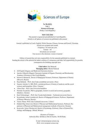
  • 4. 4 Sciences of Europe # 90, (2022) Phytopathogenic microorganisms cause signifi- cant economic damage to agriculture. Pathogens con- stantly infect seeds and all plant organs during the growing season. Pathogens, penetrating plants, dis- rupted physiological and biochemical processes and caused stunted growth, reduced assimilation surface, spotting, premature drying of leaves, impaired root de- velopment, plaques, rot, which led to a significant re- duction in yield and deterioration. In plants affected by phytopathogens, grain quality deteriorates and yields decrease [10, 12]. Monitoring of the phytosanitary condition of sor- ghum phytocenoses showed that during 2018–2021 the most common pathogens were fungal diseases: Helmin- thosporium turcicum (Luttr.) K.J. Leonard & Suggs, Alternaria alternata (Fr.) Keissl., Magnaporthe grisea (T. T. Hebert) M. E. Barr, fungi of the genus Fusarium sp., Bipolaris sp., Rhizoctonia sp., Cercospora sorghi Ellis & Everh., Ascochyta sorghi Sacc. and bacterial: Pseudomonas syringae pv. syringae, Xanthomonas va- sicola pv. holcicola, Robbsia andropogonis (Fig. 1, Fig. 2). Fig. 1. The structure of pathogens of fungal diseases of sorghum in Polissіa of Ukraine, 2018–2021 It was studied that the main share in the structure of pathogens of fungal diseases of sorghum in Polissіa were: Helminthosporium turcicum (29.6 %), Mag- naporthe grisea (22.3 %) and Alternaria alternata (12.4 %). Other pathogens Cercospora sorghi, Asco- chyta sorghi, fungi of the genus Bipolaris sp., Fusarium sp., Rhizoctonia sp. – 10.1, 8.6, 7.7, 5.5, 4.2 % respectively. The first manifestations of Helminthosporium tur- cicum appeared in the phase of 2–3 leaves (light green spots were observed, which gradually turned brown). The disease developed intensively on the leaves of adult plants in the form of elongated, elliptical, brown- ish spots with a border. In wet weather, the spots formed a gray-brown plaque, the leaves gradually with- ered and died. The affected grain was formed thin with blackening of the germinal end of the seed and reduced germination. Magnaporthe grisea develops over a wide range of temperatures (15–35 °C) and humidity (77–82 %). The first symptoms of plant damage were observed at the 29th stage of development on the BBCH scale. Small (1–2 mm) light brown spots with a pronounced brown border appeared on the leaves. Over the next 10– 12 days, the spots increased in size and reached 10 cm. They had a rounded or elliptical shape and necrotized inside, which led to premature drying and death of leaves Symptoms of Alternaria alternata have been re- ported in all years of research. The disease was detected in the ripening phase of the grain. Dark spots were formed on the scales, and during the grain ripening the embryo turned black. The formed grain affected by the pathogen was almost indistinguishable from the healthy one: large in size, well filled. However, the affected seeds had physiological abnormalities, namely: low germination energy and germination. Helminthosporium turcicum, 29,6 % Alternaria alternata, 12,4 % Magnaporthe grisea, 22,3 % Fusarium sp., 5,5 % Bipolaris sp., 7,7 % Rhizoctonia sp., 4,2 % Cercospora sorghi, 10,1 % Ascochyta sorghi, 8,6 %
  • 5. Sciences of Europe # 90, (2022) 5 Fig. 2. The structure of pathogens of bacterial diseases of sorghum in Polissіa of Ukraine, 2018–2021 Sorghum was affected by phytopathogenic bacte- ria in all years of research. The most common signs of phytopathogenic bacteria were leaf spots and less often stems. The first symptoms of phytopathogenic bacteria were observed in July. Pseudomonas syringae pv. syringae іs dominant in the structure of pathogens of sorghum bacterial dis- eases. 43.5 %, and Xanthomonas vasicola pv. holcicola and Robbsia andropogonis were 31.3 and 25.2 % re- spectively. The first signs of damage by the pathogen Pseu- domonas syringae pv. syringae is the appearance of water-saturated spots on the leaves. Eventually, they dry out and turn brown (from beige to dark brown). The spots had a characteristic red border, the color of which differs in different varieties of sorghum. In favorable for the development of Xanthomonas vasicola pv. holcicola pathogen can affect up to 60 % of plants, with losses of green mass reaching 25 %. The first symptoms of the lesion were observed in July. In- itially, water-saturated streaks appeared along the leaf blade. Gradually the strips necrotize and acquire a brownish-red color. With the strong development of the disease, the bands could expand, covering most of the surface of the leaves. Subsequently, the fabric dried and the leaf cracked. The maximum development of the pathogen was observed in the phase of milk-wax ripe- ness. A characteristic feature of Robbsia andropogonis is the appearance on the leaves and stems of sorghum of red stripes ranging in size from a few millimeters to several centimeters. Over time, under favorable condi- tions for the development of the pathogen, the size of the bands increased. The stripes of Robbsia andropogo- nis may be light brown to dark purple (the color is de- termined by the sorghum variety), but they never have a border that is characteristic of P. syringae pv. syrin- gae. Conclusion. Thus, modern climate transfor- mations are forcing agricultural producers to reconsider concepts and practical approaches to the formation of a range of phytocenosis crops capable of ensuring stable and cost-effective crops in increasingly stringent hy- drothermal conditions. Knowledge of the species composition of sorghum pathogens, as well as the peculiarities of their develop- ment and biology is key to establishing effective measures to limit their spread and development. Our further research will be aimed at improving the conservation system of sorghum, which will be based on a rational combination of organizational and economic, agronomic, immunological, biological and other methods, taking into account ESP and technology of cultivation. References 1. The Occurrence and Distribution of Sorghum Diseases in Major Production Regions of Senegal, West Africa / Prom L. K., Sarr M. P., Diatta C. et al. Plant Pathology J. 2021. № 20. P. 1–10. 2. Vyroshchuvannia zernovoho sorho v umovakh Ukrainy / Lapa O. M. ta in. Kyiv: Hlobus-Prynt, 2008. S. 52–59. 3. Makarov L. Kh. Sorhovi kultury: monohrafiia. Kherson: Ailant, 2006. 264 s. 4. Bоikо М. О. The impact of crop density and sowing time on the yield structure of grain sorghum hy- brids. Sciences of Europe: Global science center LP. 2016. Vol. 4. № 5. P. 62–65. 5. Stoliar S. H., Bardin Ya. B. Sorho – kultura velykykh mozhlyvostei. Trofolohiia (vchennia pro zakonomirnosti zhyvlennia bioty ta pravylnoho kharchuvannia liudei) – novitnii mizhdystsyplinarnyi napriam v Ukraini: mater. I Vseukr. nauk.-osvitno- prakt.konf., 25–26 kvit. 2019 r. Zhytomyr: ZhNAEU. S. 93–96. 6. Anitha K., Das I.K., Holajjer P. еt al. Sorghum Diseases: Diagnosis and Management. In: Tonapi V.A., Talwar H.S., Are A.K., Bhat B.V., Reddy C.R., Dalton T.J. (eds) Sorghum in the 21st Century: Food — Fodder — Feed — Fuel for a Rapidly Changing World. Pseudomonas syringae pv. syringae, 43,5 % Xanthomona s vasicola pv. holcicola, 31,3 % Robbsia andropogonis, 25,2 %
  • 6. 6 Sciences of Europe # 90, (2022) Springer, Singapore. 2020. P. 565– 619. doi: 10.1007/978-981-15-8249-3_23 7. Kliuchevych M. M., Stoliar S. H. Sorho zernove – nova ta perspektyvna kultura dlia Polissia. Suchasni tendentsii rozvytku haluzi zemlerobstva: problemy ta shliakhy yikh vyrishennia: materialy Mizhnarodnoi naukovo-praktychnoi konferentsii, 13–14 cherv. 2019 r. Zhytomyr: ZhNAEU, 2019. S. 129–130. 8. Performance of Grain Sorghum and Forage of the Genus Brachiaria in Integrated Agricultural Produc- tion Systems / S. Oliveira et all. Agronomy. 2020. Vol. 10. Р. 1–10. doi: 10.3390/agronomy10111714. 9. Perspektyvy vyroshchuvannia sorho v Polissi Ukrainy / S. H. Stoliar, M. M. Kliuchevych. Naukovi chytannia–2021: zbirnyk tez dopovidei naukovo- praktychnoi konferentsii naukovo-pedahohichnykh pratsivnykiv, doktorantiv, aspirantiv ta molodykh vchenykh ahronomichnoho fakultetu, 28 trav. 2021 r. Zhytomyr: Poliskyi natsionalnyi universytet, 2021. S. 45–47. 10. Minimassom P. Nikiema. Sorghum mutation breeding for tolerance to water deficit under climat change. Journal of Plant Breeding and Crop Science. 2018. Vol. 12(3). Р. 192–199. doi: 10.5897/JPBCS2020.0886. 11. Oblik shkidnykiv i khvorob silskohospodarskykh kultur, za red. V. P. Omeliuty. Kyiv: Urozhai. 1986. 288 s. 12. A highly specific tool for identification of Xanthomonas vasicola pv. musacearum based on five Xvm-specific coding sequences / Nakato G.V., Wicker E., Coutinho T.A. еt al. Heliyon. 2018. Vol. 4(12). Р. 1–11. doi: 10.1016/j.heliyon.2018. e01080.
  • 7. Sciences of Europe # 90, (2022) 7 ART STUDIES РОЛЬОВА ГРА І 3D-ТЕХНОЛОГІЇ ЯК ТВОРЧІ МЕТОДИ БАЛЕТМЕЙСТЕРІВ УІЛЬЯМА ФОРСАЙТА І МЕРСА КАННІНГЕМА Погребняк M.M. Бердянський державний педагогічний університет доктор мистецтвознавства, доцент ROLE–PLAY AND 3D-TECHNOLOGY AS CREATIVE METHODS OF CHOREOGRAPHERS OF WILLIAM FORSYTHE AND MERCE CUNNINGHAM Pogrebnyak M. Berdyansk State Pedagogical University Doctor of Art Criticism, Associate of Professor, Docent АНОТАЦІЯ Стаття присвячена творчості Уільяма Форсайта і Мерса Каннінгема – представників нових напрямів театрального танцю. В результаті аналізу творчих робіт У. Форсайта і М. Каннінгема виявлені особливості творчих методів згаданих митців. Ними стають: 1) використання рольової гри, як однієї з форм (методів) інтерактивних технологій, при побудові композиції сучасного театрального танцю, що сприяє наро- дженню нестандартних пластичних рішень на відміну від академічного балету; зокрема у поєднанні з «ме- тодом випадковостей» М. Каннінгема; 2) використання У. Форсайтом інтерактивної комп’ютерної ін- сталяції «Improvisation Technologies» для створення різноманітної геометрії танцю та несподіваних лек- сичних новоутворень; а саме, «grand battement» і «grand rond de jambe jete» зі зміщенням осі рівноваги, академічних «sisson fermeе», «пози attitude», «заноски», «fuette» з закінченням в невиворотне «developpe» і т. ін.; 3) використання М. Каннінгемом комп’ютерної програми під назвою «Life Forms» («Форми життя»), за допомогою якої можна по-перше, «оживляти» фігуру і створювати танець; по-друге, здійсню- вати взаємодію живих перформерів з 3D моделями людських силуетів. ABSTRACT Relevance of research. The article is devoted to the works of William Forsythe and Merce Cunningham – the representatives of new directions of theatrical dance. The relevance of the article is due to the need for theo- retical support of artists, ballet masters and the educational process in the field of new phenomena of choreographic art, in particular new ideas of choreographers – representatives of contemporary ballet, gradually penetrating into the world theatrical space. The article is considered the using of interactive methods in the work of choreographers William Forsythe and Merce Cunningham. Concretized features of creative methods of the mentioned artists; lex- ical neoplasms of neo-classical dance by W. Forsythe and post-modern dance by M. Cunningham. The purpose of this scientific exploration is a studing of the using of role playing game and 3-D technologies as creative methods by example of choreographers of modern dance theatre of W. Forsythe and M. Cunningham. The research methodology is based on the using of the biographical and source method to study the creative works of choreographers William Forsythe and Merce Cunningham; analysis and synthesis – to identify the fea- tures of creative methods of the mentioned artists; lexical neoplasms of neo-classical dance by W. Forsythe and post-modern dance by M. Cunningham. The scientific novelty lies in the discovery and concretization of innovative creative methods of choreogra- phers W. Forsythe and M. Cunningham. Results. As a result of the analysis of W. Forsythe and M. Cunningham’s creative works the features of cre- ative methods of mentioned artists are identified. These are: 1) using of a role-play, as one of the form (methods) of interactive technologies, when building of a composition of modern theatrical dance, that contributes to the birth of non-standard plastic solutions on unlike academic ballet; in particular in combination with M. Cunning- ham’s «method of chance»; 2) the using of W. Forsythe of an interactive computer installation «Improvisation technologies» for creation of a variety geometry of dance and unexpected lexical innovations; namely «grand battement» and «grand rond de jambe jete» with offset of the axis of balance, academic «sisson fefmee», «atti- tude», «footnotes», «fuette» with ending in irreversible «developpe» and ect.; 3) M. Cunningham’s using of a computer program is called «Life Forms», with which you can first «revive» the figure and create a dance; sec- ondly, to interact with living performers with 3D models of human silhouettes. Ключеві слова: сучасний танцтеатр, інтерактивні методи, балетмейстер, творчий метод, рольова гра, 3D-технології, індивідуальний стиль, авторські хореографічні твори. Keywords: сучасний танцтеатр, інтерактивні методи, балетмейстер, creative method, рольова гра, 3D- технології, individual style, author’s choreographic works.
  • 8. 8 Sciences of Europe # 90, (2022) Постановка проблеми. Минуло майже 100 років з моменту виникнення явища сучасного ав- торського танцтеатру. Актуальність статті обумов- лена необхідністю теоретичної підтримки митців і навчального процесу у галузі нових явищ хореогра- фічного мистецтва, нових ідей хореографів – пред- ставників світового сучасного балету, які посту- пово просотуються у світовий театральний простір, зокрема Уільяма Форсайта і Мерса Каннінгема. А саме, використання інтерактивних методів у твор- чості балетмейстерів, що сприяють виникненню нових прийомів композиції сучасного театрального танцю, його лексичних новоутворень та ін. Аналіз досліджень та публікацій. Проблемам теорії та історії сучасного танцтеатра і нових напря- мів театрального танцю ХХ–ХХІ ст. присвячені не- чисельні праці дослідників (Джека Андерсон, Дон Макдонах, Ендрю Марк Вентинк, Агнесса де Міль, Ізі Парш-Бергсон, Еврістін Стодел, Маринелла Гваттеріні, Марина Погребняк, Олександр Чепалов та ін.). Серед яких Дж. Андерсон, Дон Макдонах і Е. Марк Вентінк висвітлюють естетичні ідеї М. Каннінгема [1, с. 201–202; 2, с. 1–243; 3, с. 280– 297]. М. Гваттеріні на сторінках своєї книги торка- ється історії створення, сюжету і хореографії ба- лету М. Каннінгема «Океан» [4, с. 223–229]. М. По- гребняк у монографії обґрунтовує естетичні ідеї М. Каннінгема, як підґрунтя постмодерного танцю [5, с. 196–204]. Творчої діяльності У. Форсайта то- ркаються на сторінках своїх праць О. Чепалов [6, с. 263–273], М. Гваттеріні [4, с. 215–223]; М. Пог- ребняк аналізує неокласичний танець та творчий метод балетмейстера [5, с. 175–179]. Але дослі- дження і конкретизація рольової гри та 3-D техно- логій як творчих методів на прикладі балетмейсте- рів сучасного танцтеатру У. Форсайта і М. Каннін- гема залишаються поза межами існуючих розробок, що і є метою даної наукової розвідки. Методологія. Методологія дослідження ґрун- тується на використанні біографічного та джерело- знавчого методів для вивчення творчої діяльності балетмейстерів У. Форсайта і М. Каннінгема; ана- лізу і синтезу – для виявлення особливостей твор- чих методів згаданих митців; лексичних новоутво- рень неокласичного танцю У. Форсайта та постмо- дерного танцю М. Каннінгема. Виклад основного матеріалу. Перш за все хочу нагадати, що явище авторського танцтеатру тісно пов’язане з «ідеєю свободи» в теорії драми, яку привнесли німецькі драматурги школи «бурі та натиску» і яка на межі ХIX–початку ХХ ст. стала провідною ідеєю сучасного театру і танцтеатру зо- крема. Крім того, сама «система виразності» Ф. Дельсарта, що стала естетико-теоретичним підґ- рунтям нових напрямків сценічного танцю сприяла виникненню нових творчих методів у роботі балет- мейстерів сучасного танцтеатру. Так, авторкою досліджено, що рольова гра, як одна з форм (методів) інтерактивних технологій: 1) по-перше, використовується при побудові компози- ції сучасного театрального танцю, що сприяє наро- дженню нестандартних пластичних рішень на від- міну від академічного балету; 2) по друге, метод ро- льової гри застосовується в прийомах (методах) ко- нтактної імпровізації (підготовчої, імпровізації у просторі і часі), які скасовують хореографа-лідера, допомагають народженню лексичних новоутворень і ритмопластичних вибудувань у творчій роботі ба- летмейстера. Наприклад: віддзеркалення, накопи- чення, активний і пасивний дует або група, партер- ний рисунок, «охоплений простір», групові конс- трукції симетричної, асиметричної форми та інші. Розглянемо використання інтерактивних техноло- гій у творчій роботі балетмейстерів Уїльяма Фор- сайта і Мерса Каннінгема. У. Форсайт так висловлюється про свій твор- чий метод: «Я даю танцівнику думку, а не результат … техніка імпровізації не довідник, а відкрита інте- рактивна система» [7, с. 13]. Такі три його одноактні балети як: «Steptext» [8], «The Vertiginous Thrill of Exactituole» («Запамо- рочлива насолода точністю») і «In the Middle, Somewhat Elevated» («Всередині, щось підвішене») – найяскравіше ілюструють творчий метод У. Фор- сайта [9–11]. Так, наприклад, композиція балету «В середині, щось підвішене», на музику Тома Ві- ллемса, створеного у 1988-му р. для Паризької опери виглядає як ка- лейдоскопічне миготіння 4-х соло і 4-х дуетів. Створена Т. Віллемсом музика з чистими шумами, аранжована за допомогою комп’ютерного модифікатора підкреслює «па» лю- дей-машин, використані у танці. В композиції танцю виникає кілька центрів ро- звитку дії за рахунок використання прийомів аси- метрії і антиунісону. При побудові хореографічного тексту «меха- ніка» танцю виходить на перший план. Тіло танців- ника знаходиться у стані «колапсу», що дозволяє створювати нескінченні нові лінії руху у просторі. Імпульс руху може зароджуватися в будь-якій час- тині тіла (у лікті, коліні, тазі, нозі і т. ін.). Зміщення центру ваги і дестабілізація тіла перетворюється на стратегію. Імпровізація групи танцівників, залучених до гри в створення сценічного образу, що вимагає і свободи володіння тілом, і свободи мислення – стає основою його творчого методу роботи з виконав- цями. «Покажи мені свою ідею» – вимагає У. Фор- сайт від танцівника, як згадує роботу з балетмейс- тером соліст балету Маріїнського театру Костянтин Звєрєв [12]. Як результат, в дуетній формі танцю характерною особливістю стає ясно виражена кру- гова динаміка і постійна трансформація різних під- тримок класичного танцю (двома руками за талію, падаючі пози за обидві руки, за одну руку). При пі- дйомах як швидких так і плавних, звертають на себе увагу «grand battement» і «grand rond de jambe jete» зі зміщенням осі рівноваги [12]. А академічні «sisson fermeе», «пози attitude», «заноски», «fuette» з закінченням в невиворотне «developpe» і т. ін. зна- ходяться у постійному перетворенні, створюючи каскад лексичних новоутворень [9–11]. Крім того, створена У. Форсайтом інтеракти- вна комп’ютерна інсталяція «Improvisation
  • 9. Sciences of Europe # 90, (2022) 9 Technologies» спочатку планувалася як допоміжний засіб для професійного тренінгу артистів балету франкфуртської трупи і, з часом, стала виконувати універсальні функції, що дозволяють аналізувати будь-який рух танцівників. У цьому проекті всі рухи танцюристів вписані у віртуальний простір. Несподівані лексичні новоутворення на основі елементів класичного танцю У. Форсайт створює, починаючи малювати уявні фігури в повітрі, вико- ристовуючи всі частини тіла – ноги, руки, голову, коліна, вуха, підборіддя і т. ін. Так він створює різ- номанітну геометрію танцю, «яка графічними засо- бами креслить повний об’єм потенціальних рухів людського тіла…» [7, с. 12–13]. Досліджено, що рольова гра стає творчим ме- тодом і Мерса Каннінгема. Так, з самого початку 1940-х років особливо провокативними аспектами хореографічної теорії М. Каннінгема стали: 1) ви- користання випадковості та невизначеності; 2) ста- влення до простору сцени як до відкритого прос- тору; 3) схильність розглядати компоненти танцю- вальної постановки як самостійні сутності. Бажаючи, щоб його танці мали частину непередба- чуваності самого життя, балетмейстер почав вико- ристовувати метод «випадковостей» з використан- ням рольової гри танцівників. Тому спектаклі М. Каннінгема вражали несподіваними компози- ційними рішеннями. Він розглядав вистави, як спі- льність незалежно створених самостійних елемен- тів. Часто різні компоненти танцювального вечора вперше поєднувались на прем’єрі, дивуючи і танці- вників і аудиторію. Це стосувалося і музичного оформлення, що мало звільняти танцюристів від «рабського» підкорення, або протиставлення танцю музиці [1, с. 185–186]. Незважаючи на деякі невідповідності ритму, тону, кольору з танцем, музикою та декораціями, балетмейстер створював ефект цілісності спекта- клю. Паралельне, не пов’язане одне з одним буття музики та руху виглядало яскравим, композицій- ним прийомом. Так, за словами Джека Андерсона, плаваючі срібні подушки у якості декорації Енді Вархола до вистави «Дощовий ліс» здаються доре- чними. Вистава «Зимові обійми» містить у собі сті- льки образів боротьби і пригнічення, що нагадує глядачам жах і війни. Композицію «Звучання» («Sounddance») можна вважати хореографічний криком. «Квартет», не зважаючи на свою назву, є танцем для п’ятьох, у якому одна стороння людина даремно намагається приєднатися до групи з чоти- рьох людей, що асоціюється з явищем соціального остракізму або прірвою між поколіннями [1, с. 187]. У 1991-му році він почав використовувати у своїй хореографії комп’ютерну програму під на- звою «Life Forms» («Форми життя»), за допомогою якої можна «оживляти» фігуру і створювати танець [13]. Результатом таких експериментів можна вва- жати постановки балетмейстера «Beach Birds for Camera» (1991-й р.) з жорсткими та незграбними рухами рук та ніг і «Biped» (1999-й р.). Балет «Biped», складається з двох частин: живе виконання хореографічної партитури танцівниками і відеопроєкція, яка варіювалася від абстрактних фі- гур до анімованих відео з мальованими танцівни- цями. Для створення анімації на трьох виконавиць навішували сенсори, які фіксували відеокамери. Всі рухи переводились у 3D моделі людських силуетів, а потім виводились на сцену, де з ними взаємодіяли живі перформери. Танцівники-перформери виконують модифіко- вані «tombe», «tour» з зігнутою попереду ногою, різ- номанітні «attitude» з «flat backe», «tour channe», «grand battement», акробатичні підтримки в позу «І arabesques» (3 партнера і 1 танцівниця) і т. ін. [14]. Основне завдання балетмейстера – створення хореографії, де кожний виконавець має авторський рух і власний ритм. Продовжуючи педагогічні тради- ції хореографів танцю «модерн», він, за його ж сло- вами, не прагне зробити виконавців схожими на нього, а намагається через свою техніку надати їм тієї сили, що дозволила б відкрити власну індивідуаль- ність у рухах та думках. Висновки. В результаті аналізу творчих робіт У. Форсайта і М. Каннінгема виявлені особливості творчих методів згаданих митців. Ними стають: 1) використання рольової гри, як однієї з форм (мето- дів) інтерактивних технологій, при побудові компо- зиції сучасного театрального танцю, що сприяє наро- дженню нестандартних пластичних рішень на від- міну від академічного балету; зокрема у поєднанні з «методом випадковостей» М. Каннінгема; 2) викори- стання У. Форсайтом інтерактивної комп’ютерної інсталяції «Improvisation Technologies» для ство- рення різноманітної геометрії танцю та несподіваних лексичних новоутворень; а саме, «grand battement» і «grand rond de jambe jete» зі зміщенням осі рівноваги, академічних «sisson fermeе», «пози attitude», «за- носки», «fuette» з закінченням в невиворотне «developpe» і т. ін.; 3) використання М. Каннінгемом комп’ютерної програми під назвою «Life Forms» («Форми життя»), за допомогою якої можна по-пе- рше, «оживляти» фігуру і створювати танець; по- друге, здійснювати взаємодію живих перформерів з 3D моделями людських силуетів. Література 1. Anderson J. Ballet. Modern dance. A concise history. New Jersey: Princeton Book Company, Publishers, 1992. 235 p. 2. Merce Cunningham: dancing in space and time: essays 1944–1992 / by Jack Anderson ; edited by Richard Kostelanets. Chicago: Chicago Review Press, Incorporated, 1992. 243 p. 3. МсDonagh D., Wentink A. M. The Complete Guide to Modern Dance. Washington: Library of Congress Cataloging-in-Publication Data Mcdonagh, 1976. 639 p. 4. Гваттерини М. Азбука балета / пер. с ит. Ю. Лисовского. Москва: БММАО, 2001. 240 с. 5. Погребняк М. М. Нові напрями театрального танцю ХХ – поч. ХХІ ст.: історико-культурні переду- мови, крос-культурні зв’язки, стильова типологія: монографія. Полтава: ПП Астрая, 2021. 327 с.
  • 10. 10 Sciences of Europe # 90, (2022) 6. Чепалов О. І. Хореографічний театр Західної Європи ХХ ст.: монографія. Харків: ХДАК, 2008. 344с. 7. Чепалов А. Сага о Форсайте. Танец в Украине и мире. №2(10). 2015. С. 12–13. 8. Steptex / William Forsythe: веб-сайт. URL: https://youtu.be/ja5gyP0XjPs (дата звернення 24.01.2020). 9. In the Midlle, Somewhat Elevated» – Marta Romagna, Roberto Bolle, Zenaida Yanowsky: веб-сайт. URL: https://youtu.be/NghGmjtxeak (дата звернення 24.01.2020). 10. In the Midlle, Somewhat Elevated» – Sylvie & Laurent Pas De Deux: веб-сайт. URL: https://youtu.be/HqS4Gh1lMGA (дата звернення 24.01.2020). 11. In the Midlle, Somewhat Elevated»/William Forsyth: веб-сайт. URL: https://youtu.be/3knW29Yad8Q (дата звернення 24.01.2020). 12. ЦЛ о возвращении балетов Уильяма Фор- сайта на сцену Мариинского театра: веб-сайт. URL: https://youtu.be/1l1Kd-p1ouk (дата звернення 24.01.2020). 13. Кибербалет – современный танец и цифро- вые технологии – Biletsofit.ru: веб-сайт. URL: https:// https://biletsofit.ru/blog/kibberbalet-tanec-i-cifrovye- resheniya (дата звернення 20.02.2020]. 14. Merce Canningham Dance Company at BAM: Biped: веб-сайт. URL: https://youtu.be/YHeoYdDMbLI (дата звернення 20.02.2020).
  • 11. Sciences of Europe # 90, (2022) 11 BIOLOGICAL SCIENCES МОЖЛИВІ МЕХАНІЗМИ БІОЛОГІЧНОЇ АКТИВНОСТІ ПОЛІГЕКСАМЕТИЛЕНГУАНІДИНУ ЩОДО МЕМБРАН КЛІТИН Лисиця А.В. Рівненський державний гуманітарний університет Мандигра Ю.М. Дослідна станція епізоотології Інституту ветеринарної медицини НААН, м. Рівне, Україна Кривошия П.Ю. Дослідна станція епізоотології Інституту ветеринарної медицини НААН, м. Рівне, Україна Нечипорук Б.Д. Рівненський державний гуманітарний університет POSSIBLE MECHANISMS OF THE BIOLOGICAL ACTIVITY OF POLYHEXAMETHYLENE GUANIDINE ON CELL MEMBRANES Lysytsya A., Rivne State University of Humanities Mandygra J., Research Station of Epizootology, Institute of Veterinary Medicine NAAS, Rivne, Ukraine Kryvoshyya P., Research Station of Epizootology, Institute of Veterinary Medicine NAAS, Rivne, Ukraine Nechyporuk B. Rivne State University of Humanities АНОТАЦІЯ Мета досліджень: запропонувати і обґрунтувати можливі механізми дії дезінфектантів які містять по- лімерні похідні гуанідину, зокрема полігексаметиленгуанідин, на мембрани клітин. Методи: мас-спектро- метрії, культивування культур клітин, мікробіології, штучних бішарових ліпідних мембран. Наведено ре- зультати біофізичного і біохімічного аналізу можливих механізмів взаємодії полімерних похідних гуані- дину з цитоплазматичними мембранами клітин прокаріот та еукаріот. Встановлено, що основною мішенню для цих сполук є фосфоліпіди цитоплазматичної мембрани. Відмінності в дії препарату на клі- тинні мембрани залежать, перш за все, від їх ліпідного складу. Запропоновано можливі теоретичні моделі, які пояснюють специфіку біоцидного ефекту дезінфектантів, виготовлених на основі полігексаметиленгу- анідину, наприклад Епідез для ветеринарної медицини. При відносно низьких концентраціях препарату (10-4 %) і дозованому часі експозиції відбувається зміна ліпідного складу мембрани (через видалення час- тини фосфоліпідів або полігексаметиленгуанідин-ліпідних везикул). З цим пов'язані неогенез ліпідів і ви- явлені нами ростостимулюючі та цитопротекторні ефекти. Бактеріостатичні дози полігексаметиленгуані- дину (10-3 -10-2 %) гальмують проліферацію клітин еукаріот (фібробласти курячого ембріону), бактерицидні дози (10-2 -10-1 %) викликають значні порушення структури і функцій цитоплазматичних мембран. Мем- брани досить швидко пошкоджуються, найбільш ймовірним є килимовий механізм дії. В результаті лізису клітина гине. Отримані результати дозволяють краще зрозуміти механізми високої бактерицидної актив- ності полігексаметиленгуанідину щодо більшості мікроорганізмів і, в той же час, його відносну безпеч- ність для людини, тварин та вищих рослин. Ці дані сприятимуть розробці як нових ефективних і безпечних засобів для дезінфекції, так і стимуляторів або засобів захисту рослин. ABSTRACT To propose and substantiate possible mechanisms of action of disinfectants, containing polymer derivatives of guanidine, in particular, polyhexamethylene guanidine (PHMG), onto cell membranes. We used the following methods: mass spectrometry, cell culture cultivation, microbiology, artificial bilayer lipid membranes. The results of biophysical and biochemical analysis of possible mechanisms of interaction between polymeric guanidine de- rivatives and cytoplasmic membranes of prokaryotic and eukaryotic cells have been presented. It has been estab- lished that the main targets for these compounds are phospholipids of the cytoplasmic membrane. The differences in the action of the drug on different kind of the cell membranes depend, above all, on their lipid composition. Possible theoretical models have been proposed to explain the specificity of biocide effect of disinfectants, made on the basis of PHMG, for example it is Epidez for veterinary medicine. At relatively low concentrations (10-4 %) of the drug and the metered exposure time (1-2 min) there is a change in the lipid composition of the membrane (via the removal of some phospholipids or PHMG-lipid vesicles), which is associated with neogenesis of the phos- pholipids and the growth-stimulating and cytoprotective effects from viruses, detected by us. Bacteriostatic or sublethal concentrations (10-3 -10-2 %) of PHMG inhibit the proliferation of eukaryotic cells (chicken embryo fi-
  • 12. 12 Sciences of Europe # 90, (2022) broblasts), and bactericidal doses (10-2 -10-1 %) result in considerable perturbations which of the structure and func- tions of its cytoplasmic membranes. The membranes are rather rapidly damaged via, most probably, the carpet mechanism. It is the most common cause of cell death. The results obtained by us explain the high bactericidal activity of PHMG regarding most microorganisms and, at the same time, its relative safety for humans, animals and higher plants. These data will facilitate the development of new effective and safe means of disinfection, and stimulants or plant protection products. Ключові слова: полігексаметиленгуанідин, дезінфектанти, загибель клітин, фосфоліпіди, біофізичні моделі. Keywords: polyhexamethylene guanidine, disinfectants, cell death, phospholipids, biophysical models. Introduction. Polyhexamethylene guanidine (PHMG) is known since the 1950s as a cationic biocide with a wide spec- trum of action, impacting the cell membrane and its me- tabolism [1, 2]. Due to the specific structure of the mol- ecule, containing hydrophobic hexamethylene areas (spacers) and positively charged guanidine groups, it has antibacterial, antiviral and antifungal activity [3, 4, 5]. It is proved that PHMG may be capable of impairing the stability of cytoplasmic membrane (CPM) of the cell via electrostatic interaction with acid phospholipids [2, 6]. At present, veterinary medicine uses many guan- idine-based preparations for disinfection, including Ep- idez [7, 8]. Its main active substance is polyhexameth- ylene guanidine hydrochloride, whose characteristics and advantages were frequently discussed already [9, 10, 11, 12, 13]. At the same time, biochemical and bio- physical specificities of PHMG impact on CPM of pro- karyotic and eukaryotic cells are not fully understood [14, 15]. This issue is urgent for the elaboration of new disinfectants, which would be highly efficient and at the same time have low toxicity for humans and ani- mals. Mass spectrometry research of PHMG [16, 17, 18] requires proper interpretation and continuation. The aim of research is to investigate and analyse possible mechanisms of action of polyhexamethylene guanidine on cytoplasmic membranes of different cells. Materials and methods The results of our own experimental studies, ob- tained via mass-spectrometry methods, were used in the work [19, 20]. Mass spectrometry research was made by time-of-flight plasma desorption (TOF-PDMS) and matrix assisted (by 2.5-dihydroxybenzoic acid or 3,5- dimethoxy-4-hydroxy-cinnamic acid) laser desorp- tion/ionization time-of-flight (MALDI-TOF). PDMS mass spectra of PHMG samples were acquired by MSBC-01 spectrometer (SELMI, Ukraine) with 252 Cf nuclei fragments ionization. MALDI-TOF mass spectra of samples were acquired by Voyager DE PRO spec- trometer (Applied Biosystems, USA) with Н+ -matrix ionization. The results were analysed by MSBC pro- gram, version 4.0/m, and Data Explorer 4.0 software systems, respectively. Hereafter, m/z values of the mono-isotopic peaks of the ion distribution have been reported. The molar concentration of PHMG was cal- culated by the molecular weight of hexamethylene guanidine monomer residue, 141 Da. The method of cell cultures [21, 22, 23] was ap- plied at the Research Station of Epizootology, the Insti- tute of Veterinary Medicine of the NAAS (Rivne, Ukraine), using the primary culture of fibroblasts of the chicken embryo and interweaved culture of the tracheal cells of calf. The cells were grown in the solution, con- taining a mixture of 199 medium (45 %), a minimum Eagle medium or MEM (45 %) and blood serum of cat- tle (10 %). The monolayer was grown after seeding cell suspensions in 96-well plastic plates at 0.1 ml per well. The method of artificial bilayer lipid membranes (BLM) formed of different lipid composition involved the application of lipid bilayers [21, 24]. The membrane washing solution contained 10mM Tris–HCl (pH 7.4) and the required quantities of potassium chloride, so- dium chloride (USB, Cleveland, OH, USA), lithium chloride and cesium chloride. The membrane separated chambers were stirred when required. The biological test objects were the cultures of Escherichia coli (strain АТСС 055 К59 No. 3912/41), Staphylococcus aureus (strain АТСС No. 25923 F 49), Bacillus cereus (reference strain DNKIBSHM, Kyiv), Mycobacterium bovis (strain Vallee), field strains of Leptospira interrogans, vegetative forms and spores of American foulbrood Paenibacillus larvae subsp. lar- vae, micromycetes of the fungal species Aspergillus fu- migatus, A. flavus, A. niger (field strains), herpes vi- ruses of equine rhinopneumonitis Equine herpesvirus type 1 (strain SV-69, Moscow, RF) NB: No literature reference to this strain, no culture collection number and bovine rhinotracheitis Rhinotracheitis infectiosa bovine (strain ТК-А, Kharkiv), retrovirus – equine in- fectious anaemia virus (field strain). Polyhexamethylenebiguidined hydrochloride was synthesized in PE “Termite” (Rivne, Ukraine) by poly- condensation of hexamethylenediamine and dicyandi- amide with the addition of ammonium chloride (Si- nopharm Chemical Reagents Co. Ltd., Shanghai, China). The molecular weights of PHMG polymers de- termined by the viscosity of PHMG-containing solu- tions exhibited their distribution within the range of about 1000–2000 Da (8–16 repeat units). The estimates of kinematic and reduced viscosities were carried out by Ostwald viscometer (VPZH-2) with a capillary di- ameter of 0.56 mm. Research results We used the mass-spectrometry to determine the oligomeric composition of PHMG preparations [19]. It was established that in most cases it was rather inho- mogeneous. For instance, when the composition of four most typical oligomers of linear structure was deter- mined, they were shown to differ both in the number of monomer parts and the content of terminal groups. The mass-spectra clearly demonstrate the difference Δ m/z = 141, which corresponds to the mass of one monomer. The work [20] analysed the interaction be- tween the preparation of PHMG with such lipids as lec- ithin and cholesterol, which are main components of
  • 13. Sciences of Europe # 90, (2022) 13 cytoplasmic membranes of mammalian eukaryotes. The analysis of mass-spectra demonstrated that no sta- ble intermolecular complexes of PHMG oligomers with lipids were formed. Based on this fact, an assumption was made that during the adsorption of PHMG on the negatively charged bacterial membrane there may be either electrostatic interaction or the formation of loop- like structures. Such a stereochemical mechanism en- sures adsorption stability on the membrane, related to the plurality of the bonds, formed with phospholipids, and enhances along with the increase in the molecular mass of the polymer. The biocide activity of the prepa- ration decreases in case of poor availability of mem- brane phospholipids. In particular, this is true about bacterial spores and mycobacteria with wax-like enve- lopes. This fact is also confirmed with microbiological investigations on differential sensitivity of microorgan- isms to different PHMG salts. The experiments, con- ducted with test objects being E. coli, S. aureus, B. ce- reus, M. bovis, L. interrogans, P. larvae, A. fumigatus, A. flavus, A. niger demonstrated that the sensitivity of microorganisms to the preparation is firstly defined by the total share of lipids in the membrane and the avail- ability of their phosphate groups [7]. There is also a re- markable regularity: the increase in the relative share of acidic lipids in the external layer of CPM and thus a higher value of the negative external superficial electric potential of the membrane and the decrease in the length of fatty acid tails of phospholipids is in clear cor- relation with the increase in the sensitivity of microor- ganisms to PHMG. It is also relevant what type of anion is present in PHMG salts, for instance, the biocidal ac- tivity of PHMG chloride is generally higher compared against PHMG salts with organic acids, such as PHMG valerate, PHMG maleate and PHMG succinate. In the context of studying possible mechanisms of PHMG connecting to CPM the adsorption of the former is practically irreversible; we have studied the dynam- ics of stereochemical changes in polycation molecule depending on pH. During titrating of aqueous PHMG solutions, there are considerable changes in the optic density and viscosity, in the degree of polycation mol- ecule ionization and its conformation. As there are con- siderable gradients of pH and concentrations of cations, like Са2+ , Mg2+ , when a PHMG molecule binds to the lipids of the external CPM monolayer, there are local changes in pH and conformation of polycation mole- cules. The change in conformation of polycation mole- cule during adsorption promotes strong fixation of PHMG on the membrane, its penetration into the lipid bilayer, the change in the position of phospholipids in CPM (segregation of anionic and zwitterionic phospho- lipids), including the facilitation via their lateral diffu- sion. Another direction was the investigation on growth-stimulating and cytoprotective effects of PHMG. In particular, the cell cultures of bovine trachea (calves) and fibroblasts of chicken embryo were used to determine the toxicity of PHMG salts and their stim- ulating and protective effect. PHMG salts impact the rate of cell monolayer formation, in particular, PHMG hydrochloride concentrations in the growth medium, equalling and exceeding 10-6 –10-5 %, inhibit the for- mation of the monolayer culture of fibroblasts. But PHMG in nanomolar concentrations (0.07–7.0 nM or 10-8 –10-7 %) stimulates the proliferative activity of eu- karyotic cells and accelerates the formation of the mon- olayer. In addition, it was first discovered that prelimi- nary treatment of eukaryotic cells with PHMG salts in the concentrations of 10-5 –10-2 % for 10–15 min pre- vents their being damaged with retroviruses (RNA- viruses) and herpes viruses (DNA-viruses). The cyto- protective effect depended on the anionic composition of PHMG salts, the presence of lipids in the viral enve- lope, the stage of the cellular cycle [25]. The experi- ments with seeds of several species of agricultural crops demonstrated that PHMG salts both disinfect the seeds and may stimulate the germination and energy of sprouting. The highest growth-stimulating effect was manifested for pre-sowing treatment of the seeds of beets and legumes (peas, kidney beans, soybeans.) For instance, in some experiments the maximal values of germination and the energy of sprouting exceeded the control more than twice. PHMG succinate was found to be more efficient than PHMG chloride, the optimal concentrations of the former for the seeds of fodder beet were 0.1–0.5 %, and for peas – 0.001–0.01 %. The en- ergy of sprouting increased by 50 % for kidney beans, and the germination – by 30–35 % at the preparation concentration of 0.01 %. As for the study of PHMG effect on bilayer phos- pholipids membranes [21], it was determined that after a long-term period of membrane stability, when its con- ductivity had almost no changes, there is a sharp in- crease in the ionic current a few seconds prior to BLM breakage. The time, required for PHMG polycation ad- sorption on BLM and the rate of membrane breakage depend on the polarity of electrode charge in cis-cham- ber and phospholipids composition of the membrane. The active concentration of PHMG from the external side of the membrane was 0.0001 %, or ≈ 7 µM; the po- tential from the cis-side of BLM changed from +100 mV to -100 mV; the solution, surrounding the membrane from both sides, was 100 mM КСl. The dif- ference in the rate of BLM breakage on condition of different electric potentials on the electrode demon- strated that the electrostatic interaction of the poly- cation and the membrane is relevant in the general mechanism of adsorption and destruction of CPM, but this relevance is not decisive. Even on condition of neg- ative potential of the electrode (-100 mV), the adsorp- tion of PHMG on BLM and its destruction occur, albeit at a slower rate. The investigation on the possible negative impact of PHMG preparations (disinfectants, plant protection products or stimulators of seed germination) on zoo- and phyto-constituents of biocenoses demonstrated the results, presented below [26, 27]. For insects (bees), when coming with sugar syrup, the toxic action was manifested at PHMG concentration of ≥ 0.66 %, LD50 per os for mammals (white laboratory mice) – 2000±100 mg/kg of bodyweight. The mentioned con- centrations are practically unavailable in normal condi- tions. The minimal toxic concentration of PHMG hy- drochloride for hydrobionts (fish, shellfish, flatworms,
  • 14. 14 Sciences of Europe # 90, (2022) crustaceans) is 0.0001 % (or 1 mg/l), that for ciliates – 0.001 %. The concentrations, starting with 0.00001 % or 0.1 mg/l and below, are safe for the formed mono- layers of eukaryotic cells. The toxicity of the prepara- tion depends considerably on its chemical purity, avail- ability and amount of low molecular admixtures of hex- amethylenimine, hexamethylendiamine, methylenimine, etc. The plant components of bioceno- ses are more tolerant to the effect of PHMG, higher plants are resistant to the treatment with 0.1–0.3 % aqueous solution of the preparation. The biocide or in- hibiting effects for algae are manifested at the concen- tration of ≥ 0.0001 %. The transfer coefficient in the “soil-plant” system is < 0.01 %, “water-plant” (algae) – < 0.1 %. In general, the potential threats for ecosystems from PHMG preparations, penetrating therein, are min- imal – they are quickly adsorbed onto organic and in- organic components of soil, and in water they bind par- ticulate matters, organic substances, surface active sub- stances etc. The migration along food chains is almost absent due to the polymer structure of PHMG and its fast decomposition. No negative consequences were determined if chronic exposure was absent. The prepa- ration has no considerable impact on the ability of bio- cenoses to self-purify, self-regulate and self-restore. At the same time, taking into consideration the fact that bactericidal concentrations of PHMG hydrochlo- ride for most gram-positive and gram-negative bacteria are 0.005–0.1 %, and the bacteriostatic ones – 0.0001– 0.005, one may not state categorically that eukaryotic cells should be more resistant to PHMG preparations. Discussion of research results Therefore, the generalization and analysis of the results obtained by us and other authors led us to the conclusion that the main target for PHMG molecules is a cell membrane, and the specificity of the interaction between the preparation and cytoplasmic membranes of cells is related to several key issues. Firstly, the main target for PHMG in the cell membrane is its phospholipids, although the interaction with negatively charged groups of membrane proteins, glycolipids, glyco- and lipoproteins is also possible. The share of glycoproteins and glycolipids is known to take about 25 % of the surface potential of CPM. The mass content of lipids in cell membranes usually fluc- tuates from 25 to 70 %. The effect of PHMG polycation on other membrane components may be considered secondary and auxiliary, or indirect. Surely, all the models, presented below, are intentional simplifica- tions because the specificities of functioning of the liv- ing systems cannot be reproduced exactly in any of the simplified model systems [28]. Modern methods of molecular modelling demon- strate that the surface of even a simple single-compo- nent lipid membrane (for instance, with 1,2-diphyt- anoyl-sn-glycero-3-phosphocholine or di- oleoylphosphatidylcholine) is not polar homogeneous as it could be assumed judging by the schematic presen- tation of lipids in the form of balloons with tails. Some of these tails surface on the water-membrane boundary and form hydrophobic areas, i.e. there is an emerging mosaic-like, mostly polar surface with some hydropho- bic isles with the size of several square nanometers. Somewhat more complicated multicomponent models demonstrate the presence of more liquid lamellar phases or Ld-phases in the membranes (with prevailing phospholipids with unsaturated acid tails) and solid, or Lo-phases (with saturated fatty acids). If such a model membrane is added transmembrane spiral peptides, they are distributed between phases, getting mostly lo- cated in the liquid Ld-phase and avoiding the orderly Lo- phase. Thus, the areas from Lo-phase are more accessi- ble for the adsorption of PHMG molecules. The absence of cholesterol in the membranes of bacteria does not allow for confident assertions about the formation of rafts, as in case of CPM of eukaryotes, but bacterial membranes are also laterally inhomogene- ous. The lateral heterogeneity of the membrane struc- ture of pro- and eukaryotes, their different transmem- brane, dipole and surface potentials have implicit effect on the specificities of adsorption of PHMG molecules. Secondly, PHMG polycations may get adsorbed on any phospholipid membrane. A sufficient prerequi- site is the availability of negatively charged phosphate groups of phospholipids. In our experiments on BLM, PHMG salts are quickly and irreversibly adsorbed on comparatively “neutral” membrane with PC± (phospha- tidylcholine) and cholesterol [19, 21]. Contrary to Са2+ or Mg2+ ions, PHMG interacts both with charged and zwitterionic phospholipids. Obviously, while contact- ing even acidic phospholipids like PC- (phosphatidyl- serine), polycation iminogroups bind not only carboxyl groups of serine, but also phosphate groups, similarly to Са2+ . However, the ratio of acidic and neutral lipids in the external layer of CPM is relevant. For instance, some authors believe that bacteria, whose external layer of CPM contains a higher percentage of acidic lipids, are more sensitive to the effect of the preparation than eukaryotic cells [29]. In addition, prokaryotic mem- branes differ considerably from plasmatic membranes of eukaryotes both in their lipid composition, for in- stance, the presence of cardiolipin (CL2- ) in CPM, and superficial potential, non-lipid components, etc., thus, the rate of adsorption and its consequences should be also different. The adsorption of PHMG on CPM oc- curs unevenly, the preparation gets mostly concentrated in the areas, enriched with lipids, especially anion ones [1]. Thirdly, the properties of the lipid membrane change after PHMG adsorption. During the interaction of guanidine groups and polar heads of phospholipids, there may be the re-distribution of ions in the outer sub- membrane layer, for instance, forcing out counter ions Са2+ and Mg2+ , which usually stabilize CPM. This is accompanied with local changes in pH. Similar to other polycation antimicrobial preparations, there is possible segregation of acidic and neutral phospholipids and for- mation of membrane domains with different superficial electric potential. It is known that both the charge of phospholipid heads and ions, bound to them (in this case, it is polycation PHMG), define the value of trans- membrane potential. The example of the effect of acellisine oligomers on the membranes can be used as an analogue [30]. It is also a polycation, binding anion phospholipids mostly. In this case, the membrane should contain both
  • 15. Sciences of Europe # 90, (2022) 15 anion and zwitterionic lipids. After the adsorption of polycation, there is segregation with the formation of domains, enriched with anion phospholipids in the ar- eas of the highest accumulation of acellisine oligomers (Fig. 1). Fig. 1. The theoretical scheme of possible segregation of anion (in a darker colour) and zwitterionic phospholipids in the membrane after polycation adsorption (Source of Figure: Epand et al., 2008) However, the reason for segregation and accumu- lation of anion lipids on the internal side of the mem- brane is not quite clear in the abovementioned scheme for acyllysine. Probably, there is correlation between the interaction of lipids of the external and internal monolayers of the membrane or excessive positive charge in the areas of polycation adsorption. For in- stance, in the model membranes, where the separation of Ld and Lо phases can be observed, the clusters of these phases coincide for both monolayers. As for PHMG, it was noted [1, 31, 32] that after its adsorption there is segregation of phospholipids, along with their removal from the CPM. A similar segregation of phos- pholipids takes place after adsorption of antimicrobial peptides (AMP) on the membranes. For instance, ac- cording to the data of computer simulation of a bacte- rial membrane, containing 70 % PE± (phosphatidyleth- anolamine) and 30 % PG- (phosphatidylglycerol), there is possible formation of nanodomains (molecules of PE± have efficient interaction and force out the “unfavourable” partner PG- ), and the adsorption of AMP leads to the increase in PG- domains and the oc- currence of phase separation of lipids. Thus, the adsorption of a considerable number of PHMG molecules on CPM promotes global separation of Lo and Ld phases of lipids and segregation of neutral and acidic phospholipids. This is a thermodynamically favourable process. It is generally known [28] that the composition of the lipid matrix of native membranes has evolutionarily been formed so as to be always near the phase transition in physiological conditions. This is a condition for the formation of a mesophase (rafts) in the membranes, and the adsorption of PHMG poly- cations shifts natural equilibrium and “pushes” the pro- cess of phase transition. The step, following the segregation of lipids, is the transition of the membrane in some areas from the la- mellar L into hexagonal (cylindrical) HII phase. Gener- ally, most purified membrane phospholipids in aqueous medium are known not to form bilayers, but be situated predominantly in the hexagonal phase НІІ. As for the membranes, first of all this is notable for those, contain- ing a considerable percentage of lipids, asymmetrical in their form. For instance, the form of PE± or CL2- mole- cules resembles a cone-type form rather than the cylin- dric one, similar to PC± or PS- . Noteworthy is the fact that, compared to eukaryotic membranes, the bacterial membranes contain both a higher percentage of nega- tively charged lipids and a higher amount of such lipids of negative curvature. The places of their accumulation in CPM may serve as sources of formation for HII phase and impairment to the integrity of the external lipid monolayer. PHMG-lipid hexagonal or vesicle-like structures are formed and are likely to leave CPM sur- face rather fast (detach themselves). In case of BLM, after the adsorption of PHMG thereon, in about 5–10 min there is rather fast polariza- tion of a conditionally neutral PC± membrane. As for the membrane, containing acidic PG- or CL2- in its ex- ternal layer, there may be depolarization from “-” to “+” and a local positive surface potential may be formed. The changes in the dipole potential of the membrane promote the hydration and decompression of lipids. Re-charging (depolarization) of the external lipid monolayer of the membrane is accompanied with the increase in flip-flop transitions, thus the internal lipid layer of CPM undergoes changes as well. Similarly to the interaction between synthetic polyampholytes and anionic liposomes, when the lipids of both layers of the membrane participate due to flip-flops in the mi- crophase distribution or in case of the interaction with liposomes of synthetic polycation, its adsorption leads to the migration of anionic CL2- from the internal lipo- some layer into the external one. Flip-flop transition is especially relevant for eukaryotic CPM, as the highest number of acidic lipids therein is located on the internal side of the membrane. Wilfully flip-flops occur rather slowly (hours, days), so they are not significant for ar- tificial BLM, whereas in the native cells there are a number of enzymes, ensuring the structural asymmetry of CPM, and these transitions may occur in a matter of minutes. It is possible that growth-stimulating effect of low PGHM doses [25, 32] is related to the neogenesis of acidic lipids proper, the number of which on the in- ternal side of CPM decreases due to flip-flops, irre- versible binding to polycation and further removal.
  • 16. 16 Sciences of Europe # 90, (2022) Due to the depolarization of the membrane during the adsorption of polycation, there is an impairment of CPM asymmetry and the physical properties of the ex- ternal and internal monolayers of the membrane. In eu- karyotes, the release of anionic lipids usually is known to occur only in specific functional states of the cell (apoptosis, activation of platelets). It is clear that the abovementioned changes in CPM depend on the dose of the preparation, the dura- tion of the exposure and concentration of cells in the sample. Fourthly, during the adsorption and binding of PHMG molecules with polar phospholipids heads there is a change in both the conformation of lipids and the polycations molecule. There are changes in the value of the charge of guanidine groups and, as a result, the form of the whole molecule. It conditions the changes in the position of some phospholipid molecules in CPM (seg- regation). Similarly to some AMP, which are usually also polycations, PHMG molecules in the aqueous so- lution have mostly an unorganized structure, and while interacting with lipid membranes they become more protruded, their form approximates the linear one. For instance, the molecules of latarcin in the solution have unorganized globule-like structure, and, while adsorb- ing onto lipid structures (micelles, liposomes, bilayer membranes) acquire the form of α-helices [34]. One may assume that the perturbation of the lipid bilayer of the membrane is somewhat conditioned by conformational transformations of the very PHMG molecules. It is possible that the form of the polymer molecule depends on the anion considerably [29]. In practice, there is the widest application of PHMG chlo- ride, less frequently – PHMG phosphate, even less fre- quently – PHMG succinate or salts with other organic acids. In the first case, the anion is Cl- . The positive charge of the guanidine is delocalized on three nitrogen atoms and is additionally delocalized in the system of σ-bonds of carbohydrate (hexamethylene) area. Pulling electrons onto itself, Cl- promotes the increase in “+” potential on guanidine cation, due to the internal elec- trostatic forces of repulsion a polymer molecule ac- quires a more linear form. On the contrary, the electron density in salts with anions of phosphoric, succinate or other organic acids shifts towards the guanidine group. Here its positive charge decreases. Due to his fact, the redistribution of electron density on guanidine groups spreads along the whole polymer chain, the intramolec- ular interactions of functional groups, distant in the chain, are enhanced. As a result, the form of the macro- molecule approximates the globule-like one. In the aqueous solution it is energetically more favourable and is stabilized with hydrogen bonds and van der Waals’ interactions of hexamethylene areas. Thus, the type of the anion affects the degree of delocalization of the positive charge and the hexameth- ylene area promotes the redistribution of electron den- sity in the macromolecule. During the adsorption of PHMG on CPM there is a change in the anionic com- position, pH, localization of charges along the polymer molecule and thus its form. In their essence, PHMG salts are supramolecular complexes where the involved anions of acids (guests) affect the properties of the whole substance. The fact that the conformational changes in PHMG molecules have relevance during the impair- ment of CPM functions is also confirmed by the in- crease in the antimicrobial activity of polycation along with the increase in the molecular mass [29]. The effi- cient antimicrobial effect is inherent to PHMG oligo- mers, whose molecular mass is at least 800 Da [35]. These are oligomers with the polymerization degree of n ≥ 6, the molecules with smaller mass have low activ- ity. It is clear that the larger a polycation molecule is, the more places of its binding to phospholipids there are, and the higher perturbation of the membrane is. Fifthly, what ensures fast, irreversible binding of PHMG to the phospholipid membrane? First of all, this is the plurality of forming non-covalent bonds of poly- cation iminogroups and phospholipid polar heads. In addition, it is probable that the fixation of PHMG mol- ecules on comparatively electro-neutral lecithin-cho- lesterol lipid bilayer and on CPM may be related to the formation of peculiar “loops”. This mechanism of ad- sorption on the surface of liposomes has been described for some polycations. In our opinion, it may look for PHMG in the way, schematically presented in Fig. 2. This process is promoted by flip-flop transitions of phospholipids [11, 36], local changes in the flow (flu- idity) of the membrane in the places of polycation ad- sorption, the change in the form of PHMG molecule during adsorption. These loops may be one of the fac- tors, conditioning the perturbation in the lipid bilayer of CPM, and cell death effect causes of the disinfectant. Fig. 2. The scheme of one of the possible ways of PHMG molecule fixation on the phospholipid bilayer, 1-3 – the stages (own model)
  • 17. Sciences of Europe # 90, (2022) 17 The results, obtained by us on BLM, may serve as indirect evidence, proving this scheme [21]. In particu- lar, the adsorption of the polycation and the breakage of the membrane occur much faster on the evener and thinner flat surface of BLM from synthetic 1,2-diphyt- anoyl-sn-glycero-3-phosphocholine compared against the application of a thicker and uneven (inhomogene- ous in its phospholipids composition) BLM, prepared from lecithin or phosphatidylcholine of the yoke. It is also known that PHMG has comparatively weak effect on Mycobacteria tuberculosis [29]. This is explained not only by the fact that it is more poorly ad- sorbed on the cell surface due to its wax-like envelope, and there are many mycolic acids with long hydropho- bic tails (С ≈ 78–95), but also by the fact that aliphatic chains of membrane lipids of mycobacteria are longer (С ≈ 22–24) compared to most other microorganisms. Therefore, the formation of “loops” after PHMG ad- sorption and the perturbation of lipids get complicated. In addition, it is known that the longer carbohydrate chains of fatty acids are the denser and more compact layer, formed by such phospholipids, is. Sixly, the mechanism of PHMG action is most likely to be multiple-factor and to depend on the prep- aration concentration. It seems that different PHMG concentrations have different effect on the membrane. For instance, as for E. coli, low concentrations damage the external envelope and change the permeability of the internal membrane, whereas high concentrations cause the disorganization of the membrane in some lo- cal areas and the formation of through pores [37]. At the same time, the authors are not sure that the only rea- son of bactericide effect of PHMG was the interaction with CPM, they assume that the effect of polycation on cellular DNA and proteins may also be relevant. At comparatively low concentrations of PHMG the membrane is still capable of “self-treatment”, though its structure and permeability change. The ex- ample may be found in the fact that at the inhibiting concentrations of PHMG chloride of 2×10⁻⁵ %, A. niger have a smaller general number of lipids, especially po- lar phospholipids, required to build CPM. The compo- sition of fatty acids of these polar lipids has an increas- ing number of the saturated ones, probably, to increase the “rigidity” of the membrane under breakage. At a twice lower “growth-stimulating” concentration of 1×10⁻⁵ %, A. niger has a contrary effect of the increas- ing percentage of non-saturated fatty acids in both polar and neutral lipids. Here the membrane becomes more “liquid” and permeable. The proliferative activity of the fungus increases (growth stimulation). At “growth-stimulating” and subbacteriostatic concentrations there may start the mechanism of the cell “pushing-out” or rejecting the phospholipids, which bound to PHMG and lost their functionality. Contrary to a bilayer lipid membrane (BLM), in case of CPM this is promoted by high intracellular osmotic pressure, typical for any bacteria. There is rejection proper, not pulling-out of lipids, like in case of bacteri- cide or bacteriostatic concentrations [1]. During the ionic interaction of PHMG iminogroups and hydro- philic heads of phospholipids, their amphiphilic prop- erties decrease, and the hydration and decompression of the lipid bilayer, occurring due to the penetration of hydrophobic areas of the polymer thereto, promote the removal of “defective” lipids from the membrane (Fig. 3).
  • 18. 18 Sciences of Europe # 90, (2022) 1 2 3 4 5 Fig. 3. The scheme of PHMG molecules removing phospholipids from the bilayer, not containing steroids, and imitating the bacterial CPM, acidic lipids are indicated with dark heads; 1-5 – the stages of destruction of bilayer phospholipid membrane, acidic lipids are indicated with dark heads (own model) Due to the presence of cholesterol in eukaryotic CPM, the latter are stronger and more resistant to PHMG effect, here the removal of phospholipids is somewhat more complicated. Probably, therefore the stimulating effect of the preparation was noted only for eukaryotic cells including micromycetes (whose CPM contained ergosterol). In addition, PHMG binding to phosphoglycerides, which actually are organic anions, causes both the change in polymer conformation and folding of the pol- ymer chain, and the aggregation of anions of the formed complex. Mostly, acidic phospholipids are removed from CPM along with PHMG molecules. This reaction occurs on the edge of phase division, and many factors, which are sometimes impossible to consider, impact the rate of such heterogeneous processes. PHMG- phospholipid vesicles leave the surface of the cell. Noteworthy is the fact, that the transition of PHMG concentrations from bacteriostatic to bacteri- cide (or from stimulating to inhibiting for eukaryotic cell) is often rather abrupt and similar to phase transi- tion. It is known that in case of destroying the surface of phase division there is a jump-like change in the properties of CPM. For instance, it has been described that at the effect of the preparation on E. сoli in the con- centration of 1.3×10⁻³ % there are only slightly visible impairments in the external membrane structure of bac- teria, the permeability of CPM increases and the mor- phology of cells remains without any obvious changes. When the concentration got slightly higher, 2.3×10⁻³ %, the authors noted complete destruction of the external membrane structure, the local through membrane pore got formed, considerable damage of the internal struc- ture of the cells became evident, and the intracellular components came out, the bacterium perished [37].
  • 19. Sciences of Europe # 90, (2022) 19 Thus, the transition from the bacteriostatic to bacteri- cide concentration occurs rather quickly. There may be a need for some minimal additional critical number of polycation molecules for the energy, released during their adsorption on CPM to be sufficient to ensure the endothermic process of their penetration (or pushing through) into the depth of the lipid bilayer. The cooper- ative transition occurs due to the fact that a hydropho- bic mechanism (PHMG penetration into the hydropho- bic part of the bilayer) gets involved to substitute the ionic mechanism (the interaction of PHMG imino- groups with phospholipid heads on the first stage of ad- sorption). From the standpoint of thermodynamics, it looks profitable because due to the binding of hydro- phobic alkyl areas of PHMG to fatty acid tails of phos- pholipids, there is a release of counter ions and solvent molecules (with the increase in entropy) [38]. In case of further increase in PHMG concentration it may affect the membrane according to the “carpet” mechanism, it is similar to AMP of latarcins [34]. It de- pends on the phospholipid composition of CPM to some extent. The matter is that the formation of hexag- onal structures of phospholipids or vesicles for PS- , for instance, is remarkable only when it is in the acidic form, and as for the neutralized form (whether it is neu- tralization with metal cations or with PHMG poly- cation) lamellar structures are also rather stable. Global impairment of the membrane structure occurs due to multiple superficial binding of polymer molecules. As a result of neutralizing guanidine groups of PHMG with phosphate groups of lipids, the general hydrophobicity of the formed PHMG-lipid supramolecular complex in- creases and the properties of the polycation change. Hy- drophobic forces push (or press) PHMG-lipid for- mations inside the lipid bilayer, and the hexamethylene areas of PHMG promote it. The hydrophobic areas of PHMG interact with cholesterol (or ergosterol in yeast). The increase in the partial pressure of polycations due to the increase in their concentration from cis-side of PCM does not promote the removal of PHMG-lipid vesicles. There is phase layering of lipids in the mem- brane plane with the formation of structurally rigid clusters, forming hexagonal lipid or PHMG-lipid struc- tures in the middle hydrophobic part of the bilayer (Fig. 4). 1 2 Fig. 4. The scheme of PHMG molecules destroying the lipid bilayer, which imitates bacterial CPM; 1, 2 – consecutive stages of the process (own model) In addition, not all the iminogroups of PHMG get “neutralized” by phospholipids, some of them may re- main non-involved during adsorption and preserve its charge. Taking high hydration enthalpy of polymer de- rivatives of guanidine [39], which is also confirmed by their good solubility in water (for instance, the solubil- ity of PHMG exceeds 40 %), the intramolecular forces counteract entropy and direct the polycation molecule to “folding” in the aqueous medium. It also promotes its immersion into the hydrophobic part of the lipid bi- layer and the destruction of the latter. The PHMG pol- ycation transforms into an amphiphilic compound with considerable detergent properties. Thus, there is a the- oretical possibility that some iminogroups in the glob- ule-like area of PHMG molecule remains non-in- volved, and some water molecules may penetrate the hydrophobic part of the membrane along with them. The ability of the membrane to “self-cure” decreases rapidly. It loses its integrity and relative homogeneity. As for CPM, it undergoes depression (carpet mechanism) and fast formation of one (or several) transmembrane pores, while the cell goes through lysis. In addition, in case of BLM, the breakage occurs rela- tively quickly (1–2 sec), without the long period of gradual increase in the transmembrane ionic current [21], compared against a complicatedly organized and non-homogeneous CPM. As for the superficial tension (σ), it is clear that the adsorption is positive, if PHMG concentration in the near-surface layer is higher than in the volume. With the increase in the volume of the preparation concen- tration (с), the excess of concentration on the surface (Δс) decreases and is according to the Gibbs formula:
  • 20. 20 Sciences of Europe # 90, (2022) Δс = с/RT × (dσ/dc)T. Thus, the superficial tension de- creases considerably as well, which also promotes the implementation of the “carpet” mechanism. Therefore, while agreeing in many respects with other authors [1, 3, 29, 30, 31, 32, 37, 38, 39, 40, 41, 42], we think that in dynamics the process of damaging CPM of the cell may look as follows below. After the adsorption of the first PHMG molecules on the mem- brane due to lateral diffusion of lipids in the external monolayer (besides, the bactericidal activity of PHMG increases with the rise in temperature), mostly acidic phospholipids start getting concentrated (or held) near the polycation molecule of PHMG. Here mostly the li- pids of liquid Ld are moving, not Lo of the CPM phase or those, composing rafts. The changes of the form of disinfectant molecules and their peculiar folding also promote the segregation of lipids. The areas with dif- ferent value of electric potential are formed in the ex- ternal layer of the membrane. Due to the effect of elec- trostatic forces, the places of accumulation of acidic li- pids accumulate more and more polycation molecules. The hydrophobicity and curvature of the membrane in- crease in the adsorption areas for PHMG molecules. The depression or bulging of some membrane areas oc- curs according to the “carpet” mechanism (for high concentrations of the preparation). The hydration and decompression of lipids lead to the transition of some areas of the external layer of the membrane from the lamellar into the hexagonal phase. This is promoted by the “loop-like” mechanism of fixing a PHMG molecule on the membrane. Phase transition of lipids starts af- fecting the work of membrane proteins. The internal layer of CPM is getting damaged due to depolarization of the membrane, there is a change in dipole potential and flip-flop transitions of lipids. PHMG also belongs to the group of comparatively weak cation surface ac- tive substances, but the detergent properties of the formed amphiphilic PHMG-lipid complexes are en- hanced. The vesicles, leaving CPM, are formed (more frequently for comparatively low concentrations of the preparation). The cell gets rid of some phospholipids, which have become dysfunctional. Further fate of the cell depends on the ability of the lipid layer of its CPM to “self-cure”, on the rate of including the neogenesis (synthesis) of new lipids, the number of PHMG mole- cules and the duration of its effect. During further in- crease in the preparation concentration due to the dila- tation of the lipid bilayer the cell loses К+ ions, there are changes in the transmembrane gradients of other ions, the increase in the sizes of hydrophobic areas on CPM surface, and there is partial plasmolysis. The functioning of proteins, bound to the membrane, is im- paired. The continued increase in PHMG concentration or prolongation of exposure time leads to irreversible changes in the membrane. The depression of a rather dilated membrane occurs further according to the “carpet” mechanism, one or several transmembrane pores are formed therein, and CPM loses its barrier, transportation and other functions completely. The first pore is likely to form in the part of CPM, where the sizes of the hydrophobic area in the external monolayer are maximal. Some authors also believe that direct con- tact between PHMG and cell membranes is necessary for PHMG-induced toxicity [43]. Conclusions. Therefore, the results of our investi- gations and the analysis of the data, obtained by other authors, allow for the assumption that the main targets for PHMG are phospholipids of the cytoplasmic mem- brane. With comparatively low concentrations of the preparation and metered exposure time, there is a change in the lipid composition of CPM (via the re- moval of some phospholipids or PHMG-lipid vesicles), this is related to neogenesis of lipids and growth-stim- ulating and cytoprotective effects, observed by us. In case of bacteriostatic or sublethal doses for eukaryotes, the growth cells is inhibited, in case of bactericide or lethal doses – the lipid bilayer of the membrane under- goes considerable perturbation, is damaged quickly (carpet mechanism) and the cell perishes due to lysis. It should be also added that a biological membrane is first and foremost an anisotropic structure in all three di- mensions. This is an unbalanced structure, where con- centration gradients are created and constantly main- tained. Due to this fact and the inclusion of various pro- tein and non-protein components, the external and internal monolayers of this bilayer structure differ sig- nificantly in their composition, in the electrostatic po- tential of the surface and the binding of ions. The plas- matic membrane interacts with the cytoskeleton. In ad- dition to the transmembrane transfer of molecules, the transfer of functionally important signals is carried out through coordinated structural changes in the mem- brane itself. The adsorption of some PHMG molecules on the membrane may have a relevant effect on its in- tegrity and functioning. References 1. Gilbert, P., Moore, L. (2005). Cationic antisep- tics: diversity of action under a common epithet. Jour- nal of Applied Microbiology, 99(4), 703–715. doi: 10.1111/j.1365-2672.2005.02664.x 2. Kim, H. R., Hwang, G. W., Naganuma, A., Chung, K.-H. (2016). Adverse health effects of humid- ifier disinfectants in Korea: lung toxicity of polyhexa- methylene guanidine phosphate. The Journal of Toxi- cological Sciences, 41(6), 711–717. doi: 10.2131/jts.41.711 3. Oule, M. K., Azinwi, R., Bernier, A.-M., Ka- blan, T., Maupertuis, A.-M., Mauler, S., Nevry, R. K., Dembele, K., Forbes, L., Diop, L. (2008). Polyhex- ametylene guanidine hydrochloride-based disinfectant: a novel tool to fight methicillin-resistant Staphylococ- cus aureus and nosocomial infections. Journal of Med- ical Microbiology, 57, 1523–1528. doi: 10.1099/jmm.0.2008/003350-0 4. Vitt, A., Sofrata, A., Slizen, V., Sugars, R. V., Gustafsson, A., Gudkova, E. I. et al. (2015). Antimicro- bial activity of polyhexamethylene guanidine phos- phate in comparison to chlorhexidine using the quanti- tative suspension method. Annals of Clinical Microbi- ology and Antimicrobials, 14(36). doi: 10.1186/s12941-015-0097-x 5. Dias, F. G. G., Pereira, L. K., Parreira, R. L. T., Veneziani, R. C. S., Bianchi, T. C., Fontes, V. F. N. P.