
Empfohlen
Weitere ähnliche Inhalte
Was ist angesagt?
Was ist angesagt? (20)
Ähnlich wie Malignant bone tumor
Ähnlich wie Malignant bone tumor (20)
Mehr von Saurabh Chahar
Mehr von Saurabh Chahar (15)
Kürzlich hochgeladen
Kürzlich hochgeladen (20)
Malignant bone tumor
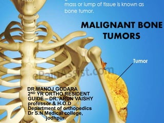
- 1. MALIGNANT BONE TUMORS DR.MANOJ GODARA 2ND YR ORTHO RESIDENT GUIDE – DR. ARUN VAISHY professor & H.O.D Department of orthopedics Dr S.N Medical college, jodhpur
- 2. DEFINATION – . Malignant bone tumors are those that are biologically more aggressive not only they cause rapid tissue destruction but also have greater incidence of spread and metastasis . Malignant tumor of bone and cartilage constitute about 0.5% of all malignant tumor . Primary bone tumors are relatively rare . Metastatic bone tumors are the most common malignant tumors of skeleton system. 70%of all malignant tumors are metastatic in nature
- 3. Characteristics of malignant bone tumors . They have aggressive spread, metastasis and invasiveness to surrounding tissue . More rapid increase in size. . less differentiation . They have wide zone of transition and interrupted periosteal reaction. . ill-defined and irregular bone margins
- 5. CLASSIFICATION OF MALIGNANT BONE TUMORS
- 8. Enneking staging for malignant Tumors It is based on histological grade ,local tumor extent (whether confined to anatomical compartment or not) and the presence and absence of metastasis
- 9. Malignant bone tumor Primary bone tumors 30% Secondary bone tumors 70% Multiple myeloma -most commo Osteosarcoma – 2ndmost common . Chondrosarcoma . Ewing's sarcoma . Chordoma . Fibrosarcoma Secondaries are metastasis of other primary tumors of body. They arises from Lungs, breast or prostrate cancer. Most common site of deposition of secondaries are vertebra and pelvis
- 10. Osteosarcoma • Def :-Primary malignant tumor in which malignant mesenchymal cell produces osteoid or immature bone . most common primary malignant tumor of the bone excluding those of hematopoietic origin like multiple myeloma . Incidence :- 1:3 per 1 million /year peak incidence at second decade of life more prevalent in male then in female.
- 12. • Classified as Primary and secondary osteosarcoma PRIMARY OSTEOSARCOMA . High grade – Conventional osteosarcoma , Telangiectatic osteosarcoma small cell osteosarcoma .low grade – central osteosarcoma perosteal osteosarcoma .intermediate grade – periosteal osteosarcoma
- 15. Clinical features • .Most patients usually present with persistent pain . Deep ,firm , fixed mass . Swelling usually develops at the end of long bones and its consistency varies from soft ,almost fluctuant to firm and indurated to bony hard . Overlying skin may become stretched, thin and glossy . Motion of adjacent joint is unimpaired until the muscle acting on the joint became involved
- 17. Diagnostic • Plain x-ray finding variable mixture of radio-opacity due to osteogensis and radiolucency due to destructive changes .
- 18. • . Irregular destruction in metaphysis . .New bone formation . Periosteal reaction .sunburst appearance :-> fine lines of increase densities , representing newly formed spicule of bone ,radiate laterally from bone at the right angle from the shaft
- 20. Sometimes pathological fractures may be seen on X-RAY imaging
- 21. . MRI help to determine the level of resection and preoperative planning Used to know the soft tissue extent • to screen the skip lesion and find out the involvement of the medullary canal,epiphysis and physeal plate CT-SCAN Not much of use in diagnosis of osteosarcoma H RCT chest to detect lung metastasis which is the most common site of metastasis.
- 22. Angiogram
- 23. Bone scan
- 28. • No pain and no tenderness • Wide sx excision is the t/t
- 29. Prognostic factor • Poor prognostic factor includes -Mets at the time of presentation -Primary tumor located in axial skeleton -large tumor -increase ALP and LDH -Poor response to prior chemo
- 31. .For high grade osteosarcoma - surgical excision of primary tumor with chemotherapy .Multi agent chemotherapy is the standard care in osteosarcoma .Most current protocol incorporate these agent in three or four drug combinations
- 32. CHEMOTHERAPY
- 34. Surgery
- 37. ROTATIONPLASTY
- 41. CHONDROSARCOMA • Chondrosarcoma is a malignant tumor of cartilage producing cells. • Most common sarcoma of bone in patient over 20 yrs of age. 9 percent of all primary malignancy of bone… .Usually beyond 3rd decade of life but peak incidence in the 5th to 7th decade .M.F= 3:2 .Pelvic is the most common site (ilium) followed by proximal humerus ,proximal femur and ribs are the other site
- 44. Clinical features . Deep ,dull pain (worsen at night ,insidious progression over months )is most common presentation . local swelling .pathological fractures (in high grade chondrosarcoma)rare presentation .Neurovascular disturbance with loss of motion
- 45. Diagnostic • On Plain radiograph (X-RAY) . Expansion of medullary portion of bone with cortical thickening is typical finding. . Periosteal reaction is scant/absent . Irregular matrix calcification- punctuate , popcorn, comma shaped calcification( rings and arcs appearance) •
- 49. • CT SCAN Differentiate benign from malignant lesion • For characterization of lesion in anatomically complex areas- sacrum and spine MRI .Depict high water content of lesion with lobulation at margins .Best to see intramedullary extent , Cortical erosion, bone destruction, reactionary edema, soft tissue extention are well depicted. .It helps in differentiating Enchondromas with chondrosarcoma
- 50. MRI
- 52. Bone biopsy and histological findings • Histology Composed of malignant cells with abundant cartilaginous matrix, cartilage permeates the host bony trabeculae Histologically three grades Grade 1- less cellular and less nuclear atypia. Grade 2 - ↑ cellularity and nuclear atypia Grade 3- ↑ ↑ cellularity and pleomorphic anaplastic cells
- 53. Gross appearance
- 54. Differential Diagnosis Enchondroma .enchondroma and low grade chondrosarcoma radiologically as well as histologically appear similar Both located in metaphysis with stippled calcification and endosteal scalloping • But in chondrosarcoma endosteal scalloping is present in >2/3 rd of area and aggressive changes such as bone destruction ,cortical erosion ,periosteal reaction , soft tissue mass is more marked
- 56. Treatment . These tumor generally not sensitive to chemotherapy and radiotherapy and surgery is the only reliable treatment .for grade 1 chondrosarcoma- if endosteal scalloping and cortical erosion absent on x-ray ,no contrast uptake and no mass on MRI then extended intralesional curettage can be done but if present then we goes for wide resection .for grade 2 & 3 – wide resection is the only treatment modality 10 yr survival rate grade 1 – 90% grade 2 – 40-50% grade 3 – 15-20%
- 57. SECONDARY CHONDROSARCOMA • Chondrosarcoma arising in a known benign precursor lesion ex- osteochondroma , enchondroma olliers disease, Maffuccic syndrome-25-30% .develops at somewhat earlier age then primary .usually low grade malignancy and have favorable prognosis .plain radiograph shows increase thickness in cartilaginous cap in osteochondroma and destructive permeation of bone and soft tissue mass in enchondroma T/t- complete wide surgical excision
- 59. INTRODUCTION • 2ND most common malignant tumor of late childhood and early adulthood • incidence 0.3/100000/yr • This small blue round cell tumor is arise from mesenchymal cells • more common in males • Peak incidence during first two decade of life • cytogenetic- most commonly a reciprocal translocation with FLI1 gene on chromosome 11 t(11;22)
- 64. • CT scan- to evaluate the pattern of bone destruction • MRI- to identify extra osseous and bone marrow involvement • v
- 73. CHORDOMA • Low to intermediate grade malignant tumor of cranium and spine • Arise from the embryonic remnant of notochord • account 1-4% of all bone malignancy • Age- 5th -6th decade of life • M>F • Site- sacrococcygeal region (40-50%) base of skull(35-40%) vertebrae (15-20%) C/F- mild and intermittent pain in rectal and anal region constipation and urinary difficulty neurological spine in chordoma of spine cranial nerve palsy in skull base chordoma
- 74. • Plain x-ray shows destructive bony lesion with in the vertebral body and surrounding soft tissue mass.
- 75. Complete en bloc surgical resection is the treatment of choice • local recurrence varies from 15-20% • 5% of chordoma show lung ,skin and brain Mets
- 76. MULTIPLE MYELOMA
- 82. • MRI – for detecting thoracic and lumber spine lesion • Bone scan – more then 50% lesion can be missed • plain radiography shows diffuse osteoporosis and lytic lesion • no reactive bone formation