
Empfohlen
Weitere ähnliche Inhalte
Was ist angesagt?
Was ist angesagt? (20)
Ähnlich wie parathyroid scan
Ähnlich wie parathyroid scan (20)
Mehr von @Saudi_nmc
Mehr von @Saudi_nmc (20)
Kürzlich hochgeladen
Kürzlich hochgeladen (20)
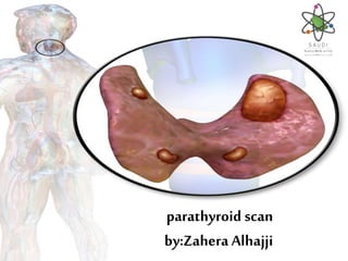
parathyroid scan
- 2. Introduction •-There are four parathyroid glands located alongside and sometimes within the thyroid gland. •-There are usually two parathyroid glands near the left lobe of the thyroid and two near the right lobe. •-They are small, oval-shaped glands each approximately 6 mm and weighing approximately 30–50 mg.
- 3. Indication •Localization of primary and secondary parathyroid cancer •Localization of hyperfunctioning parathyroid tissue (adenoma/ hyperplasia). •Localization of hyperfunctioning parathyroid tissue in patients with persistent or recurrent disease
- 4. Contraindication •-Pregnant women •-patient on calcium medication •- patient on thyroid medication (off for 5 days) •Precaution •Precaution: Discontinuation of breast feeding for nursing mothers for 24 hours after imaging.
- 5. Radiopharmaceutical •99mTechnetium(sestamibi-tetrofosmin) adult: 15 mCi •99mTechnetium-pertechnetate adult: 5-12 mCi •Child: As per body weight •Technique of administration •Intravenous Injection
- 6. Examination Time • Initial injection and images at 15-20 min • Delayed images at 2 hours .
- 7. EQUIPMENT CAMERA Small or Large field of view COLLIMATOR (pinhole or low energy all purpose or low energy high Resolution ) .
- 8. Patient position& Imaging field • Patient position: Supine. • Imaging field: Neck and mediastinum
- 9. PROCEDURE • Prepare the camera and Place patient in supine position, pillow under shoulders, head back, neck extended. • • Position camera anterior over extended neck and mediastinum. • Remove any attenuating necklaces or pendants. • • Instruct patient to remain motionless during acquisitions.
- 10. • Acquire ANT dynamic images. • Oblique views may be requested by the physician • Repeat all images at approximately 2hours after injection. Use the same camera, and as close as possible to the original patient positions and collimator positions.