
Empfohlen
Weitere ähnliche Inhalte
Was ist angesagt?
Was ist angesagt? (20)
Andere mochten auch
Andere mochten auch (20)
Ähnlich wie Osteomyelitis seminar
Ähnlich wie Osteomyelitis seminar (20)
Kürzlich hochgeladen
Kürzlich hochgeladen (20)
Osteomyelitis seminar
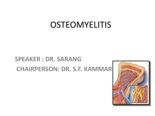
- 1. OSTEOMYELITIS SPEAKER : DR. SARANG CHAIRPERSON: DR. S.F. KAMMAR
- 2. vascular anatomy Infants : Metaphysis and diaphyseal vessels penetrate the physis(growth plate) upto 1 year. So high incidence of septic arthritis with epiphyseal involvement in osteomyelitits in infants Children: No metaphyseal blood vessels penetrate the physis and the epiphyseal blood supply is distinct and seperate.
- 3. Adults • Metaphyseal vessels gradually penetrate the vanishing physis, establish the communication between metaphysis and subarticular end of the bone(previously epiphysis) • thus septic arthritis secondary to osteomyelitis occurs in adults.
- 4. • The outer one-third of the cortex is supplied primarily by the periosteal system, and the inner two-thirds is supplied by the medullary arteries. • The metaphyseal complex primarily supplies the physis and metaphyseal regions of long bones. It merges with the medullary and epiphyseal arterial system following physeal closure • The three systems are interconnected such that if one is injured, another can compensate to provide revascularisation.
- 5. Definition • Osteomyelitis is defined as an inflammation of the bone caused by an infecting organism. • it is due to a single organism, but polymicrobial infections can occur, especially in the diabetic foot.
- 6. Osteomyelitis is classified based on Duration • Acute • Subacute and • Chronic osteomyelitis Mechanism of infection • Haematogenous- from bacteremia • Exogenous -open fractures -contiguous spread from infected local tissues
- 8. • Rapidly destructive pyogenic infection • Hematogenous origin • Starts in metaphysis of a actively growing long bone • Usually in infants and children(M>F) • May occur in adults due to spread of infection directly from another site or through compound fractures,hematogenous spread is rare but present in immunocompromised patients.
- 9. Causative Organisms • Infants – Staph. aureus most common – Streptococcus group B – Coliform organisms • 6 months to 4 years – Staph. Aureus – H. influenzae (not common nowadays due to immunization)
- 10. Causative Organisms • Older children & adults – Staph . aureus • Sickle cell disease – Salmonella paratyphi • IV drug abusers - pseudomonas • Fungal osteomyelitis –in chronically ill patients on long term intravenous therapy or parenteral nutrition
- 11. Etiology Pre-disposing factors- 1. Disease is common in active bone growth 2. Male > female 3. Poor nutrition 4. Trauma 5. Skin, dental, respiratory, GI, urinary tract infection 6. Burns 7. Iv drug abuse 8. Sickle cell anaemia 9. Immuno compromised state 10.Old age, debilitated state
- 12. METAPHYSIS is MC site .. Why? 1. Growing end, increased blood supply 2. Hairpin bend of blood vessels 3. Immature cells of metaphysis due to high cell turnover 4. Defective phagocytosis 5. Dead & dying tissue of cartilage, good nutritive material for organisms 6. End arteries, arteries open into sinusoids 7. Common site for Trauma – haemorrhage
- 13. Pathogenesis • Bacterial seeding Inflammatory reaction local ischemic necrosis of bone
- 14. Abscess formation as abscess enlarges intramedullary pressure increases cortical ischemia
- 15. • Purulent material escape through cortex into subperiosteal space • subperiosteal abscess • if left untreated • sequestra formation and finally chronic osteomyelitis
- 16. Physical examination • Fever, • Pain at the site of infection, • Limited use of the affected extremity. • Constitutional symptoms, such as lethargy and anorexia • Local signs of inflammation • Cellulitis • LNpathy
- 17. Physical examination Signs are often age-dependent. • Neonates – have a thin periosteum that is easily penetrated by infection – will present with swelling at the affected site & adjacent joint and irritability on movement of the limb. • Older children, – due to thicker metaphyseal cortex and densely adherent periosteum, – will have point tenderness
- 18. • In infants , elderly and immunocompromised pts clinical findings may be minimal
- 19. Investigations • WBC count – often normal • Erythrocyte sedimentation rate (ESR), usually raised • C reactive protein (CRP) usually raised & useful in monitering the course of treatment.
- 20. Bacterial Cultures From sites of bone infection • in order to focus antibiotic treatment • Aspiration performed through thin metaphyseal bone with an 18-gauge spinal needle under local infiltration • Positive in 48% to 85% cases Blood culture • Positive in 50% cases
- 21. Radiography • Soft tissue swelling will be evident within 48 hours of the onset of infection. • Periosteal new-bone formation or bony destruction may be evident by 10 to 12 days.
- 22. Acute osteomyelitis The first x-ray, 2 days after symptoms began, is normal . metaphysealmottling and periosteal changes were not obvious until the second film, taken 14 days later. eventually much of the shaft was involved.
- 23. Bone scan . • can confirm diagnosis within 24 to 48 hrs after onset •Highly sensitive but lacks specificity •Examples •Tecnetium99m bone scan •Gallium scanning •Indium-111 tagged WBCs
- 24. Magnetic resonance imaging • sensitivity of 96% to 100% and a specificity of 75% in the detection of osteomyelitis. • Can show early inflammatory changes in bone marrow & soft tissue • Useful for detecting intraosseous and subperiosteal abscess
- 25. Computed tomography • Identification of sequestra Newer methods: FDG-PET SPECT
- 26. TREATMENT
- 27. NADE proposed five principles for the treatment of acute hematogenous osteomyelitis (1) an appropriate antibiotic is effective before abscess formation. (2) antibiotics do not sterilize avascular tissues or abscesses, and such areas require surgical removal. (3) if such removal is effective, antibiotics should prevent their re-formation, and primary wound closure should be safe. (4) surgery should not damage further already ischemic bone and soft tissue. (5) antibiotics should be continued after surgery.
- 28. Treatment • General supportive care – Intravenous fliuds – Analgesics – Comfortable positioning & immobilization of limb
- 29. Antibiotic therapy • If started before significant bone and soft-tissue necrosis, it is more likely to be successful without the need for surgical. • According to culture sensitivity report • Before culture – according to Gram staining • If Gram staining –ve then empirical treatment should cover all possible pathogens • CRP level monitored to know response to therapy
- 30. Antibiotic therapy • Intravenous therapy ranging from 4 to 8 weeks (Success of treatment correlates most closely with an adequate serum level of the antibiotic, rather than the route of administration) • Transition to oral antibiotics once clinical improvement was observed • Treatment continuing until normalization of the ESR & CRP.
- 31. Antibiotic therapy Infants – recommended that the entire course of treatment be intravenous as • more prone to generalized sepsis, • have less consistent oral antibiotic absorption, • have a less predictable radiographic and serologic response to treatment,
- 32. Surgery • Indications – Abscess – No improvement despite of appropriate antibiotic therapy • Principle of surgery is to remove infected nonviable necrotic bone and soft tissue, decompression of abscess cavity facilitating antibiotic delivery.
- 33. Surgery • If subperiosteal abscess – multiple drill holes through cortex • If intramedullary pus – cortical window
- 34. Complications • Septicemia • Dehydration & electrolyte imbalance • Recurrence of infection • Chronic osteomyelitis • Growth arrest
- 36. • Subacute osteomyelitis has a more insidious onset and lacks the severity of symptom. • Because of the indolent course of subacute osteomyelitis, diagnosis typically is delayed for more than 2 weeks.
- 37. • Systemic signs and symptoms are minimal. • Temperature is only mildly elevated • Mild-to-moderate pain is one of the only consistent signs suggesting the diagnosis. • Wbc counts are normal. • ESR is elevated in only 50% of patients, and blood cultures are negative. • Even with an adequate bone aspirate or biopsy specimen, a pathogen is identified only 60% of the time.
- 38. • The indolent course of subacute osteomyelitis is thought to be the result of increased host resistance, decreased bacterial virulence, or the administration of antibiotics before the onset of symptoms.
- 39. classification • Radiographic classification of subacute hematogenous osteomyelitis was described by Gledhill and was modified by Roberts et al.
- 40. Type Gledhill Classification Robert et al. Classification Differential Diagnosis I Solitary localized zone of radiolucency surrounded by reactive new bone formation Ia—Punched-out radiolucency Langerhans' cell histiocytosis Ib—Punched-out radiolucent lesion with sclerotic margin Brodie abscess II Metaphyseal radiolucencies with cortical erosion — Eosinophilic granuloma; osteogenic sarcoma III Cortical hyperostosis in diaphysis; no onion skinning Localized cortical and periosteal reaction Osteoid osteoma IV Subperiosteal new bone and onion skin layering Onion skin periosteal reaction Ewing sarcoma V — Central radiolucency in epiphysis Chondroblastoma VI — Destructive process involving vertebral body Tuberculosis; osteogenic sarcoma
- 41. type 1,central metaphyseal lesion;type2,eccentric metaphyseal lesion with cortical erosion; type 3, diaphyseal cortical lesion; type 4,diaphyseal lesion with periosteal new bone formation, but without definite bony lesion; type 5, primary subacute epiphyseal osteomyelitis; and type 6, subacute osteomyelitis crossing physis to involve metaphysis and epiphysis. Classification of subacute osteomyelitis:
- 42. • The diagnosis often must be established by an open biopsy and culture. • Purulent material is not always obtained on biopsy, but granulation tissue is a common finding. • S. aureus and Staphylococcus epidermidis are the predominant organisms identified in subacute osteomyelitis.
- 43. • Biopsy and curettage followed by treatment with appropriate antibiotics ( IV anibiotics for 48 hrs followed by 6 weeks of oral antibiotics ) generally are recommended for all lesions that seem to be aggressive For lesions that seem to be a simple abscess in the epiphysis or metaphysis, biopsy is not recommended.
- 44. BRODIE ABSCESS • localized form of subacute osteomyelitis that occurs most often in the long bones of the lower extremities of young adults. • In adults, the metaphyseal- epiphyseal area is involved. • Intermittent pain of long duration is the presenting complaint, along with local tenderness over the affected area.
- 45. • On plain radiographs, appears as a lytic lesion with a rim of sclerotic bone . • S. aureus is cultured in 50% of patients; in 20%, the culture is negative. • It requires an open biopsy with curettage to make the diagnosis. The wound should be closed loosely over a drain. • iv antibiotics 2days f/b oral for 6 weeks
- 46. • More recently, Jones et al. described a based on radiographic appearance: Type A - Brodie abscess. Type B - sequestrum involucrum. four subtypes: • B1 localized cortical sequestrum. • B2 sequestrum with structural involucrum. • B3 sequestrum with sclerotic involucrum and • B4 sequestrum without structural involucrum. Type C -sclerotic • Physeal damage is indicated by the addition of “P” (proximal) or “D” (distal) to the classification.
- 48. Introduction • Chronic osteomyelitis is often defined as the presence of ongoing bone infection for longer than 1 month in the presence of devitalized bone • Infection mainly involves - Marrow spaces -Haversian canals -Subperiosteal Spaces
- 49. Hematogenous osteomyelitis of Tubular bone in child
- 50. Hairpin Bend vessels flow becomes considerably slower and more turbulent
- 51. Factors That Turn Acute Bone Infection to Chronic Osteomyelitis: Trauma (orthopaedic surgery or open fracture) Prosthetic orthopaedic device Diabetes Peripheral vascular disease Alcoholism Intravenous drug abuse Chronic steroid use Immunosuppression Tuberculosis HIV and AIDS Sickle cell disease
- 52. • The hallmark of chronic osteomyelitis is infected dead bone within a compromised soft-tissue envelope • The infected foci within the bone are surrounded by sclerotic, relatively avascular bone covered by a thickened periosteum and scarred muscle and subcutaneous tissue. • This avascular envelope of scar tissue leaves systemic antibiotics essentially ineffective
- 53. • Bacteria settle down in metaphysis • Primary focus in metaphysis (form abscess in metaphysis) • Subperiosteal abscess • Sequestrum formation (dead bone ) • Involucrum formation (new bone formed by cambium layer of periosteum) • Pus eventually perforates periosteum and forms abscess in soft tissues. • Ultimately abscess burst on surface and forms discharging sinus
- 54. Chronic osteomylitis involving metaphysis of tibia
- 55. SEQUESTRUM • Dead piece of bone separated from living bone by a layer of unhealthy granulation tissue and lying freely in the cavity • Types 1) Tubular – in pyogenic infections below 2 yrs 2) Feathery – pyogenic infections 3) Course sandy – tuberculosis 4) Dense ivory – syphilis
- 56. 5) Coloured ( Black) – in ulna & tibia osteomyelitis 6) Ring – Amputation stump & pin track infections 7) Bombay – Calcaneal OM 8) Corolliform – Pyogenic infections 9) Buttonhole – Pott’ puffy tumour, Tuberculosis of skull bones
- 57. INVOLUCRUM: it is a immature,subperiosteal,reactive,living new bone formation around dead bone
- 59. CLASSIFICATION OF CHRONIC OSTEOMYELITIS • Cierny and Mader classification system is based on physiological and anatomical criteria, to determine the stage of infection • The physiological criteria are divided into three classes based on three types of hosts
- 60. • Class A hosts have a normal response to infection and surgery • Class B hosts are compromised and have deficient wound healing capabilities • When the results of treatment are potentially more damaging than the presenting condition, the patient is considered a class C host
- 61. • Anatomical criteria consist of four types • Type I, a medullary lesion, is characterized by endosteal disease • In type II, superficial osteomyelitis is limited to the surface of the bone
- 62. • Type III is a localized infection characterized by full-thickness cortical sequestration and cavitation (in this type, complete débridement of the area would not lead to instability) • Type IV is a diffuse osteomyelitic lesion that creates mechanical instability, either at presentation or after appropriate treatment
- 63. Stage Characteristic Features I Medullary Endosteal disease II Superficial Cortical surface infected because of coverage defect III Localised Cortical sequestrum that can be excised without compromising stability IV Diffuse I, II and III plus mechanical instability before or after debridement. Anatomical Type Cierney and Mader staging system of Chronic Osteomyelitis
- 64. Class Host’s immune system Features A host Normal Immunocompetent with good local vascularity B host Compromised Local or systemic factors that compromise immunity or healing C host Prohibitive Minimal disability, prohibitive morbidity anticipated, poor prognosis for cure, treatment worse than disease Physiological classification
- 65. Clinical Staging (Cierny-Mader, 1985) Anatomic Type + CLINICAL STAGE Physiologic Class Example: IV B tibial osteomyelitis = diffuse tibial lesion in a systemically compromised host
- 66. • USE OF THIS CLASSIFICATION • - To decide whether treatment should be • 1) Simple or Complex • 2) Curative or Palliative • 3) Limb sparing or Ablative
- 67. CLINICAL FEATURES • DURING THE PERIOD OF INACTIVITY - Usually no symptoms - Skin over the focus is dusky, thin, scarred - Break in the skin causes ulceration that heals slowly - Muscles are scarred & leads to contractures of the adjacent joints
- 68. • DURING ACUTE EXACERBATION - Aching pain worsening at night, overlying soft tissue becomes edematous, warm redddened & tender - patient is febrile - As infection progresses, sinus may open up & drain extruding small sequestrum at intervals
- 69. - Intervals between flare ups may be months or years - Flare ups may be due to poor general condition & lowered resistance - Recurrent toxemia will eventually cause debilitary & sometimes fatal amyloidosis
- 70. DIAGNOSIS • The diagnosis of chronic osteomyelitis is based on CLINICAL, LABORATORY & IMAGING studies
- 71. • POSSIBLE CLINICAL FINDINGS - Asymptomatic - Pain - Fever - Tenderness --Erythema - Swelling - Sinus Tract - Limp - Drainage
- 72. • LAB. INVESTIGATIONS - Nonspecific - Gives no indication of severity of infection Raised ESR & CRP Raised WBC ( Raised Lymphocytes) in 35% patients Biopsy with Culture & Sensitivity (Gold Standard)
- 73. IMAGING
- 74. It takes from 10 to 21 days for an osseous lesion to become visible on conventional radiography, because a 30–50% reduction of bone density must occur before radiographic change is apparent - earliest radiographic changes appears after 8 to 10 days - In early stages, bone appears moth eaten & osteoporotic due to CORTICAL DESTRUCTION with sclerotic areas - PERIOSTEUM IS ELEVATED by subperiostel laminations of new bone -grdually each necrotic dense area becomes surrounded by white ring representing new bone formation, the INVOLCRUM
- 75. Ssequestrum with normal structural involucrum of a proximal humerus.
- 77. SINOGRAPHY Helpful if the sinus is present in locating focus of the infection in chronic osteomyelitis
- 78. Sinogram
- 79. Scintigraphy They are more useful in acute om than chronic • TC 99m Poly Phosphate scan • Gallium citrate scan • Indium • Sensitivity (84 to 100 percent) and specific (70 to 96 percent)
- 80. 1)99M Tc Scan - Shows increased uptake in the areas of increased blood flow or oseoblastic activity
- 81. 2) 67Ga Scan - shows increased uptake in areas where leucocytes or bacteria accumulate - normal Ga scan excludes presence of osteomyelitis - useful as follow up examination after surgery
- 82. These leukocytelabeled images suggest multiple areas of bilateral pedal osteomyelitis.
- 83. CT SCAN - Provides excellent definition of cortical bone - especially useful in identification of Sequestra
- 84. CT Scan
- 85. MRI SCAN - for evaluation of soft tissue involvement - reveals a well defined rim of high signal intensity surrounding the focus of active disease ( RIM SIGN)
- 86. A case of chronic osteomylitis of fibula
- 87. MR images chronic osteomyelitis of right distal femur.
- 88. TREATMENT .
- 89. • Chronic OM generally cannot be eradicated without surgical treatment • Surgery for Chronic OM consists of sequestrectomy & resection of scarred & infected bone & soft tissue
- 90. • THE GOALS OF SURGERY - Eradication of infection by achieving a viable & vascular environment -Radical debridement -Prevent recurrences
- 91. SEQUESTRECTOMY & CURETTAGE All sinus tracts are excised completely along with sequestra, purulent material & scarred and necrotic tissue if sclerosis bone seals off a cavity within the medullary canal, it is opened into the canal in both directions to allow blood vessels to grow into the cavity
- 92. • When medullary canal is infected intramedullary reaming has shown favourable results in the treatment of medullary osteomyelitis.
- 93. • AFTER TREATMENT - Limb is splinted until the wound is healed & then it is protected to prevent pahological fracture - prolonged antibiotic therapy is given usually for 6 weeks - bony & soft tissue defects must be filled to reduce chances of continued infection & loss of function
- 94. • METHODS TO ELIMINATE THIS DEAD SPACE 1) Bone grafting with primary or secondary closure 2) use of PMMA beads 3) local muscle flaps & skin grafting with or without bone grafting 4) microvascular transfer of muscle, myocutaneos, osseous & osteocutaneous flaps 5) use of bone transport( Ilizarov’s technique)
- 95. • OPEN BONE GRAFTING( PAPINEAU TECHNIQUE) This procedure is based on the following principles: - granulation tissue markedly resists infection - autogenous cancellous bone grafts are rapidly revascularized and are resistant to infection
- 96. • DONE IN 3 STAGES 1)Excision of infected tissue without or with stabilization using an external fixator or an intramedullary rod 2) Cancellous autografting 3) skin closure
- 97. PMMA BEAD CHAINS • -The rationale is to deliver levels of antibiotics locally in concentrations that exceed the minimal inhibitory concentrations • Primary wound closure is done • Suction drains are not recommanded • Aminoglycosides are mc used -most commercially available bone cements have a prepackaged form available with GENTAMICIN (500mg /40g) - In short-term implantation, the beads are removed within 10 days and in long-term implantation, they may be left for 80 days
- 98. Preparation of antibiotic beads
- 99. Polymethylmethacrylate (PMMA) beads connected together in a chain are the most widely used drug delivery system.
- 101. BIODEGRADABLE ANTIBIOTIC DELIVERY SYSTEMS • -Biodegradable substrates contain osteoconductive and osteoinductive materials, which can be used to promote new bone formation - Bioabsorbable substrates (calcium sulfate or calcium phosphate) that can be mixed with antibiotics ( Vancomycin or Tobramycin) -These beads typically resorb by about 8 weeks after surgery
- 102. • Advantages - 2nd procedure is not required to remove the implant - Calcium in the substrate can be used in new bone formation
- 103. CLOSED SUCTION DRAINS • -For resistant focal infections, topical instillation of solution containing mild detergent ( eg. Alevaire) & one or more antibiotic seems to be effective
- 104. -Closed suction antibiotic ingress and egress high- volume irrigation systems(Lautenbach continuous irrigation method) can be used over 3 to 21 days - the material collected through suction tube is cultured every day when 3 successive negative cultures are obtained, the antibiotic detergent solution is discontinued
- 106. SOFT TISSUE TRANSFER • - Soft-tissue transfer is done to fill dead space left behind after extensive debridement - transfer of vascularized muscle tissue improves the local biological environment by bringing in a blood supply for antibiotic delivery and osseous and soft-tissue healing.
- 107. Most commonly, a local muscle flap is used in the treatment of chronic osteomyelitis of the tibia The gastrocnemius muscle is used for defects around the proximal third of the tibia the soleus muscle is used for defects around the middle third A microvascular free muscle transfer is required for defects around the distal third of the tibia
- 108. - When a microvascular free muscle flap is used, and segmental bone loss has occurred, autogenous cancellous bone grafting can be done about 6 weeks after the initial free flap transfer - free fibular graft can be used for segmental bone loss of the tibia - If chronic osteomyelitis involves segmental bone loss of the tibia and the fibula, the results of a free fibular graft are not good, and amputation or reconstruction by the Ilizarov technique is advised
- 109. ILIZAROV’S TECHNIQUE The Ilizarov technique has been helpful in the treatment of chronic osteomyelitis and infected nonunion -
- 110. • BELFAST TECHNIQUE 2 Stage Technique - in 1st stage, all necrotic & infected tissues are debrided & is covered with soft tissue transfer - in 2nd stage , at later time autogenous cancellous grfating is done after infection has been completely subsided
- 111. Adjunctive techniques • Silver iontophoresis • hyperbaric oxygen therapy • Pulsed electromagnetic fields and ultrasound
- 112. AMPUTATION FOR CHRONIC OSTEOMYELITIS • - Malignant change of sinus tract - Madura Mycosis - Joint contractures & stiffness
- 113. COMPLICATIONS • Growth disturbances • Pathological fractures • Muscle contractures • Secondary septicemia • Epithelioma • Malignant changes( Squamous cell ca,Reticulum cell ca, Fibrosarcoma) • Joint stiffness • Amyloidosis
- 115. SCLEROSING OM OF GARRE • - Idiopathic cortical sclerosis - mc site Tibia -chronic form of disease in which the bone is thickened and distended, but abscesses and sequestra are absent -affects children and young adults -cause is unknown, but it is thought to be an infection caused by a low-grade, possibly anaerobic bacterium
- 117. • CLINICAL FEATURES - Intermittent pain - Swelling & Tenderness over affected bone
- 118. • INVESTIGATIONS - ESR : slightly raised - C/S : usually negative - X Ray : expanded bone with generalised sclerosis - Biopsy : chronic low grade non specific OM
- 119. • DIFFERTIAL DIAGNOSIS - Paget’s disease - Osteoid osteoma
- 120. • TREATMENT - Fenestration of sclerotic bone - Antibiotics
- 121. RESIDUAL STAGE OF OSTEOMYELITIS - characterized by a complete absence of the signs and symptoms of infection, including drainage -The bone is sclerotic, and its blood supply and strength are normal
- 122. • TREATMENT -correcting leg-length inequality or angular and joint deformities -contracted scars must be released -adherent scars must be replaced by myocutaneous flaps
- 123. PATHOLOGICAL FRACTURE IN OSTEOMYELITIS -Because the involucrum is sometimes insufficient, the shaft of a long bone may fracture during the acute or subacute stage of osteomyelitis before immobilization has been started -all operations necessary to combat the infection should be carried out thoroughly, and bone fragments are then realigned and immobilized as with any other fracture -External fixation or cast immobilization usually is preferred -If bone loss is significant, the defect can be filled with autogenous bone graft, a vascularized osseous graft, or bone transport using the Ilizarov technique
- 124. CHRONIC RECURRENT MULTIFOCAL OSTEOMYELITIS • It is characterized by an insidious onset of mild-to-moderate pain with signs of inflammation over the affected parts, which tend to recur • often affects the metaphysis of long bones
- 125. • ETIOLOGICAL FACTORS - Infectious disease : Propiobacterium acne - Autoimmune Diseases : Psoriasis & IBD - Genetic Predisposition : LPIN2 gene
- 126. • CLINICAL FEATURES - Nonspecific : pain, swelling, restricted mobility - Duration : from days to several years
- 127. • INVETIGATIONS - raised ESR & CRP with normal WBC count - Cultures : negative - Biopsy : may be useful
- 128. TREATMENT • No effective treatment for CRMO available • Cases of disease remission after treatment with IFN gamma have been reported • Resolution of symptoms followed by recurrence months later is characteristic of this disease • Generally, the symptoms continue to recur over 2 years, and the disease generally is self-limiting • The long-term prognosis seems to be good
- 129. Spine OM • Commonly affects lumbar spine < thoracic spine < cervical spine • Usual site : vertebral bodies • Simultaneous involvement of the 2 adjacent vertebral end plates with intervening discs
- 131. • ETIOLOGY - richly vascular metaphyseal bone near anterior longitudinal ligament - avascular disc - end arterial supply of interosseous artery in adults
- 132. • X Ray Findings - disc space narrowing with end plate erosion with vertebral body destruction - vertebral end plate sclerosis with increased density in subchondral bone - spinal fusion
- 133. • TREATMENT - IV Antibiotics - Rest - Spinal immobilisation - Surgery
- 134. TUBERCULOUS OM • - chronic granulomatous infection caused by AFB - usually secondary to primary elsewhere - commonly involving metaphysis in children & epiphysis in adults
- 135. • CLINICAL FEATURES -swelling - local tenderness - muscle spasm - cold abscess with chronic discharging sinus
- 136. • INVESTIGATIONS - Tubeculin test - Biopsy :typical tubercles - Blood : lymphocytosis - X Ray: osteoporosis, loss of outline of articular cortex, granular foci
- 137. • TREATMENT - Sinuses with sequestra : curretage & excision - AKT
- 138. Thank you