1. Collecting and Culturing Thermophiles at Goldstrike Canyon
Sara B. Gowin, Joshua Morgan, Ama Tran and Camille Naaktgeboren
Abstract
This focus of this project is potential thermophiles, a type of extremophile which
grows at temperatures at or above 50° C. Water samples were collected from four
hot springs in the Goldstrike canyon area and inoculated into several standard
microbiological media. Organisms were then isolated into pure cultures and each
organism’s unique characteristics were studied. Four organisms were originally
isolated from pond 3, but upon later inspection, additional organisms were found in
the cultures indicating possible associations between rapidly and more slowly
growing individuals. Grams stains and endospore stains were used to determine
morphology and cell characteristics. The optimal temperature for each isolate was
determined by growing each at various temperatures and looking for maximum cell
density. Additional experiments were performed to determine whether any of the
isolates produced antibiotic compounds that inhibited the growth of other
thermophiles.
Materials and Methods
Collection
A sample was collected from a hot spring (360 0’ 10.38” N 1140 44’ 58.19” W), also
called pond 3, south of the Hoover Dam in the Goldstrike Canyon area of Southern
Nevada (Figure 1). The temperature of the hot spring was 440 C. Water from the
surface of the hot spring as well as soil/sediment from the bottom of the spring
were collected in a metal thermos and taken to the laboratory for analysis. The
sample was maintained at 450 C while stored in the lab.
Culture
Five milliliters of the water/sediment sample were inoculated into approximately 50
mL of M17 (Difco) medium. The cultures were incubated at 450 C and were
checked for growth at 24 and 48 hours. At 72 hours, The M17 inoculated samples
were removed from incubation and used to inoculate four types of liquid media (LB,
LB-MRS, TSB, and NA). Four Kerr jars were warmed to 450 C approximately one-
fourth full with equal parts water and prepared media. One mL of culture was
transferred via standard pipette into the jars of media/water mixture. They were
incubated at 450 C with exposure to light for 18 of the 24 hour incubation period.
Once sufficient growth was established in these media the cultures from the TSB
were used to streak Chromagar orientation plates. Re-streaking was performed
until pure cultures were obtained.
Microscopic Examination
Gram-staining of the mixed cultures was performed to get an initial indication of
the species and variety present. Once the organisms were isolated, Gram staining
was preformed on the 24-hour cultures to view morphology, cellular characteristics,
and begin species classification. Endospore stains were also done using thirteen-
day-old cultures to determine whether isolates had formed endospores from low
nutrient levels.
Ideal Growth Temperature
Samples six through nine of the fifteen organisms that were isolated from all four
ponds were inoculated in six Tryptic Soy Broth tubes. Each tube was allowed to
grow at temperatures of 50°, 60°, 70°, 80°, 90°, and 100° C. Optical density at 550
nm was determined every four hours for twelve hours. Results of initial
temperature growth experiment were inconclusive, so a repeat test was run at 40°,
45°, 50°, and 55° C and light absorbance was measured every twelve hours for 72
hours.
Additional studies were performed to determine whether cells remained viable
after incubation at a temperature higher than their optimal. Broth cultures (TSB)
that had been growing at 550 C for 72 hours were streaked onto TSA plates and
allowed to incubate at 500 C. Plates were checked for growth after 24 hours.
Antibiotic activity
Organisms were screened for antibiotic activity using a disc diffusion method. The
isolated organisms were grown in 15 mL of TSB for 48 hours. These broth cultures
were then filtered using a Nalgene filter unit with a 0.45 micron filter. The resulting
fluid was placed on Whatman antibiotic activity 2017 filter paper discs. Those discs
were then placed on a lawns of each other organism grown on Tryptic Soy agar
plates and incubated at 500 C for 24 hours. Plates were analyzed for zones of
inhibition.
Conclusions and Future Research
The organisms cultured may be considered thermophiles as their ideal temperature is
between 45 and 500 C. Tests can be done to determine exact conditions they flourish in.
With proper resources, DNA analysis could be done to quantify the amount different
species present. Long time monitoring may see growth in slower growing organisms from
the pond. With the proper connections, it is possible for this experiment to be conducted
by most people if they have the time, basic biology lab skills, and access to a low tech lab.
Introduction
Extremophiles are organisms which thrive in environments outside the parameters
at which many animals and plants can tolerate. Types of extremophiles include
acidophilies (acid-loving), halophiles (salt-loving), psychrophile (cold-loving),
thermophiles (heat-loving), and xerophiles (dry-loving). Thermophiles thrive in
temperatures at or above 50° C. NASA is interested in these highly adapted
organisms due to extreme conditions on other planets. By studying the ecology and
biology of extremophiles, we can gain insight into the limits of life on Earth, and the
possibility of finding life on other planets. The Goldstrike Canyon area was chosen
as a study site due to the presence of numerous hot springs in the canyon.
The goal of this study was to determine whether there were organisms that
qualified as thermophiles in this hot spring. If there were such organisms we also
wished to determine by which methods they could be cultured and once cultured,
what their general characteristic were. Once those were established additional
information concerning these organisms could be further studied.
Acknowledgments
The funding for this project was provided by a NASA Informal Science Education grant.
College of Southern Nevada Biology Department and Planetarium
Heidi Porter, Ph.D., Pam Maher, M.A.T.
References
Bradley, J.P., Harvey, R.P. & McSween Jr., H.Y. (1996). Magnetite whiskers and platelets in the ALH84001
Martian meteorite: Evidence of vapor phase growth. Geochimica et Cosmochimica Acta, 60 (24),5149-5155.
Retrieved from http://www.sciencedirect.com/science/article/pii/S0016703796003833
Cavicchioli, R., R. Amils, D. Wagner, and T. McGenity. Life and applications of extremophiles. (2011). Environmental
Microbiology 13(8), 1903-1907. doi: 10.1111/j.1462-2920.2011.02512.x
0
0.2
0.4
0.6
0.8
1
1.2
1.4
1.6
0 12 24 36 48 60 72 84
40
45
50
55
0
0.2
0.4
0.6
0.8
1
1.2
1.4
1.6
1.8
2
0 12 24 36 48 60 72 84
40
45
50
55
0
0.2
0.4
0.6
0.8
1
1.2
1.4
1.6
0 12 24 36 48 60 72 84
40
45
50
55
Results
Prokaryotic organisms were observed in the M17 medium after 48 hours of growth and
once transferred to the additional types of media the same types of organisms were
observed. Initially four pure cultures were obtained from these primary cultures. The
analysis of these cultures demonstrated that three were bacillus and one was a
diplobacillus (Table 1). Two were Gram positive and two were Gram negative. All but one
formed endospores. All organisms had optimal growth temperatures between 45 and 500
C (Figure 2). Organisms 6 and 7 remained viable after being incubated at 550 C for 72
hours (Figure 3).
After two weeks of culturing, two of the original isolates appeared to have additional
organisms growing with them. Organism 7 had a small, Gram positive coccus growing with
it that appeared to form endospores. Organism 9 had two additional organisms—a Gram
positive rod and a Gram positive coccobacillus.
None of the organisms exhibited any antibiotic activity against any others that were
isolated from this hot spring or any isolates from other hot springs in the same canyon.
Figure 1. Hot spring from which organisms were collected. The spring is located in the
Goldstrike Canyon area of southern Nevada, just south of the Hoover Dam.
0
0.1
0.2
0.3
0.4
0.5
0.6
0.7
0.8
0.9
0 20 40 60 80
40
45
50
55
Table 1. Chromagar appearance, morphology, Gram reactions and endospore characteristics of the four
isolates from the hot spring.
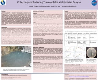
Figure 2. Results of spectrophotometric growth analysis.
Absorbance (OD 550nm) on y axis, time (in hours) on x axis.
Clockwise from upper left- organism 6, organism 7,
organism, 8, organism 9.
Figure 3. Results of plating out
broth cultures incubated at 55
0C for 72 hours. Organism 6
and 7 demonstrate growth after
24 hours.
Sample Chromagar Appearance Morphology Gram Reaction Endospore Present
6
Blue - solid color, deep
growth
diplobacillus Negative Yes
7
Blue – diffuse color
(darker center), white
outer surface
1 bacillus
1 coccus
g- rod
g+ coccus
Yes - coccus
8 Buff – surface bacillus Negative No
9
Buff – surface, granular
shape
2 bacillus
1 coccobacillus
g- rod
g+ rod
g+ coccobacillus
Yes - coccobacillus