SlideShare ist ein Scribd-Unternehmen logo
1 von 53
1
CONTENTS
2
 INTRODUCTION
 HISTORY
 MRI VS CT SCAN
 HOW MRI WORKS
 COMPONENTS OF MRI
 COMPUTER SYSTEM
 COOLING OF MAGNETS
 ADVANTAGES
 DISADVANTAGES
 SHAPE OF MRI MACHINE
Introduction
3
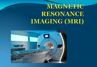
 MRI is a type of scan that uses strong magnetic fields
and radio waves to produce detailed images of insideof
the body.
 An MRI scanner is a large tube that contains powerful
magnets.You lie inside the tube duringscan.
 MRI perhaps the best application ofsuperconductivity
which directlyaffected the humanity across the globe.
Definition
Magnetic Resonance Imaging
Magnet
Radio Frequency = Resonance
Imaging
It is a non-invasive method for mapping internal
structure within the body which uses non-ionizing
electromagnetic radiation and employes radio
frequency radiation in the presence of carefully
controlled magentic fields to produce high quality
cross-sectional images of the body in any plane
Introduction
 Prof Peter Mansfield was awarded Nobel
prize in 2003 for his discovery in MRIwith
Prof Paul C Lauterbur of USA.
The concept of NMR imaging used in
present day MRI system was proposed
by Paul Lauterbur as early as1973.
5
Notes
6
 MRI is perhaps the best application of superconductivity which directly affected the
humanity across the globe. It is a common tool with the radiologist in diagnostic
hospitals for imaging various soft tissue parts of the human body and for detecting
tumors. Theconceptof NMR imaging used in presentday MRI systemswas proposed
by Paul Lauterbur as early as 1973. MRI exploits the presence of vast amount of
hydrogen (protons) in a human body as the water content in a human body is said to
be about 80 %. When protons in the tissues of the body, aligned in a static magnetic
field (B0), are subjected to resonant RF excitation, they absorb energy. Proton relaxes
back and emits resonant signal which is a characteristic of the tissue. The signal is
picked-up by a receiver located inside the magnet bore and is used to construct the
image using Fourier transform. Since the NMR signal frequency is proportional to the
magnetic field the whole tissue can be mapped by assigning different values of the
proton frequency to different proton locations in the sample using well computed
field gradient. All MRIs use proton NMR for mapping proton density which is
different in different types of tissues. The images show contrast which helps in
identifying these tissues and the changes occurring in a sample tissue. MRI turns out
to bean ideal technique forsoft tissue regionsof the bodysuch as brain, eyesand soft
tissue part of the head. Since bones have low density of protons they appear as dark
regions.
History
7
MRI VS CT SCAN
8
CT SCAN
• Uses X-rays forimaging.
• Exposure to ionizingradiation.
• Resolutionproblem.
• Injection of a contrast medium
(dye) can causekidney.
• problems or result in allergic or
injection-site reactions in some
people.
• Less cost thanMRI.
• Quick process and easilyavailable.
MRI
• Uses large external field, RFpulse
and 3 different gradientfields.
• MRI machines do notemit
ionizing radiation.
• Good resolution & 3-D
reconstruction.
• Gadolinium contrast isrelatively
nontoxic.
• Morecost.
• Lengthy process andnon
availability.
Notes
9
 Whyweare using MRI instead of CT scan , here some
comparison between thistwo.
 CT scan usesx ray technology toproduce image but MRI
uses large magnetic field to elicitimage.
 Certain advantages of CT scan over MRI as it is less
expensive, easily available ,quick process .
 Butstill weare going for MRI technology because it has no
ionization radiation so no harm to body, produce good
resolution 3D image and each n every inner injury can be
detected. Soeveryone now preferring MRI although it has
high cost.
HOW MRI WORKS
10
 MRI exploits the presence of vast amount of hydrogen in a
human body as the water content in human body is said to
be about80%.
 At the centre of each hydrogen atom is an even smaller
particle , called proton. Protons are like tiny magnets and
areverysensitive to magnetic fields and has magneticspin.
 MRI utilizes this magneticspin propertiesof protonsof
hydrogen to elicitimages.
 Then whyour bodycan’t like magnets?
HOW MRI WORKS
•The protons i.e. hydrogen ions in abody
•are spinning in a haphazard fashionand
•cancel all the magnetism.
•That is our naturalstate.
•When there is large magnetic fieldacts
•on our body, protons in our body line up in
•same direction.
•In same way that magnetcan pull the
needle of acompass.
11
Notes
12
 Human body is largely made of water molecules, which
consistsof smallerparticles i.e hydrogen and oxygen atoms.
 Protons lies at thecentreof each atom, which is sensitive to
any magnetic fields and hence this proton serves as a
magnet. Normally water molecules in our body are
randomly arranged, but upon entering on the MRI scanner
first magnet causes body’s water molecules to align in one
directionand second magnetwas then turned on and off in
a series of quick pulses, causing each hydrogen atom to
alter their alignment and quickly , switches back to their
original relaxed state, when switchedoff.
HOW MRI WORKS
13
COMPONETS OF MRI
1) Main magnet
(superconducting
magnet)
2)Gradient coils
3)RF coils(radiofrequency)
14
Schematic diagram of MRIscanner
Notes
15
 A superconducting magnet is the heart and most expensive part of an MRI
scanner.MRI magnets need high homogeneity and high temporal stability
similarto NMR spectrometers. However, the magnetic field requirement in
the present day MRI scanners for clinical use is limited to 3T only. Another
majordifferencewith NMR magnet is thatsample size is much larger.
 The main magnet is superconducting, cooled to LHe temperature and
mounted in an efficient cryostat with a horizontal bore to accommodate the
patient. Inside the main magnet is a set of gradient coils for changing the
field along the X, Y and Z directionsrequired for imaging. Inside thegradient
coils are the RF coils producing the field B1 for rotating the spin by an angle
dictated by the pulse sequence. These coils also detect the signal emitted by
the spins inside the body. At the centre is a patient table which is computer
controlled.
 The magnet, the RF bodycoil and thegradientcoil assemblyrepresentthe
three major subsystems that comprise the resonance module of the MR
scanner.
COMPONETS OF MRI
16
17
Different Types of MRI
Coils in MR Systems
• Gradient coils
• RF coil
1. Transmit Receive Coil
2. Receive Only Coil
3. Transmit Only Coil
4. Multiply Tuned Coil
COMPONETS OF MRI
19
1.Superconducting magnet
 A superconducting magnet is the heart andmost
expensive part of an MRIscanner.
 The magnetic field requirement in the present day MRI
scanners for clinical use islimited to 3T only.
 The main magnet is superconducting, cooled to LHe
temperatureand mounted in an efficientcryostat with
a horizontal bore to accommodate thepatient.
COMPONETS OF MRI
20
2.Gradientcoils
 Gradient coils are used to producedeliberatevariations
in the main magneticfield.
 Thereare usually three sets of gradient coils, one for
each direction.
 The variation in the magnetic field permits localization
of image slices as well as phase encoding and frequency
encoding.
 The set of gradient coils for the z axis are Helmholtz
pairs, and for the x and y axis paired saddle coils.
Notes
21
 It generates secondary magnetic field with in primary
magnetic field, theyare located in boreof primary magnet.
They are arranged in opposition toeach otherto +veand
–ve pulse.
 Gradientcoils are setof magnetization coils, which causeof
variation in magnetic field. They must be able to cause
spatial variation along thedirectionof man magnetic field.
 They are along with RF pulseare responsible forsliceand
voxel formation.
 Gradient is extra magnetic field which is added to the
magnetic field.
COMPONENTS OF MRI
2.Gradientcoils
X coil – create a varying
magnetic field from leftto
right.
Y coil- create a varying
magnetic field from topto
bottom.
Z coil- create a varying
Magnetic field from head totoe.
22
23
COMPONETS OF MRI
24
3. RF Coils
 Same as Radio waves – high wavelength, low energy
electromagnetic waves.
 RF coils are the "antenna" of the MRIsystem
 That transmit the RF signal and receives the return signal.
 They are simply a loop of wire either circular or
rectangular.
 Inside the gradient coils are the RF coils producing the
field B for rotating the spin by an angle dictated by the
pulse sequence. These coils also detect the signal emitted
by the spins inside the body. At the centre is a patient table
which is computercontrolled.
25
COMPONENTS OF MRI
3.RF Coils
Start RF pulses (Excitation- Protons jump to higher energy
state by absorbing radiation).
26
COMPONENTS OF MRI
3.RF coils
Stop RF pulses (Relaxation- Protons returnto
their original state emittingradiation)
27
Notes
28
 RF used to transmit RF pulses receiving signalsin MRI produce
best possible images. It can make magnetization of hydrogen
nuclei , turn it 90 degree away from magneticfield.
 Some low energy (parallel protons) flip toa high energy (anti
parallel) state decreasing longitudinal magnetization.
 Protons process in phase, at a result net magnetizationvector
turns towards the transverse plane, i.e. right angles to the
primary magnetic field = transversemagnetization.
 Each proton is rotating around itsaxis 63,000,000 rotation per
second. The 63MHz rotation is in the frequency range called
Radio frequency.
 Rotation speed α magnetic field strength
Comprehensive Receiving coils
 standard configuration:
QD head coil QD Neck Coil QD Body Coil
QD Extremity Coil Flat Spine Coil Breast Coil
Making
Images of the
NMR Signal
• Uniform magnetic field to set the stage (Main Magnet)
• Gradient coils for positional information
• RF transceiver (excite and receive)
• Digitizer (convert received analog to digital)
• Pulse sequencer (controls timing of gradients, RF, and
digitizer)
• Computer (FFT to form images, store pulse sequences,
display results, archive, etc.)
COMPUTER SYSTEM
31
Notes
32
 Receives RF signal and performsanalog todigital
conversion.
 Digital signal representing image of body part is stored in
temporary image space or case space. It store digital signal
during dataacquisition, digital signal then sent toan image
processor were a mathematical formula called Fourier
transformation is applied to imageof MRI scan is displayed
on a monitor.
COOLING OF MAGNETS
33
 MRI (magneticresonant imaging) machineswork bygenerating avery
large magnetic field using a super conducting magnet and many coils
of wires through which a current is passed. Maintaining a large
magnetic field needs a lot of energy, and this is accomplished using
superconductivity, which involves trying to reduce the resistance in the
wires to almost zero. This is done by bathing the wires in a continuous
supply of liquid helium at-269.1C.
 A typical MRI scanner uses 1,700 liters of liquid helium, whichneeds
to be topped upperiodically.
 Recently small special purpose refrigerators have been proposed for
recondensation of evaporated helium, which together with a
cryocooler forthe radiation shieldsgiveacompleteclosed refrigeration
system.
COOLING OF MAGNETS
30
9652071_orig.gif
Notes
35
 In this figure the cryostat has an outervacuumcase (OVC) made of metal, one thermal shield (usuallyat a
temperature of 40–50 K) and the helium vessel, housing the magnet assembly. Top left shows a typical
cryocooler in its vertical orientation, ready to fit into the cryocoolersleeve, as indicated.
 The liquid helium fill level to keep the magnet superconducting at 4 K is also shown. For a complete fill,
typically 1500–2000 l is used. Depending on the temperature gradient that may develop inside the magnet
(from bottom to top) and on the superconducting coil design, which defines coil stability, lowerfill volumes
may be tolerable. The minimum allowable volume may also differ between the ramping process and the
subsequent persistent operation of the rampedmagnet.
 Any advanced/alternative cryogenic concept for MRI applications needs to address all the following operating
modes:
 Energy saving pre-cooling of the magnet down tothe
 operating temperature (usuallydone with liquid nitrogen ora recoverable liquid helium facility).
 Magnet ramp up to full field, preferably with captured boil-off helium gasduring ramp.
 Normal operating condition (NOC) with extra heat loads (due to gradient heating) that reduce the cryogenic
margin, and ensure no helium loss (zeroboil-off/recovery).
 Rampdown.
 Shipping ‘ride-through’ (from factory to MRI site), optimizing losses to minimize thecost.
 Cooldown to operating temperature or refill at the customersite, with high-efficiency transfer.
 Safe ramp upat the customersite.
Cryocoolertechnology is constantlyprogressing. Currently, the dual-stage cryocoolercools the thermal shield
thermally linked to its first stage. The second stage is connected to the recondenser which re-liquefies
escaping helium gas from the heliumvessel.
COOLING OF MAGNETS
36
LASER COOLING SYSTEM(LCS)
• LCS is one of the recent technologies used tocool magnet in
MRI. The temperature of a laser system can determine its
lifetime, performance andsafety.
• In laser cooling, atomic and molecular samples are cooled down
to nearly absolute zero through the interaction with one or more
laser fields.
• The basic principleof lasercooling is Dopplereffect .
• The Dopplereffect, or Dopplershift, is thechange in wavelength
and frequency caused by the movement of an observer relative
to thesource.
LCS
37
 In Doppler effect the frequency of light is tuned slightly below an
electronic transition in theatom. Because the light is detuned to lower
frequency, the atom will absorb more photons if they move towards
the light source. If light is applied from two opposite directions, the
atom will scatter more photons. If this process continuous, the speed
of theatom reducesand hence the kineticenergyalsoreduces. Which
reduces the temperatureof theatom, and hencecooling of theatom is
achieved.
 As per Doppler cooling, if a stationary atom sees the laserneither red
shifted nor blue shifted, it does not absorb the photon. An atom
moving away from the lasersees that the laser is red shifted, then also
itdoes notabsorb photon. If an atom is moving towards the laserand
sees that it is blue-shifted, the it absorbs the photon and thus the
speed of the atom will getreduced.
LCS
38
Notes
39
 In this proposed system four temperature sensors are fixed
on the four sides of the superconducting magnet. It can
predict the temperature level at the superconducting
magnet, and transmit it to the controller. So the controller
has to be designed for making thecooling effective. And we
have to place our model in controller so that it can provide
the corresponding wavelength of laser for the predicted
temperature .
MRI Artifacts
Motion
related
artifacts
Para-
magnetic
artifacts
Phase Wrap
artifacts
Frequency
artifacts
Susceptibility
artifacts
Clipping
artifact
Chemical
Shift Artifact Spike artifact
“Zebra”
artifact
MotionArtifacts  Motion artifacts are caused by
phase mis-mapping of the protons.
Para-MagneticArtifacts  Para-magnetic artifacts
are caused by metal (~ iron
Phase WrapArtifacts  Phase wrap artifacts are
caused by mis-mapping of phase.
Frequency Artifacts  Frequency artifacts are
caused by „dirty‟ frequencies. Faulty electronics,
external transmitters, RF-cage leak, non-shielded
equipment in the scanner room, metal in the patient,
Susceptibility Artifacts  Susceptibility is theability
of substances to be magnetized, for example iron in
blood.
ClippingArtifact  Signal clipping or „over flow‟
occurs when the receiver gain is set to high during the
pre-scan.
Chemical Shift Artifact  Chemical shift artifacts are
caused by different resonance frequencies of hydrogen
in lipids and hydrogen in water
SpikeArtifact Aspike artifact is caused by one
„bad‟ data point in k-space
“Zebra” Artifact  The “Zebra” artifact may occur
when the patient touches the coil, or as a result of
phase wrap.
Indications
• Diagnosing: strokes; infections of the
brain/spine/CNS; tendonitis
• Visualising: Injuries; torn ligaments – especially in
areas difficult to see like the wrist, ankle or knee
• Evaluating: Masses in soft tissue; cysts; bone
tumours or disc problems.
Contraindications
• The strength of the magnet is 5000 times stronger
than the earth so all metals must be removed.
• People with pacemakers or metal fragments in the
eye cannot have a scan
• There has not been enough research done on babies
and magnetism, so pregnant women shouldn’t have
one done before the 4th month of pregnancy – unless
it is highly necessary.
ADVANTAGES OF MRI
46
 No ionizing radiation & no short/long-termeffects
demonstrated.
 Variable thickness, anyplane
 Bettercontrastresolution & tissuediscrimination
 Various sequences toplaywith tocharacterize the
abnormal tissue.
 Many details without I.Vcontrast.
Advantages
The MRI does not use ionizing
radiation, which is a comfort to
patients
• Also the contrast dye has a very
low chance of side effects
• ‘Slice’ images can be takenon
many planes
DISADVANTAGES
48
 Veryexpensive
 Dangerous forpatientswith metallicdevices placed within
the body.
 Difficult to be performed on claustrophobicpatients.( fear
of closed space)
 Movementduring scanning maycause blurry images.
 RF transmitters can cause severe burnsif mishandled.
 Not easilyavailable
Disadvantages
1. Claustrophobia-Patients are in a very enclosed
space.
2. Weight and size - There are limitations to how big
a patient can be.
3. Noise - The scanner is very noisy.
4. Keeping still - Patients have to keep very still for
extended periods of time.
5. Cost - A scanner is very, very expensive, therefore
scanning is also costly.
6. Medical Contraindications - Pacemakers, metal
objects in body etc.
SHAPE OF MRI MACHINE
CLOSED MRI
50
OPEN MRI
COMPARISON
51
CLOSED MRI
 High field typically 1.5T –3T.
 High image quality
 Fast imaging
 Advanced application
 Increased patient anxiety.
 Claustrophobic patients
problems.
 High acoustic noise levels.
OPEN MRI
 Low field typically 0.2T–
0.4T
 Low image quality
 Slow imaging
 Limited application
 Less patient anxiety.
 Claustrophobic patients
handling.
 Lower acoustic noiselevels.
SOME MRI IMAGES
52
53

Weitere ähnliche Inhalte

Was ist angesagt?

General Consideration of all imaging Modalities
General Consideration of all imaging ModalitiesGeneral Consideration of all imaging Modalities
General Consideration of all imaging ModalitiesMuhammad Arif Afridi
 
Magnetic resonance imaging
Magnetic resonance imagingMagnetic resonance imaging
Magnetic resonance imagingvishal gupta
 
Mri components
Mri componentsMri components
Mri componentsSyed Hammad .
 
Computed Tomography and Spiral Computed Tomography
Computed Tomography and Spiral Computed Tomography Computed Tomography and Spiral Computed Tomography
Computed Tomography and Spiral Computed Tomography JAMES JACKY
 
Basic of mri
Basic of mriBasic of mri
Basic of mriAchyut Bora
 
Diagnostic radiology
Diagnostic radiologyDiagnostic radiology
Diagnostic radiologyprasadvagal
 
Magnetic resonance imaging (mri) asit meher ppt
Magnetic resonance imaging (mri) asit meher pptMagnetic resonance imaging (mri) asit meher ppt
Magnetic resonance imaging (mri) asit meher pptAsit Meher
 
Physics of ct mri
Physics of ct mriPhysics of ct mri
Physics of ct mriLokender Yadav
 
Mri coils
Mri coilsMri coils
Mri coilsSelf
 
Basic Pulse Sequences In MRI
Basic Pulse Sequences In MRIBasic Pulse Sequences In MRI
Basic Pulse Sequences In MRIUpakar Paudel
 
Mri gradient coils
Mri gradient coilsMri gradient coils
Mri gradient coilsShahnawaz Khan
 
Computed tomography
Computed tomographyComputed tomography
Computed tomographyRituraj Mishra
 
Radiation Dosimeters
Radiation DosimetersRadiation Dosimeters
Radiation DosimetersAdel Shaher
 

Was ist angesagt? (20)

Mri safety
Mri safetyMri safety
Mri safety
 
General Consideration of all imaging Modalities
General Consideration of all imaging ModalitiesGeneral Consideration of all imaging Modalities
General Consideration of all imaging Modalities
 
Magnetic resonance imaging
Magnetic resonance imagingMagnetic resonance imaging
Magnetic resonance imaging
 
Mri hardware
Mri hardwareMri hardware
Mri hardware
 
Mri components
Mri componentsMri components
Mri components
 
Computed Tomography and Spiral Computed Tomography
Computed Tomography and Spiral Computed Tomography Computed Tomography and Spiral Computed Tomography
Computed Tomography and Spiral Computed Tomography
 
Basic of mri
Basic of mriBasic of mri
Basic of mri
 
Diagnostic radiology
Diagnostic radiologyDiagnostic radiology
Diagnostic radiology
 
MRI
MRIMRI
MRI
 
Magnetic resonance imaging (mri) asit meher ppt
Magnetic resonance imaging (mri) asit meher pptMagnetic resonance imaging (mri) asit meher ppt
Magnetic resonance imaging (mri) asit meher ppt
 
Physics of ct mri
Physics of ct mriPhysics of ct mri
Physics of ct mri
 
Mri coils
Mri coilsMri coils
Mri coils
 
Basic Pulse Sequences In MRI
Basic Pulse Sequences In MRIBasic Pulse Sequences In MRI
Basic Pulse Sequences In MRI
 
Gamma Camera
Gamma CameraGamma Camera
Gamma Camera
 
Mri basics
Mri basicsMri basics
Mri basics
 
Mri gradient coils
Mri gradient coilsMri gradient coils
Mri gradient coils
 
Radiology ppt
Radiology pptRadiology ppt
Radiology ppt
 
Computed tomography
Computed tomographyComputed tomography
Computed tomography
 
Magnetic Resonance Imaging
Magnetic Resonance ImagingMagnetic Resonance Imaging
Magnetic Resonance Imaging
 
Radiation Dosimeters
Radiation DosimetersRadiation Dosimeters
Radiation Dosimeters
 

Ähnlich wie MRI-2.pptx

Magnetic resonance imaging
Magnetic resonance imaging Magnetic resonance imaging
Magnetic resonance imaging Fiona Daley
 
MRI EQUIPMENTS.pptx
MRI EQUIPMENTS.pptxMRI EQUIPMENTS.pptx
MRI EQUIPMENTS.pptxRohit Bansal
 
MAGNETIC RESONANCE IMAGING QUESTION AND ANSWER 3 MARKS.pdf
MAGNETIC RESONANCE IMAGING QUESTION AND ANSWER 3 MARKS.pdfMAGNETIC RESONANCE IMAGING QUESTION AND ANSWER 3 MARKS.pdf
MAGNETIC RESONANCE IMAGING QUESTION AND ANSWER 3 MARKS.pdfGanesan Yogananthem
 
lec 2a- MRI.ppt
lec 2a- MRI.pptlec 2a- MRI.ppt
lec 2a- MRI.pptMISSCOM1
 
Shahid beheshti medical science unversity
Shahid beheshti medical science unversityShahid beheshti medical science unversity
Shahid beheshti medical science unversityalikhamees2
 
Basic principles of magnetic resonance imaging for beginner - Dr Sanjana Ravi...
Basic principles of magnetic resonance imaging for beginner - Dr Sanjana Ravi...Basic principles of magnetic resonance imaging for beginner - Dr Sanjana Ravi...
Basic principles of magnetic resonance imaging for beginner - Dr Sanjana Ravi...Dr. Sanjana Ravindra
 
Magnetic Resonance Imaging
Magnetic Resonance ImagingMagnetic Resonance Imaging
Magnetic Resonance ImagingOğuz Gençer
 
magnetic_resonance_imaging1.ppt
magnetic_resonance_imaging1.pptmagnetic_resonance_imaging1.ppt
magnetic_resonance_imaging1.pptHARSHIKARIZANI
 
Mripresenation
MripresenationMripresenation
MripresenationFLI
 
Magnetic Resonance Imaging
Magnetic Resonance ImagingMagnetic Resonance Imaging
Magnetic Resonance ImagingNeha Mannewar
 
MRI physics part 1: Basic principle by GKM
MRI physics part 1: Basic principle by GKMMRI physics part 1: Basic principle by GKM
MRI physics part 1: Basic principle by GKMGulshan Verma
 

Ähnlich wie MRI-2.pptx (20)

Magnetic resonance imaging
Magnetic resonance imaging Magnetic resonance imaging
Magnetic resonance imaging
 
Aftab ahmad
Aftab ahmadAftab ahmad
Aftab ahmad
 
MRI
MRIMRI
MRI
 
BASIC MRI.pptx
BASIC MRI.pptxBASIC MRI.pptx
BASIC MRI.pptx
 
MRI EQUIPMENTS.pptx
MRI EQUIPMENTS.pptxMRI EQUIPMENTS.pptx
MRI EQUIPMENTS.pptx
 
MRI Cyrogenics
MRI CyrogenicsMRI Cyrogenics
MRI Cyrogenics
 
MAGNETIC RESONANCE IMAGING QUESTION AND ANSWER 3 MARKS.pdf
MAGNETIC RESONANCE IMAGING QUESTION AND ANSWER 3 MARKS.pdfMAGNETIC RESONANCE IMAGING QUESTION AND ANSWER 3 MARKS.pdf
MAGNETIC RESONANCE IMAGING QUESTION AND ANSWER 3 MARKS.pdf
 
lec 2a- MRI.ppt
lec 2a- MRI.pptlec 2a- MRI.ppt
lec 2a- MRI.ppt
 
Shahid beheshti medical science unversity
Shahid beheshti medical science unversityShahid beheshti medical science unversity
Shahid beheshti medical science unversity
 
Basic principles of magnetic resonance imaging for beginner - Dr Sanjana Ravi...
Basic principles of magnetic resonance imaging for beginner - Dr Sanjana Ravi...Basic principles of magnetic resonance imaging for beginner - Dr Sanjana Ravi...
Basic principles of magnetic resonance imaging for beginner - Dr Sanjana Ravi...
 
MRI of Brain: Basics
MRI of Brain: BasicsMRI of Brain: Basics
MRI of Brain: Basics
 
MRI
MRIMRI
MRI
 
Magnetic Resonance Imaging
Magnetic Resonance ImagingMagnetic Resonance Imaging
Magnetic Resonance Imaging
 
NDE by magnetic resonance.docx
NDE by magnetic resonance.docxNDE by magnetic resonance.docx
NDE by magnetic resonance.docx
 
magnetic_resonance_imaging1.ppt
magnetic_resonance_imaging1.pptmagnetic_resonance_imaging1.ppt
magnetic_resonance_imaging1.ppt
 
Mripresenation
MripresenationMripresenation
Mripresenation
 
Magnetic Resonance Imaging
Magnetic Resonance ImagingMagnetic Resonance Imaging
Magnetic Resonance Imaging
 
MRI
MRIMRI
MRI
 
1_MRI.pptx
1_MRI.pptx1_MRI.pptx
1_MRI.pptx
 
MRI physics part 1: Basic principle by GKM
MRI physics part 1: Basic principle by GKMMRI physics part 1: Basic principle by GKM
MRI physics part 1: Basic principle by GKM
 

Mehr von SaniaSaeed56

haemophilia.pptx
haemophilia.pptxhaemophilia.pptx
haemophilia.pptxSaniaSaeed56
 
Critical thinking (1).pptx
Critical thinking (1).pptxCritical thinking (1).pptx
Critical thinking (1).pptxSaniaSaeed56
 
Bleeding , Coagulation.pptx
Bleeding , Coagulation.pptxBleeding , Coagulation.pptx
Bleeding , Coagulation.pptxSaniaSaeed56
 
boils and carbuncles powerpoint.pptx
boils and carbuncles powerpoint.pptxboils and carbuncles powerpoint.pptx
boils and carbuncles powerpoint.pptxSaniaSaeed56
 
Stroke CVa.pptx
Stroke CVa.pptxStroke CVa.pptx
Stroke CVa.pptxSaniaSaeed56
 
thrombosis.pptx
thrombosis.pptxthrombosis.pptx
thrombosis.pptxSaniaSaeed56
 
Wound Classifications.ppt
Wound Classifications.pptWound Classifications.ppt
Wound Classifications.pptSaniaSaeed56
 
L.L Prosthetics.pptx
L.L Prosthetics.pptxL.L Prosthetics.pptx
L.L Prosthetics.pptxSaniaSaeed56
 
Complications After Amputation.pptx
Complications After Amputation.pptxComplications After Amputation.pptx
Complications After Amputation.pptxSaniaSaeed56
 
Nuclear Medicine.pptx
Nuclear Medicine.pptxNuclear Medicine.pptx
Nuclear Medicine.pptxSaniaSaeed56
 
Reflexes-lec3.pptx
Reflexes-lec3.pptxReflexes-lec3.pptx
Reflexes-lec3.pptxSaniaSaeed56
 
REPRODUCTIVE_HEALTH_2, safemotherhood-1.ppt
REPRODUCTIVE_HEALTH_2, safemotherhood-1.pptREPRODUCTIVE_HEALTH_2, safemotherhood-1.ppt
REPRODUCTIVE_HEALTH_2, safemotherhood-1.pptSaniaSaeed56
 
theoriesofhumandevelopment-.pptx
theoriesofhumandevelopment-.pptxtheoriesofhumandevelopment-.pptx
theoriesofhumandevelopment-.pptxSaniaSaeed56
 
Reflexes-lec3.pptx
Reflexes-lec3.pptxReflexes-lec3.pptx
Reflexes-lec3.pptxSaniaSaeed56
 
ULTRASOUND(Radiology).pdf
ULTRASOUND(Radiology).pdfULTRASOUND(Radiology).pdf
ULTRASOUND(Radiology).pdfSaniaSaeed56
 
Endoscopy-1.pptx
Endoscopy-1.pptxEndoscopy-1.pptx
Endoscopy-1.pptxSaniaSaeed56
 

Mehr von SaniaSaeed56 (17)

haemophilia.pptx
haemophilia.pptxhaemophilia.pptx
haemophilia.pptx
 
Critical thinking (1).pptx
Critical thinking (1).pptxCritical thinking (1).pptx
Critical thinking (1).pptx
 
Bleeding , Coagulation.pptx
Bleeding , Coagulation.pptxBleeding , Coagulation.pptx
Bleeding , Coagulation.pptx
 
boils and carbuncles powerpoint.pptx
boils and carbuncles powerpoint.pptxboils and carbuncles powerpoint.pptx
boils and carbuncles powerpoint.pptx
 
Stroke CVa.pptx
Stroke CVa.pptxStroke CVa.pptx
Stroke CVa.pptx
 
thrombosis.pptx
thrombosis.pptxthrombosis.pptx
thrombosis.pptx
 
Wound Classifications.ppt
Wound Classifications.pptWound Classifications.ppt
Wound Classifications.ppt
 
L.L Prosthetics.pptx
L.L Prosthetics.pptxL.L Prosthetics.pptx
L.L Prosthetics.pptx
 
Complications After Amputation.pptx
Complications After Amputation.pptxComplications After Amputation.pptx
Complications After Amputation.pptx
 
Nuclear Medicine.pptx
Nuclear Medicine.pptxNuclear Medicine.pptx
Nuclear Medicine.pptx
 
Reflexes-lec3.pptx
Reflexes-lec3.pptxReflexes-lec3.pptx
Reflexes-lec3.pptx
 
CP.ppt
CP.pptCP.ppt
CP.ppt
 
REPRODUCTIVE_HEALTH_2, safemotherhood-1.ppt
REPRODUCTIVE_HEALTH_2, safemotherhood-1.pptREPRODUCTIVE_HEALTH_2, safemotherhood-1.ppt
REPRODUCTIVE_HEALTH_2, safemotherhood-1.ppt
 
theoriesofhumandevelopment-.pptx
theoriesofhumandevelopment-.pptxtheoriesofhumandevelopment-.pptx
theoriesofhumandevelopment-.pptx
 
Reflexes-lec3.pptx
Reflexes-lec3.pptxReflexes-lec3.pptx
Reflexes-lec3.pptx
 
ULTRASOUND(Radiology).pdf
ULTRASOUND(Radiology).pdfULTRASOUND(Radiology).pdf
ULTRASOUND(Radiology).pdf
 
Endoscopy-1.pptx
Endoscopy-1.pptxEndoscopy-1.pptx
Endoscopy-1.pptx
 

KĂźrzlich hochgeladen

A Call to Action for Generative AI in 2024
A Call to Action for Generative AI in 2024A Call to Action for Generative AI in 2024
A Call to Action for Generative AI in 2024Results
 
Top 5 Benefits OF Using Muvi Live Paywall For Live Streams
Top 5 Benefits OF Using Muvi Live Paywall For Live StreamsTop 5 Benefits OF Using Muvi Live Paywall For Live Streams
Top 5 Benefits OF Using Muvi Live Paywall For Live StreamsRoshan Dwivedi
 
Factors to Consider When Choosing Accounts Payable Services Providers.pptx
Factors to Consider When Choosing Accounts Payable Services Providers.pptxFactors to Consider When Choosing Accounts Payable Services Providers.pptx
Factors to Consider When Choosing Accounts Payable Services Providers.pptxKatpro Technologies
 
08448380779 Call Girls In Greater Kailash - I Women Seeking Men
08448380779 Call Girls In Greater Kailash - I Women Seeking Men08448380779 Call Girls In Greater Kailash - I Women Seeking Men
08448380779 Call Girls In Greater Kailash - I Women Seeking MenDelhi Call girls
 
Automating Google Workspace (GWS) & more with Apps Script
Automating Google Workspace (GWS) & more with Apps ScriptAutomating Google Workspace (GWS) & more with Apps Script
Automating Google Workspace (GWS) & more with Apps Scriptwesley chun
 
Handwritten Text Recognition for manuscripts and early printed texts
Handwritten Text Recognition for manuscripts and early printed textsHandwritten Text Recognition for manuscripts and early printed texts
Handwritten Text Recognition for manuscripts and early printed textsMaria Levchenko
 
Kalyanpur ) Call Girls in Lucknow Finest Escorts Service 🍸 8923113531 🎰 Avail...
Kalyanpur ) Call Girls in Lucknow Finest Escorts Service 🍸 8923113531 🎰 Avail...Kalyanpur ) Call Girls in Lucknow Finest Escorts Service 🍸 8923113531 🎰 Avail...
Kalyanpur ) Call Girls in Lucknow Finest Escorts Service 🍸 8923113531 🎰 Avail...gurkirankumar98700
 
Breaking the Kubernetes Kill Chain: Host Path Mount
Breaking the Kubernetes Kill Chain: Host Path MountBreaking the Kubernetes Kill Chain: Host Path Mount
Breaking the Kubernetes Kill Chain: Host Path MountPuma Security, LLC
 
Raspberry Pi 5: Challenges and Solutions in Bringing up an OpenGL/Vulkan Driv...
Raspberry Pi 5: Challenges and Solutions in Bringing up an OpenGL/Vulkan Driv...Raspberry Pi 5: Challenges and Solutions in Bringing up an OpenGL/Vulkan Driv...
Raspberry Pi 5: Challenges and Solutions in Bringing up an OpenGL/Vulkan Driv...Igalia
 
08448380779 Call Girls In Civil Lines Women Seeking Men
08448380779 Call Girls In Civil Lines Women Seeking Men08448380779 Call Girls In Civil Lines Women Seeking Men
08448380779 Call Girls In Civil Lines Women Seeking MenDelhi Call girls
 
Mastering MySQL Database Architecture: Deep Dive into MySQL Shell and MySQL R...
Mastering MySQL Database Architecture: Deep Dive into MySQL Shell and MySQL R...Mastering MySQL Database Architecture: Deep Dive into MySQL Shell and MySQL R...
Mastering MySQL Database Architecture: Deep Dive into MySQL Shell and MySQL R...Miguel AraĂşjo
 
The Role of Taxonomy and Ontology in Semantic Layers - Heather Hedden.pdf
The Role of Taxonomy and Ontology in Semantic Layers - Heather Hedden.pdfThe Role of Taxonomy and Ontology in Semantic Layers - Heather Hedden.pdf
The Role of Taxonomy and Ontology in Semantic Layers - Heather Hedden.pdfEnterprise Knowledge
 
04-2024-HHUG-Sales-and-Marketing-Alignment.pptx
04-2024-HHUG-Sales-and-Marketing-Alignment.pptx04-2024-HHUG-Sales-and-Marketing-Alignment.pptx
04-2024-HHUG-Sales-and-Marketing-Alignment.pptxHampshireHUG
 
Partners Life - Insurer Innovation Award 2024
Partners Life - Insurer Innovation Award 2024Partners Life - Insurer Innovation Award 2024
Partners Life - Insurer Innovation Award 2024The Digital Insurer
 
Finology Group – Insurtech Innovation Award 2024
Finology Group – Insurtech Innovation Award 2024Finology Group – Insurtech Innovation Award 2024
Finology Group – Insurtech Innovation Award 2024The Digital Insurer
 
A Domino Admins Adventures (Engage 2024)
A Domino Admins Adventures (Engage 2024)A Domino Admins Adventures (Engage 2024)
A Domino Admins Adventures (Engage 2024)Gabriella Davis
 
Developing An App To Navigate The Roads of Brazil
Developing An App To Navigate The Roads of BrazilDeveloping An App To Navigate The Roads of Brazil
Developing An App To Navigate The Roads of BrazilV3cube
 
How to convert PDF to text with Nanonets
How to convert PDF to text with NanonetsHow to convert PDF to text with Nanonets
How to convert PDF to text with Nanonetsnaman860154
 
08448380779 Call Girls In Diplomatic Enclave Women Seeking Men
08448380779 Call Girls In Diplomatic Enclave Women Seeking Men08448380779 Call Girls In Diplomatic Enclave Women Seeking Men
08448380779 Call Girls In Diplomatic Enclave Women Seeking MenDelhi Call girls
 
The 7 Things I Know About Cyber Security After 25 Years | April 2024
The 7 Things I Know About Cyber Security After 25 Years | April 2024The 7 Things I Know About Cyber Security After 25 Years | April 2024
The 7 Things I Know About Cyber Security After 25 Years | April 2024Rafal Los
 

KĂźrzlich hochgeladen (20)

A Call to Action for Generative AI in 2024
A Call to Action for Generative AI in 2024A Call to Action for Generative AI in 2024
A Call to Action for Generative AI in 2024
 
Top 5 Benefits OF Using Muvi Live Paywall For Live Streams
Top 5 Benefits OF Using Muvi Live Paywall For Live StreamsTop 5 Benefits OF Using Muvi Live Paywall For Live Streams
Top 5 Benefits OF Using Muvi Live Paywall For Live Streams
 
Factors to Consider When Choosing Accounts Payable Services Providers.pptx
Factors to Consider When Choosing Accounts Payable Services Providers.pptxFactors to Consider When Choosing Accounts Payable Services Providers.pptx
Factors to Consider When Choosing Accounts Payable Services Providers.pptx
 
08448380779 Call Girls In Greater Kailash - I Women Seeking Men
08448380779 Call Girls In Greater Kailash - I Women Seeking Men08448380779 Call Girls In Greater Kailash - I Women Seeking Men
08448380779 Call Girls In Greater Kailash - I Women Seeking Men
 
Automating Google Workspace (GWS) & more with Apps Script
Automating Google Workspace (GWS) & more with Apps ScriptAutomating Google Workspace (GWS) & more with Apps Script
Automating Google Workspace (GWS) & more with Apps Script
 
Handwritten Text Recognition for manuscripts and early printed texts
Handwritten Text Recognition for manuscripts and early printed textsHandwritten Text Recognition for manuscripts and early printed texts
Handwritten Text Recognition for manuscripts and early printed texts
 
Kalyanpur ) Call Girls in Lucknow Finest Escorts Service 🍸 8923113531 🎰 Avail...
Kalyanpur ) Call Girls in Lucknow Finest Escorts Service 🍸 8923113531 🎰 Avail...Kalyanpur ) Call Girls in Lucknow Finest Escorts Service 🍸 8923113531 🎰 Avail...
Kalyanpur ) Call Girls in Lucknow Finest Escorts Service 🍸 8923113531 🎰 Avail...
 
Breaking the Kubernetes Kill Chain: Host Path Mount
Breaking the Kubernetes Kill Chain: Host Path MountBreaking the Kubernetes Kill Chain: Host Path Mount
Breaking the Kubernetes Kill Chain: Host Path Mount
 
Raspberry Pi 5: Challenges and Solutions in Bringing up an OpenGL/Vulkan Driv...
Raspberry Pi 5: Challenges and Solutions in Bringing up an OpenGL/Vulkan Driv...Raspberry Pi 5: Challenges and Solutions in Bringing up an OpenGL/Vulkan Driv...
Raspberry Pi 5: Challenges and Solutions in Bringing up an OpenGL/Vulkan Driv...
 
08448380779 Call Girls In Civil Lines Women Seeking Men
08448380779 Call Girls In Civil Lines Women Seeking Men08448380779 Call Girls In Civil Lines Women Seeking Men
08448380779 Call Girls In Civil Lines Women Seeking Men
 
Mastering MySQL Database Architecture: Deep Dive into MySQL Shell and MySQL R...
Mastering MySQL Database Architecture: Deep Dive into MySQL Shell and MySQL R...Mastering MySQL Database Architecture: Deep Dive into MySQL Shell and MySQL R...
Mastering MySQL Database Architecture: Deep Dive into MySQL Shell and MySQL R...
 
The Role of Taxonomy and Ontology in Semantic Layers - Heather Hedden.pdf
The Role of Taxonomy and Ontology in Semantic Layers - Heather Hedden.pdfThe Role of Taxonomy and Ontology in Semantic Layers - Heather Hedden.pdf
The Role of Taxonomy and Ontology in Semantic Layers - Heather Hedden.pdf
 
04-2024-HHUG-Sales-and-Marketing-Alignment.pptx
04-2024-HHUG-Sales-and-Marketing-Alignment.pptx04-2024-HHUG-Sales-and-Marketing-Alignment.pptx
04-2024-HHUG-Sales-and-Marketing-Alignment.pptx
 
Partners Life - Insurer Innovation Award 2024
Partners Life - Insurer Innovation Award 2024Partners Life - Insurer Innovation Award 2024
Partners Life - Insurer Innovation Award 2024
 
Finology Group – Insurtech Innovation Award 2024
Finology Group – Insurtech Innovation Award 2024Finology Group – Insurtech Innovation Award 2024
Finology Group – Insurtech Innovation Award 2024
 
A Domino Admins Adventures (Engage 2024)
A Domino Admins Adventures (Engage 2024)A Domino Admins Adventures (Engage 2024)
A Domino Admins Adventures (Engage 2024)
 
Developing An App To Navigate The Roads of Brazil
Developing An App To Navigate The Roads of BrazilDeveloping An App To Navigate The Roads of Brazil
Developing An App To Navigate The Roads of Brazil
 
How to convert PDF to text with Nanonets
How to convert PDF to text with NanonetsHow to convert PDF to text with Nanonets
How to convert PDF to text with Nanonets
 
08448380779 Call Girls In Diplomatic Enclave Women Seeking Men
08448380779 Call Girls In Diplomatic Enclave Women Seeking Men08448380779 Call Girls In Diplomatic Enclave Women Seeking Men
08448380779 Call Girls In Diplomatic Enclave Women Seeking Men
 
The 7 Things I Know About Cyber Security After 25 Years | April 2024
The 7 Things I Know About Cyber Security After 25 Years | April 2024The 7 Things I Know About Cyber Security After 25 Years | April 2024
The 7 Things I Know About Cyber Security After 25 Years | April 2024
 

MRI-2.pptx

  • 1. 1
  • 2. CONTENTS 2  INTRODUCTION  HISTORY  MRI VS CT SCAN  HOW MRI WORKS  COMPONENTS OF MRI  COMPUTER SYSTEM  COOLING OF MAGNETS  ADVANTAGES  DISADVANTAGES  SHAPE OF MRI MACHINE
  • 3. Introduction 3  MRI is a type of scan that uses strong magnetic fields and radio waves to produce detailed images of insideof the body.  An MRI scanner is a large tube that contains powerful magnets.You lie inside the tube duringscan.  MRI perhaps the best application ofsuperconductivity which directlyaffected the humanity across the globe.
  • 4. Definition Magnetic Resonance Imaging Magnet Radio Frequency = Resonance Imaging It is a non-invasive method for mapping internal structure within the body which uses non-ionizing electromagnetic radiation and employes radio frequency radiation in the presence of carefully controlled magentic fields to produce high quality cross-sectional images of the body in any plane
  • 5. Introduction  Prof Peter Mansfield was awarded Nobel prize in 2003 for his discovery in MRIwith Prof Paul C Lauterbur of USA. The concept of NMR imaging used in present day MRI system was proposed by Paul Lauterbur as early as1973. 5
  • 6. Notes 6  MRI is perhaps the best application of superconductivity which directly affected the humanity across the globe. It is a common tool with the radiologist in diagnostic hospitals for imaging various soft tissue parts of the human body and for detecting tumors. Theconceptof NMR imaging used in presentday MRI systemswas proposed by Paul Lauterbur as early as 1973. MRI exploits the presence of vast amount of hydrogen (protons) in a human body as the water content in a human body is said to be about 80 %. When protons in the tissues of the body, aligned in a static magnetic field (B0), are subjected to resonant RF excitation, they absorb energy. Proton relaxes back and emits resonant signal which is a characteristic of the tissue. The signal is picked-up by a receiver located inside the magnet bore and is used to construct the image using Fourier transform. Since the NMR signal frequency is proportional to the magnetic field the whole tissue can be mapped by assigning different values of the proton frequency to different proton locations in the sample using well computed field gradient. All MRIs use proton NMR for mapping proton density which is different in different types of tissues. The images show contrast which helps in identifying these tissues and the changes occurring in a sample tissue. MRI turns out to bean ideal technique forsoft tissue regionsof the bodysuch as brain, eyesand soft tissue part of the head. Since bones have low density of protons they appear as dark regions.
  • 8. MRI VS CT SCAN 8 CT SCAN • Uses X-rays forimaging. • Exposure to ionizingradiation. • Resolutionproblem. • Injection of a contrast medium (dye) can causekidney. • problems or result in allergic or injection-site reactions in some people. • Less cost thanMRI. • Quick process and easilyavailable. MRI • Uses large external field, RFpulse and 3 different gradientfields. • MRI machines do notemit ionizing radiation. • Good resolution & 3-D reconstruction. • Gadolinium contrast isrelatively nontoxic. • Morecost. • Lengthy process andnon availability.
  • 9. Notes 9  Whyweare using MRI instead of CT scan , here some comparison between thistwo.  CT scan usesx ray technology toproduce image but MRI uses large magnetic field to elicitimage.  Certain advantages of CT scan over MRI as it is less expensive, easily available ,quick process .  Butstill weare going for MRI technology because it has no ionization radiation so no harm to body, produce good resolution 3D image and each n every inner injury can be detected. Soeveryone now preferring MRI although it has high cost.
  • 10. HOW MRI WORKS 10  MRI exploits the presence of vast amount of hydrogen in a human body as the water content in human body is said to be about80%.  At the centre of each hydrogen atom is an even smaller particle , called proton. Protons are like tiny magnets and areverysensitive to magnetic fields and has magneticspin.  MRI utilizes this magneticspin propertiesof protonsof hydrogen to elicitimages.  Then whyour bodycan’t like magnets?
  • 11. HOW MRI WORKS •The protons i.e. hydrogen ions in abody •are spinning in a haphazard fashionand •cancel all the magnetism. •That is our naturalstate. •When there is large magnetic fieldacts •on our body, protons in our body line up in •same direction. •In same way that magnetcan pull the needle of acompass. 11
  • 12. Notes 12  Human body is largely made of water molecules, which consistsof smallerparticles i.e hydrogen and oxygen atoms.  Protons lies at thecentreof each atom, which is sensitive to any magnetic fields and hence this proton serves as a magnet. Normally water molecules in our body are randomly arranged, but upon entering on the MRI scanner first magnet causes body’s water molecules to align in one directionand second magnetwas then turned on and off in a series of quick pulses, causing each hydrogen atom to alter their alignment and quickly , switches back to their original relaxed state, when switchedoff.
  • 14. COMPONETS OF MRI 1) Main magnet (superconducting magnet) 2)Gradient coils 3)RF coils(radiofrequency) 14 Schematic diagram of MRIscanner
  • 15. Notes 15  A superconducting magnet is the heart and most expensive part of an MRI scanner.MRI magnets need high homogeneity and high temporal stability similarto NMR spectrometers. However, the magnetic field requirement in the present day MRI scanners for clinical use is limited to 3T only. Another majordifferencewith NMR magnet is thatsample size is much larger.  The main magnet is superconducting, cooled to LHe temperature and mounted in an efficient cryostat with a horizontal bore to accommodate the patient. Inside the main magnet is a set of gradient coils for changing the field along the X, Y and Z directionsrequired for imaging. Inside thegradient coils are the RF coils producing the field B1 for rotating the spin by an angle dictated by the pulse sequence. These coils also detect the signal emitted by the spins inside the body. At the centre is a patient table which is computer controlled.  The magnet, the RF bodycoil and thegradientcoil assemblyrepresentthe three major subsystems that comprise the resonance module of the MR scanner.
  • 17. 17
  • 18. Different Types of MRI Coils in MR Systems • Gradient coils • RF coil 1. Transmit Receive Coil 2. Receive Only Coil 3. Transmit Only Coil 4. Multiply Tuned Coil
  • 19. COMPONETS OF MRI 19 1.Superconducting magnet  A superconducting magnet is the heart andmost expensive part of an MRIscanner.  The magnetic field requirement in the present day MRI scanners for clinical use islimited to 3T only.  The main magnet is superconducting, cooled to LHe temperatureand mounted in an efficientcryostat with a horizontal bore to accommodate thepatient.
  • 20. COMPONETS OF MRI 20 2.Gradientcoils  Gradient coils are used to producedeliberatevariations in the main magneticfield.  Thereare usually three sets of gradient coils, one for each direction.  The variation in the magnetic field permits localization of image slices as well as phase encoding and frequency encoding.  The set of gradient coils for the z axis are Helmholtz pairs, and for the x and y axis paired saddle coils.
  • 21. Notes 21  It generates secondary magnetic field with in primary magnetic field, theyare located in boreof primary magnet. They are arranged in opposition toeach otherto +veand –ve pulse.  Gradientcoils are setof magnetization coils, which causeof variation in magnetic field. They must be able to cause spatial variation along thedirectionof man magnetic field.  They are along with RF pulseare responsible forsliceand voxel formation.  Gradient is extra magnetic field which is added to the magnetic field.
  • 22. COMPONENTS OF MRI 2.Gradientcoils X coil – create a varying magnetic field from leftto right. Y coil- create a varying magnetic field from topto bottom. Z coil- create a varying Magnetic field from head totoe. 22
  • 23. 23
  • 24. COMPONETS OF MRI 24 3. RF Coils  Same as Radio waves – high wavelength, low energy electromagnetic waves.  RF coils are the "antenna" of the MRIsystem  That transmit the RF signal and receives the return signal.  They are simply a loop of wire either circular or rectangular.  Inside the gradient coils are the RF coils producing the field B for rotating the spin by an angle dictated by the pulse sequence. These coils also detect the signal emitted by the spins inside the body. At the centre is a patient table which is computercontrolled.
  • 25. 25
  • 26. COMPONENTS OF MRI 3.RF Coils Start RF pulses (Excitation- Protons jump to higher energy state by absorbing radiation). 26
  • 27. COMPONENTS OF MRI 3.RF coils Stop RF pulses (Relaxation- Protons returnto their original state emittingradiation) 27
  • 28. Notes 28  RF used to transmit RF pulses receiving signalsin MRI produce best possible images. It can make magnetization of hydrogen nuclei , turn it 90 degree away from magneticfield.  Some low energy (parallel protons) flip toa high energy (anti parallel) state decreasing longitudinal magnetization.  Protons process in phase, at a result net magnetizationvector turns towards the transverse plane, i.e. right angles to the primary magnetic field = transversemagnetization.  Each proton is rotating around itsaxis 63,000,000 rotation per second. The 63MHz rotation is in the frequency range called Radio frequency.  Rotation speed Îą magnetic field strength
  • 29. Comprehensive Receiving coils  standard configuration: QD head coil QD Neck Coil QD Body Coil QD Extremity Coil Flat Spine Coil Breast Coil
  • 30. Making Images of the NMR Signal • Uniform magnetic field to set the stage (Main Magnet) • Gradient coils for positional information • RF transceiver (excite and receive) • Digitizer (convert received analog to digital) • Pulse sequencer (controls timing of gradients, RF, and digitizer) • Computer (FFT to form images, store pulse sequences, display results, archive, etc.)
  • 32. Notes 32  Receives RF signal and performsanalog todigital conversion.  Digital signal representing image of body part is stored in temporary image space or case space. It store digital signal during dataacquisition, digital signal then sent toan image processor were a mathematical formula called Fourier transformation is applied to imageof MRI scan is displayed on a monitor.
  • 33. COOLING OF MAGNETS 33  MRI (magneticresonant imaging) machineswork bygenerating avery large magnetic field using a super conducting magnet and many coils of wires through which a current is passed. Maintaining a large magnetic field needs a lot of energy, and this is accomplished using superconductivity, which involves trying to reduce the resistance in the wires to almost zero. This is done by bathing the wires in a continuous supply of liquid helium at-269.1C.  A typical MRI scanner uses 1,700 liters of liquid helium, whichneeds to be topped upperiodically.  Recently small special purpose refrigerators have been proposed for recondensation of evaporated helium, which together with a cryocooler forthe radiation shieldsgiveacompleteclosed refrigeration system.
  • 35. Notes 35  In this figure the cryostat has an outervacuumcase (OVC) made of metal, one thermal shield (usuallyat a temperature of 40–50 K) and the helium vessel, housing the magnet assembly. Top left shows a typical cryocooler in its vertical orientation, ready to fit into the cryocoolersleeve, as indicated.  The liquid helium fill level to keep the magnet superconducting at 4 K is also shown. For a complete fill, typically 1500–2000 l is used. Depending on the temperature gradient that may develop inside the magnet (from bottom to top) and on the superconducting coil design, which defines coil stability, lowerfill volumes may be tolerable. The minimum allowable volume may also differ between the ramping process and the subsequent persistent operation of the rampedmagnet.  Any advanced/alternative cryogenic concept for MRI applications needs to address all the following operating modes:  Energy saving pre-cooling of the magnet down tothe  operating temperature (usuallydone with liquid nitrogen ora recoverable liquid helium facility).  Magnet ramp up to full field, preferably with captured boil-off helium gasduring ramp.  Normal operating condition (NOC) with extra heat loads (due to gradient heating) that reduce the cryogenic margin, and ensure no helium loss (zeroboil-off/recovery).  Rampdown.  Shipping ‘ride-through’ (from factory to MRI site), optimizing losses to minimize thecost.  Cooldown to operating temperature or refill at the customersite, with high-efficiency transfer.  Safe ramp upat the customersite. Cryocoolertechnology is constantlyprogressing. Currently, the dual-stage cryocoolercools the thermal shield thermally linked to its first stage. The second stage is connected to the recondenser which re-liquefies escaping helium gas from the heliumvessel.
  • 36. COOLING OF MAGNETS 36 LASER COOLING SYSTEM(LCS) • LCS is one of the recent technologies used tocool magnet in MRI. The temperature of a laser system can determine its lifetime, performance andsafety. • In laser cooling, atomic and molecular samples are cooled down to nearly absolute zero through the interaction with one or more laser fields. • The basic principleof lasercooling is Dopplereffect . • The Dopplereffect, or Dopplershift, is thechange in wavelength and frequency caused by the movement of an observer relative to thesource.
  • 37. LCS 37  In Doppler effect the frequency of light is tuned slightly below an electronic transition in theatom. Because the light is detuned to lower frequency, the atom will absorb more photons if they move towards the light source. If light is applied from two opposite directions, the atom will scatter more photons. If this process continuous, the speed of theatom reducesand hence the kineticenergyalsoreduces. Which reduces the temperatureof theatom, and hencecooling of theatom is achieved.  As per Doppler cooling, if a stationary atom sees the laserneither red shifted nor blue shifted, it does not absorb the photon. An atom moving away from the lasersees that the laser is red shifted, then also itdoes notabsorb photon. If an atom is moving towards the laserand sees that it is blue-shifted, the it absorbs the photon and thus the speed of the atom will getreduced.
  • 39. Notes 39  In this proposed system four temperature sensors are fixed on the four sides of the superconducting magnet. It can predict the temperature level at the superconducting magnet, and transmit it to the controller. So the controller has to be designed for making thecooling effective. And we have to place our model in controller so that it can provide the corresponding wavelength of laser for the predicted temperature .
  • 41. MotionArtifacts  Motion artifacts are caused by phase mis-mapping of the protons. Para-MagneticArtifacts  Para-magnetic artifacts are caused by metal (~ iron Phase WrapArtifacts  Phase wrap artifacts are caused by mis-mapping of phase.
  • 42. Frequency Artifacts  Frequency artifacts are caused by „dirty‟ frequencies. Faulty electronics, external transmitters, RF-cage leak, non-shielded equipment in the scanner room, metal in the patient, Susceptibility Artifacts  Susceptibility is theability of substances to be magnetized, for example iron in blood. ClippingArtifact  Signal clipping or „over flow‟ occurs when the receiver gain is set to high during the pre-scan.
  • 43. Chemical Shift Artifact  Chemical shift artifacts are caused by different resonance frequencies of hydrogen in lipids and hydrogen in water SpikeArtifact Aspike artifact is caused by one „bad‟ data point in k-space “Zebra” Artifact  The “Zebra” artifact may occur when the patient touches the coil, or as a result of phase wrap.
  • 44. Indications • Diagnosing: strokes; infections of the brain/spine/CNS; tendonitis • Visualising: Injuries; torn ligaments – especially in areas difficult to see like the wrist, ankle or knee • Evaluating: Masses in soft tissue; cysts; bone tumours or disc problems.
  • 45. Contraindications • The strength of the magnet is 5000 times stronger than the earth so all metals must be removed. • People with pacemakers or metal fragments in the eye cannot have a scan • There has not been enough research done on babies and magnetism, so pregnant women shouldn’t have one done before the 4th month of pregnancy – unless it is highly necessary.
  • 46. ADVANTAGES OF MRI 46  No ionizing radiation & no short/long-termeffects demonstrated.  Variable thickness, anyplane  Bettercontrastresolution & tissuediscrimination  Various sequences toplaywith tocharacterize the abnormal tissue.  Many details without I.Vcontrast.
  • 47. Advantages The MRI does not use ionizing radiation, which is a comfort to patients • Also the contrast dye has a very low chance of side effects • ‘Slice’ images can be takenon many planes
  • 48. DISADVANTAGES 48  Veryexpensive  Dangerous forpatientswith metallicdevices placed within the body.  Difficult to be performed on claustrophobicpatients.( fear of closed space)  Movementduring scanning maycause blurry images.  RF transmitters can cause severe burnsif mishandled.  Not easilyavailable
  • 49. Disadvantages 1. Claustrophobia-Patients are in a very enclosed space. 2. Weight and size - There are limitations to how big a patient can be. 3. Noise - The scanner is very noisy. 4. Keeping still - Patients have to keep very still for extended periods of time. 5. Cost - A scanner is very, very expensive, therefore scanning is also costly. 6. Medical Contraindications - Pacemakers, metal objects in body etc.
  • 50. SHAPE OF MRI MACHINE CLOSED MRI 50 OPEN MRI
  • 51. COMPARISON 51 CLOSED MRI  High field typically 1.5T –3T.  High image quality  Fast imaging  Advanced application  Increased patient anxiety.  Claustrophobic patients problems.  High acoustic noise levels. OPEN MRI  Low field typically 0.2T– 0.4T  Low image quality  Slow imaging  Limited application  Less patient anxiety.  Claustrophobic patients handling.  Lower acoustic noiselevels.
  • 53. 53