
Empfohlen
Weitere ähnliche Inhalte
Was ist angesagt?
Was ist angesagt? (20)
Ähnlich wie poster sandy final
Ähnlich wie poster sandy final (20)
poster sandy final
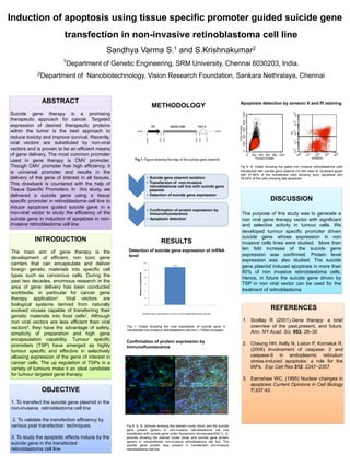
- 1. Induction of apoptosis using tissue specific promoter guided suicide gene transfection in non-invasive retinoblastoma cell line Sandhya Varma S.1 and S.Krishnakumar2 1Department of Genetic Engineering, SRM University, Chennai 6030203, India. 2Department of Nanobiotechnology, Vision Research Foundation, Sankara Nethralaya, Chennai ABSTRACT Suicide gene therapy is a promising therapeutic approach for cancer. Targeted expression of desired therapeutic proteins within the tumor is the best approach to reduce toxicity and improve survival. Recently, viral vectors are substituted by non-viral vectors and is proven to be an efficient means of gene delivery. The most common promoter used in gene therapy is CMV promoter. Though CMV promoter has high efficiency, it is universal promoter and results in the delivery of the gene of interest in all tissues. This drawback is countered with the help of Tissue Specific Promoters. In this study, we delivered a suicide gene using a tissue specific promoter in retinoblastoma cell line to induce apoptosis guided suicide gene in a non-viral vector to study the efficiency of the suicide gene in induction of apoptosis in non- invasive retinoblastoma cell line. INTRODUCTION The main aim of gene therapy is the development of efficient, non toxic gene carriers that can encapsulate and deliver foreign genetic materials into specific cell types such as cancerous cells. During the past two decades, enormous research in the area of gene delivery has been conducted worldwide, in particular for cancer gene therapy application1. Viral vectors are biological systems derived from naturally evolved viruses capable of transferring their genetic materials into host cells2. Although non viral vectors are less efficient than viral vectors3, they have the advantage of safety, simplicity of preparation and high gene encapsulation capability. Tumour specific promoters (TSP) have emerged as highly tumour specific and effective in selectively allowing expression of the gene of interest in cancer cells. The up regulation of TSPs in a variety of tumours make it an ideal candidate for tumour targeted gene therapy. OBJECTIVE 1. To transfect the suicide gene plasmid in the non-invasive retinoblastoma cell line 2. To validate the transfection efficiency by various post transfection techniques. 3. To study the apoptotic effects induce by the suicide gene in the transfected retinoblastoma cell line • Suicide gene plasmid isolation • Transfection of non-invasive retinoblastoma cell line with suicide gene plasmid • Detection of suicide gene expression • Confirmation of protein expression by immunofluorescence • Apoptosis detection METHODOLOGY RESULTS Fig 1: Graph showing the over expression of suicide gene in transfected non-invasive retinoblastoma cell line (~10fold increase). DISCUSSION The purpose of this study was to generate a non viral gene therapy vector with significant and selective activity in tumour cells. We developed tumour specific promoter driven suicide gene whose expression in non invasive cells lines were studied. More than ten fold increase of the suicide gene expression was confirmed. Protein level expression was also studied. The suicide gene plasmid induced apoptosis in more than 60% of non invasive retinoblastoma cells. Hence, in future the suicide gene driven by TSP in non viral vector can be used for the treatment of retinoblastoma REFERENCES 1. Scollay R (2001).Gene therapy: a brief overview of the past,present, and future. Ann. NY Acad. Sci. 953, 26–30 2. Cheung HH, Kelly N, Liston P, Korneluk R. (2006) Involvement of caspase- 2 and caspase-9 in endoplasmic reticulum stress-induced apoptosis: a role for the IAPs. Exp Cell Res 312: 2347−2357 3. Earnshaw WC. (1995) Nuclear changes in apoptosis Current Opinions in Cell Biology 7:337:43. Fig 8: A, B: pictures showing the stained nuclei (blue) and the suicide gene protein (green) in non-invasive retinoblastoma cell line transfected with suicide gene under fluorescent microscope(40X) C, D: pictures showing the stained nuclei (blue) and suicide gene protein (green) in untransfected non-invasive retinoblastoma cell line. The suicide gene protein was present in transfected non-invasive retinoblastoma cell line Fig 9: A: Graph showing the gated non invasive retinoblastoma cells transfected with suicide gene plasmid (10,000 cells) B: Quadrant graph with 61.84% of the transfected cells showing early apoptosis and 35.02% of the cells showing late apoptosis Fig 1: Figure showing the map of the suicide gene plasmid Confirmation of protein expression by immunofluorescence Detection of suicide gene expression at mRNA level Apoptosis detection by annexin V and PI staining