
Empfohlen
Weitere ähnliche Inhalte
Was ist angesagt?
Was ist angesagt? (20)
Ähnlich wie Brain psychology
Ähnlich wie Brain psychology (20)
Mehr von Sana butt
Mehr von Sana butt (13)
Kürzlich hochgeladen
Kürzlich hochgeladen (20)
Brain psychology
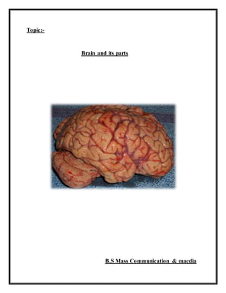
- 1. Topic:- Brain and its parts B.S Mass Communication & maedia
- 2. Central nervous system Brain Central nervous system The central nervous system (CNS) is the processing center for the nervous system. It receives information from and sends information to the peripheral nervous system. The two main organs of the CNS are the brain and spinal cord, But we study only brain. Brain The brain processes and interprets sensory information sent from the spinal cord. The brain is the control center of the body. It consists of three main components: the forebrain, the midbrain and the hindbrain. Forebrain Main regions of forebrain are: Thalamus Hypothalamus Limbtic system Cerebrum Thalamus: The thalamus is a sensory way station. All sensory information except smell-related data must go through the thalamus on the way to the cerebrum. Hypothalamus: The hypothalamus lies under the thalamus and helps to control the pituitary gland and the autonomic nervous system. The hypothalamus plays an important role in regulating body temperature and biological drives such as hunger, thirst, sex, and aggression. Limbic System: The limbic system includes the hippocampus, the amygdala, and the septum. Parts of the limbic system also lie in the thalamus and the hypothalamus. The limbic system processes emotional experience. The amygdala plays a role in aggression and fear, while the hippocampus plays a role in memory.
- 3. Cerebrum The cerebrum, the biggest part of the brain, controls complex processes such as abstract thought and learning. The wrinkled, highly folded outer layer of the cerebrum is called the cerebral cortex. The corpus callosum is a band of fibers that runs along the cerebrum from the front of the skull to the back. It divides the cerebrum into two halves, or hemispheres. Each hemisphere is divided into two deep grooves, known as the fissure of Rolando and the fissure of Sylvius.There are four sections or lobe in each hemisphere: the occipital lobe, the parietal lobe, the temporal lobe, and the frontal lobe: The occipital lobe: It is located in the very back of our brain, the occipital lobes are responsible for our eyesight. They contain the primary visual cortex which helps us interpret the information sent to us by our eyes. The parietal lobe: It is located above the fissure of sylvius but back the fissure of Rolando. It contains Sensory Cortex which is located in the front of the parietal lobe (directly behind the sensory cortex in the frontal lobe), this structure is responsible for us feeling touch sensations from our body. Every time you feel a type of touch sensations (both pleasurable and pain) the information is sent up by sensory neurons to the thalamus and sent to the sensory cortex so we can feel it. The bottom of the cortex is responsible for the top of our body and the top of the cortex responsible for the bottom of our body. The temporal lobe: It is located below the fissure of Sylvius,lying just inside the temples. It contains the primary auditory cortex, which is involved in processing auditory information. The left temporal lobe also contains Wernicke’s area. Wernicke's area is located in our left temporal lobe and is responsible for interpreting both written and spoken speech. You use Wernicke's area both the read and to listen. If you damage Wernicke's area (Wernicke's Aphasia) you would be unable to understand what you are reading or hearing. The frontal lobe: It is located in front of the brain. There are two specific areas in the frontal lobe, Broca's Area and Motor Cortex. The left frontal lobe contains Broca’s area, (at least for most people- in some left handed people, Broca's area is on the right
- 4. side) which influences speech production. If you damage Broca's area (called Broca's Aphasia) you will be unable to talk. Motor Cortex is located in the back of the frontal lobe this thin strip of tissue sends signals (motor neurons) to tell our body to move. The things we move more (fingers) have more space devoted on the motor cortex, than parts of us we do not move much (pinky toes). The top of the motor cortex controls the bottom of our body and the bottom of the cortex controls the top of our body. The frontal lobe also processes memory, planning, goal-setting, creativity, rational decision making, and social judgment. Brain Hemispheres Lateralization refers to the fact that the right and left hemispheres of the brain regulate different functions. The left hemisphere specializes in verbal processing tasks such as writing, reading, and talking. The right hemisphere specializes in nonverbal processing tasks such as playing music, drawing, and recognizing childhood friends. Because of the organization of the nervous system, the left hemisphere of the brain controls the functioning of the right side of the body. Likewise, the right hemisphere controls the functioning of the left side of the body. Vision and hearing operate a bit differently. What the left eye and right eye see goes to the entire brain. However, images in the left visual field stimulate receptors on the right side of each eye, and in-formation goes from those points to the right hemisphere. Information perceived by the right visual field ends up in the left hemisphere. In the case of auditory information, both hemispheres receive input about what each ear hears. However, information first goes to the opposite hemisphere. If the left ear hears a sound, the right hemisphere registers the sound first. The fact that the brain’s hemispheres communicate with opposite sides of the body does not affect most people’s day-to-day functioning because the two hemispheres constantly share information via the corpus callosum. However, severing the corpus callosum and separating the hemispheres causes impaired perception. Hindbrain The hindbrain is composed of the medulla, the pons, and the cerebellum. The medulla lies next to the spinal cord and controls functions outside conscious
- 5. control, such as breathing and blood flow. In other words, the medulla controls essential functions. The pons: It is a Latin word which means bridge. It affects activities such as sleeping, waking, and dreaming. The cerebellum: It is a Latin word which means little brain. It controls balance and coordination of movement. Damage to the cerebellum impairs fine motor skills, so a person with an injury in this area would have trouble playing the guitar or typing a term paper. Midbrain The midbrain is the part of the brain that lies between the hindbrain and the forebrain. The midbrain helps us to locate events in space. It also contains a system of neurons that releases the neurotransmitter dopamine. The reticular formation runs through the hindbrain and the midbrain and is involved in sleep and wakefulness, pain perception, breathing, and muscle reflexes. Methods to study brain function A variety of procedures are used by physiological psychologists to study the functions of different areas of the brain. The methods help us to study the localization of the functional areas of the brain. Traditional Methods lesion A lesion is the removal or destruction of part of the brain. Doctors will lesion a patients brain during brain surgery (usually to remove some type of tumor). By removing parts of the brain we were able to learn what different parts of the brain do. For example, if a doctor removed a tumor in your left temporal lobe of the brain and you were then unable to speak, we could assume that speech comes from that area of the brain. Brain lesions were commonly used in the mid 1900s to control mentally unstable patients. Part of the frontal lobe was removed (frontal lobotomy) and drastic behavioral changes occurred after surgery (we learned that our personalities are strongly centered in the front of our brains).
- 6. Stimulation Stimulation The brain can be stimulated either electrically or chemically. It is stimulated electrically when electrodes send small electrical pulses to certain parts of the brain.When certain chemicals are introduced to the brain, they can affect certain things like hunger or thirst. EEG (Electroencephalogram) The brain is just like an electrical battery. An EEG machine measures brain waves. If you are awake it measures what we call alpha waves (short active waves) and when you are asleep it measures other waves like delta waves (long inactive waves). If the EEG measures no activity then you are either brain dead or watching Jerry Springer. It is used commonly in sleep research. Scanning and Imaging Techniques The advances in modern technology have made it possible to study the internal workings of the brain without having to cut surgically into a person's skull. The brain scanning are the mechanical and electrical measurements of biochemical and electrical activities of specified brain areas. Some of these techniques are discussed below. CAT Scan (Computerized Axial Tomography) A CAT scan is just a really sophisticated x-ray of the brain. It gives us a 3D picture of the brain which is great for locating tumors, but it does not show brain activity or function. PET Scan (Positron Emission Tomography) A PET scan is the best way for us to see activity in the brain. The patient will usually swallow a substance (like glucose) and the PET scan will see what parts of the brain are using the substance. If a patient seems to be using alot of the substance in a certain part of the brain, we can tell what part of the brain is working. Magnetic Resonance Imaging (MRI) It produces a strong magnetic field in which the person's head is positioned. The radio waves directed at the brain cause the hydrogen atoms to emit signals, which are analyzed by a computer. The details of the MRI are superior to CAT scan, because it can distinguish between closely related brain structures.