SlideShare ist ein Scribd-Unternehmen logo
1 von 63
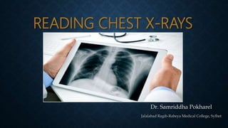
READING CHEST X-RAYS
Dr. Samriddha Pokharel
Jalalabad Ragib-Rabeya Medical College, Sylhet
X-RAYS
X-rays are a form of electromagnetic radiation.
Wavelength(λ) : 10 picometres to 10 nanometres (10×10−12 m) to (10×10−9)
(Shorter than visible and UV Rays and longer than Gamma
Rays)
Frequencies(f) : 30 petahertz to 30 exahertz (3×10¹⁶ Hz to 3×10¹⁹ Hz)
Energy(E) : 100 eV to 200 keV
Discovered by: Wilhelm Conrad Roentgen (Father of Radiology) in 1895A.D.
RADIOGRAPHIC DENSITIES/GRAYSCALE
• Different body tissues absorb X-Rays at different extents.
 Gas (air in the lungs)  Least dense/Least absorption of X-Rays  Black(Radiolucent)
 Metal/Bone  More dense/Absorb more radiation  White(Radiopaque)
White – Metal
Off White – Bone
Light grey – Soft
Tissue
Dark Grey – Fat
Black - Air
BEFORE INTERPRETING THE X-RAY….
• Patient’s Details and Site Determination(left side and right side)
• View : Postero-Anterior or Antero-Posterior
• Exposure of the film to radiation
• Rotation of the patient
• Breath : Inspiration or Expiration
Mnemonic- P-VERB
o In exam, the X-ray provided will be an inspiratory film with adequate exposure and
usually of posteroanterior view(children’s x-ray may be anteroposterior)
4 MAJOR VIEWS OF THE CHEST RADIOGRAPH
PosteroAnterior- 1.Most commonly preferred.
2. Standard view for Chest X-rays.
3.Patient stands upright with the chest placed on the film after
full inspiration.
AnteroPosterior- 1.Used in debilitated , very ill , uncooperative patients and in
children.
Lateral - 1. Usually done in conjunction with PA view Chest X-Ray
2.Lung lobes and lobar pathology, Mediastinum and its pathology e.g.
Mediastinal mass, Thoracic wall and basal consolidation can be better
visualized.
Lateral Decubitus - 1.Specialized projection used to demonstrate small pleural
effusions or pneumothorax.
A-P VIEW FILM VS P-A VIEW FILM
Points PA view AP view
Clavicle Over the lung fields Above lung apex
Scapulae Away from lung fields Over lung fields
Ribs Posterior ribs distinct Anterior ribs distinct
Heart Close to the anatomical size Relatively enlarged
LATERAL DECUBITUS VIEW
 200ml or more fluid is needed to see blunting of costophrenic angle on
Postero-Anterior view.
 Lateral view X-ray can show blunting of costophrenic angle when there is
100ml of pleural fluid.
 Lateral decubitus view can show free flowing fluid in the pleura <50ml
EXPOSURE OF THE FILM TO
RADIATION/PENETRATION
• On an adequately exposed chest radiograph ,the lower thoracic vertebrae are visible
through the heart and the Broncho-vascular markings(trachea ,aortic arch , etc.)
must be seen.
GOOD INSPIRATORY FILM:
• On a proper Inspiratory chest radiograph:
 First 6 Anterior ribs are visible.
 First 10 Posterior ribs are visible.
ROTATION
• If the spinous process of vertebral body is equidistant from the medial ends of clavicle, there is NO
rotation.
• Rotation results in reduced distance on the side in front.
Here, reduced
distance is on
the left
meaning the
left side is in
front. Hence,
the patient is
rotated
towards right.
NO Rotation
INTERPRETING CHEST X-RAYS
ABCDEFGHI APPROACH
• Airway
• Bones and Soft tissue
• Cardiac Shadow
• Diaphragm
• Effusions(Pleura)
• Fields(Lungs)
• Gastric Bubble (Fundic gas)
• Hila and Mediastinum
• Impressions (of tubes or pacemakers)
For studying it is easier to follow ABCDEFGHI approach however for exams Outside to inside approach
will be a faster method.
FIRST LET’S LOOK AT DIFFERENT STRUCTURES AND THEIR
NORMAL ANATOMY WITHIN THE RADIOGRAPHIC FILM
POSTERO-ANTERIOR
VIEW
A- Costo-phrenic
angle
B-Diaphragm
C-Heart
D-Aortic knob
E-Trachea
F- Hilum and
Pulmonary artery
G-Carina
H- Fundic gas
J-SVC
LATERAL VIEW
A- Costo-phrenic
angle
B-Diaphragm
C-Heart
D-Aortic knob
E-Trachea
F- Hilum and
Pulmonary artery
G-Carina
H- Fundic gas
J-SVC
INTERFACES IN THE CHEST RADIOGRAPH
• An interface is formed when two structures of significantly variable densities are in front
of one another .
• In Chest X-ray , interface lines are seen on the lung fields due to variable densities of the
lung(gas) and other organs(soft tissues).
SILHOUETTE SIGN:
• The X-ray image will depend on the sum of various densities encountered by
the X-ray beam as it courses through the body.
• If the structures of similar densities are juxtaposed then the anatomical soft
tissue border(interface lines) will not be visible. This is called SILHOUETTE
SIGN.
Here, there is juxtaposition of heart and
consolidated lung which are of similar
densities. Hence, the left heart border is
not visible.
AIRWAY
PA
VIEW
LATERAL
VIEW
CARIN
A
AIRWAY
• Trachea lies centrally and appears as a vertical black rectangle.
Slight tracheal deviation towards right is Normal.
Extension : Larynx(C6) to Carina(T4/T5) Length: 10-12cm
Bifurcates at the level of sternal angle.
Transverse diameter : approx. 19.5mm in male and 17.5mm in
female
Deviation towards the lesion Deviation away from lesion
Lobar collapse(esp. upper
lobe),Pneumonectomy
Large Pleural Effusion
Pulmonary Fibrosis Tension Pneumothorax
Some
Mediastinal
Masses may also
cause Tracheal
Deviation.
• Carina is an important landmark during endotracheal intubation.
• The Endotracheal tube should end 5mm(+/-2mm) above Carina
• Sub carina angle should be less than 90º.
AIRWAY
INTUBATION
End of ETT
Carina
SUB CARINA
ANGLE
RIGHT AND LEFT PRINCIPAL(MAIN
STEM) BRONCHI AND THEIR
BRANCHES
BONES AND SOFT TISSUE
• Bones
1. Look at each rib in turn and look for any pathologies.
2. Count the ribs (From posterior to anterior following the arc).
3. Look at the clavicles.
4.Look at the spine.
5. Look for pathologies in other surrounding bones i.e scapula and humerus.
Counting Ribs Right Lateral
Scoliosis
RIB NOTCHING
• Deformation in Superior or Inferior surface of the ribs is known as rib notching.
Notice the notches in the inferior aspect of
the ribs shown by arrows.
Superior Rib Notching Inferior Rib Notching
1.Osteogenesis
Imperfecta
2.Poliomyelitis
3.Hyperparathyroidism
4.Collagen Vascular
disease
5.Large
Neurofibromatosis
1.Coarctation of Aorta
2.Superior Vena Caval
Obstruction.
3.Arteriovenous Fistula
4. Following Blalock
Taussig Shunt
5.Neurofibromatosis
Type1
CAUSES:
• Soft tissue
1. Thick soft tissue may obscure lung markings
2. Breast tissue may obscure cost-phrenic angle
Breast tissue
LOOK FOR:
1.Enlarged nodes in
Supraclavicular
fossa.
2.Surgical
Emphysema in the
lateral thoracic
wall.
3.Pneumoperitoneum
Under the diaphragm.
• Pneumoperitoneum Subcutaneous Emphysema
(Notice the air within the lateral
thoracic wall)
CARDIAC SHADOW
• Right and Left radiological heart borders
 The Radiological right heart border is formed by:
1. Right Atrium 2. Part of Superior Vena Cava
 The Radiological left heart border is formed by:
1. Left Atrium 2. Left Ventricle 3. Aortic knuckle(knob) 4. Pulmonary
trunk
 Inferior Radiological border of heart is formed by :
1. Right Ventricle
CHAMBERS OF THE HEART ON CHEST X-RAY PA VIEW
CARDIO-THORACIC RATIO
• (CR+CL)<(T/2)
• Normal Cardio-Thoracic ratio is less than 0.5 in adults [(CR+CL)/CT<0.5]
T
CARDIOMEGALY
• LEFT ATRIAL ENLARGEMENT:
Causes:
1. Mitral Stenosis, Mitral
Regurgitation
2. Left Ventricular Failure
3. Ventricular Septal Defect
4. Patent Ductus Arteriosus
5. Left Atrial Myxoma
RIGHT ATRIAL ENLARGEMENT:
Causes:
1.Pulmonary Hypertension
2. Tricuspid stenosis, Pulmonary
Stenosis
3. Tetralogy of Fallot
4. Cor Pulmonale
5. Rt. Ventricular failure
RIGHT VENTRICULAR ENLARGEMENT:
Causes:
1. Pulmonary Hypertension
2. Tricuspid insufficiency
3. Atrial Septal Defect
LEFT VENTRICULAR ENLARGEMENT:
Causes:
1. Hypertension
2. Aortic Stenosis
3. Ventricular Septal Defect
4. Aortic Regurgitation, Mitral
LEFT ATRIAL ENLARGEMENT AND IT’S SIGNS
SEEN IN MITRAL STENOSIS
SIGNS:
1. Cardiothoracic ratio is greater than 0.5 in adult.
2. Double Right Heart Border (Double density) { blue and white lines}
3. Straightening of the left heart border{ red line} [ Later, the straight border may
turn convex outward(third mogul sign)
4. Splaying of carina
( sub carinal angle >90Âş) due to
elevation of left main stem bronchus
{ yellow }
RIGHT ATRIAL ENLARGEMENT
• Cardiomegaly with enlargement towards the right and posteriorly
• Prominent right superior border
• Right Atrial Margin is 5.5cm(or more) away from midline.
RIGHT VENTRICULAR HYPERTROPHY IN
TETRALOGY OF FALLOT
• Right Ventricular hypertrophy with upturned cardiac apex. BOOT SHAPED
HEART
• Oligaemic(decreased pulmonary vascular marking) lung fields.
LEFT VENTRICULAR ENLARGEMENT
• Cardiomegaly with downturned cardiac apex.
• Depressed left hemi-diaphragm.
SOME RADIOLOGICAL SIGNS SEEN IN CARDIAC
DISEASES
Total Anomalous Pulmonary
Venous Drainage
(Snowman Sign)
Partial Anomalous
Pulmonary Venous Drainage
(Scimitar sign)
Transpostion of great arteries
(Egg sign)
Ebstein’s Anomaly
(Box sign)
Coarctation of Aorta
(3 sign on PA view)
(Reverse 3 on Lateral view)
Thoracic Aortic Aneurysm Tubular heart in COPD.
Also, notice the hyperinflated
lung and lowered down
diaphragm.
DIAPHRAGM
• Both the domes of the diaphragm should from a sharp contour with the lateral chest wall.
• Costo-phrenic angle must be sharp and usually around 30º.
• Most common cause of blunting of costo-phrenic angle is pleural effusion. Blunting may also
be caused by basal consolidation.
• Pleural effusion first obliterates costo-phrenic angle then cardio-phrenic angle.
EFFUSIONS(PLEURA)
• Pleura is only visible on a radiology film when there is a pathology.
• Some common pathologies of pleura are:
1.Pleural Effusion 2.Pneumothorax 3.Pleural thickening
4.Hydropneumothorax
PLEURAL EFFUSION
RADIOLOGICAL FINDINGS:
• This is a Chest X-ray PA View showing dense homogeneous opacity on the left lung field throughout
the lower and part of middle zone with a concave margin upwards.
The costo-phrenic angle, cardio-phrenic angle and heart border on the left are obscured.
‘Dense Homogeneous’ is used when the radiographic density of the opacity is same as that of liver.
DIAGNOSIS: Left sided Pleural Effusion.
RADIOLOGICAL FINDINGS:
• This is a Chest X-ray showing dense homogeneous opacity throughout the left lung field with
obliterated cardio-phrenic, costo-phrenic angles and left heart border. There is Tracheal and
Mediastinal Deviation towards the right.
DIAGNOSIS: Left Sided Massive Pleural Effusion(with trachea and mediastinal deviation towards
right)
Tracheal
Shift
Exudative causes (having
protein rich fluid)
Transudative causes
1.Pneumonia
2.Tuberculosis
3.Malignancies
4.Pulmonary Embolism
1.Congestive heart
failure
2.Cirrhosis
3.Nephrosis
CRITERIA FOR EXUDATIVE PLEURAL FLUID:
(any 1 of the following criteria must be met)
o Pleural Fluid protein/Serum protein>0.5
o Pleural fluid LDH/Serum LDH>0.6
o Pleural fluid LDH>2/3rd of upper normal serum limit
PNEUMOTHORAX
The radiological film shows a Hypertranslucent area on the left lung field near the
apex without any Bronchovascular margin. On close inspection, a visible pleural
margin is seen infero-medially to this area.
DIAGNOSIS: Left sided Pneumothorax
An, expiratory film should be
ordered if someone is suspected of
Pneumothorax which shows the
area clearly.
TENSION PNEUMOTHORAX (ONE WAY VALVE)
• When excessive amount of air is trapped within the pleural spaces under positive
pressure causing mediastinal and tracheal shift, it is called tension pneumothorax.
Deviated Trachea with ETT
Tension Pneumothorax
PLEURAL THICKENING
• Notice the Peripheral shadowing on the right side with decreased lung field.
Some causes of pleural
thickening:
1. Chronic lung infections
like TB
2. Asbestosis, Silicosis
3. Malignancies such as
Mesothelioma,
Metastasis
4. Post Radiation
HYDROPNEUMOTHORAX
• Notice the homogeneous dense opacity on the right lung field with horizontal upper
border and the lack of any bronchovascular markings above it. This is called an Air-fluid
level.
• Most common cause of Hydropneumothorax is Iatrogenic (air is accidently introduced
during drainage of pleural effusion)
FIELDS
• ZONES
Upper: superior to the lower margin of 2nd rib anteriorly
Middle: lower margin of 2nd rib to lower margin of 4th rib anteriorly
Lower : Below lower margin of 4th rib anteriorly
Lungs can also be divided by 2 vertical lines into 3 areas .
Medial1/3rd
Middle1/3rd
Lateral1/3rd
Notice, the braonchovascular
markings are clear and well
defined in the medial 1/3rd
Become smaller in the
middle 1/3rd and appear as
fine patterns of branching
lines in the medial most part
of lateral 1/3rd .
LOBAR ANATOMY
HIDDEN AREAS IN THE LUNG FIELDS
• Some areas in the lung fields are hidden due to the soft tissues or bones
superimposing on them.
NORMAL CHEST X-RAY (RED) COMPARED TO
RADIOPAQUE (BLUE) AND HYPERTRANSLUSCENT
(GREEN) FILMS
PLETHORIC AND OLIGEMIC LUNG FIELDS
• Plethoric lung field means increased
bronchovascular markings due to increased
pulmonary blood flow.
Causes:
1.Left to right shunts (ASD,VSD,PDA)
2. Transposition of great vessels
3. Partial or Total anomalous pulmonary
venous return
• Oligemic lung field means decreased
bronchovascular markings due to decreased
pulmonary blood flow.
Causes:
1. Tetralogy of fallot
2. Right to left shunt in Pulmonary stenosis or
atresia, Tricuspid atresia
3. Pulmonary Embolism ( Westermark sign)-
CONSOLIDATION
Consolidation is an airspace disease that involves filling of the alveolar space with fluid(pulmonary edema), pus( as
in pneumonia), blood or even cells(in carcinomas).
Areas of consolidation appear white most often with ill defined margins.
Air Bronchograms : Air spaces in the alveoli become opacified while the bronchi remain air filled making them
appear as small black thin tubular structures within the white area of consolidation.
Notice the small black
streaks running through
the white area.
CONSOLIDATION
• Patterns to look for:
1. Diffuse/ Patchy/ Focal
2. Perihilar/ Peripheral
3.Unilateral/ Bilateral
4.Segmental/ Lobar
• Identify the zones that the consolidation covers.
• For lobe identification on PA View:
1. Upper lobe consolidation lies superior to the major
fissure often producing a sharp margin. It silhouettes with the
superior mediastinum.
2. Middle lobe consolidation silhouettes the right heart
margin.
3. Lower lobe consolidation silhouettes the hemi-
diaphragm.
4. Lingular consolidation appears close to the left heart
border.
Small focal area of
consolidation on the right
lung field in the right
lower zone and peri-hilar
consolidation on the left
lung field typical of
Bronchopneumonia
Patchy consolidation
In the right lower
zone .
SOME PRESENTATIONS OF TUBERCULAR CONSOLIDATION
Chronic TB
presenting as
calcified
lesions in the
right middle
and lower
zones and left
middle and
lower zones
close to the
cardiac
shadow.
Patchy reticular
opacities in the
right upper and
middle zones .
• INTERSTITIAL PULMONARY EDEMA
Findings:
1.Septal lines( Kerley B Lines)
2. Peribronchial cuffing (small doughnut shaped
rings representing fluids in the thickened bronchial
wall)
3. Pleural effusions and Fluid between fissures
can also be seen.
With increase in extravascular fluid from
pulmonary capillaries to the interstitium the fluid
moves centrally making these signs more
prominent.
PULMONARY EDEMA
• ALVEOLAR PULMONARY EDEMA is caused by fluid leaking from the interstitial tissues
into the alveoli and presenting as consolidation.
Alveolar edema radiates symmetrically from hilar regions in a ‘bat’s wing’ appearance.
Cardiogenic causes of alveolar edema most often show enlarged heart shadow.
SEPTAL LINES
• Kerley A lines:(White arrows) linear
opacities from periphery to hila caused by
distension of anastomotic channels
between peripheral and central lymphatics
• Kerley B lines:(white arrow heads) short
horizontal lines situated perpendicularly
to the pleural surface close to the lung
base.
• Kerley C lines : (black arrow heads) radial
opacities away from hilum
CAVITARY LESION
• Cavitary lesions are seen as an area of radiopaque margin with hypertransluscent
area within it.
• Lung Abscesses are cavitary lesions with radiopaque margin and having an air-fluid
level within it.
• Cavitary lesions can be seen in Malignancies, TB, etc.
Lung abscessCavitary
Lesion
GASTRIC BUBBLE
HILA
• Hilum is the area on the medial aspect of lungs through which Bronchi, vessels and
nerves enter and exit the lungs.
PULMONARY VASCULAR PATTERN
• Normal lung vascular pattern has following features:
1. Arteries and Veins branching vertically to upper and lower lobes.
2. The Upper lobe vessels have smaller diameter than lower lobe vessels on an erect Chest X-
ray.
 In Pulmonary Venous Hypertension, vessels branching upwards have a larger diameter than
the vessels branching downwards. This is known as ‘Cephalization’.
IMPRESSIONS OF TUBES OR DEVICES
Chest X-Ray with Left
Ventricular Assist DeviceChest x-ray showing metal suture
wires after Sternotomy
PRACTICE QUESTIONS
Thank You

Weitere ähnliche Inhalte

Was ist angesagt?

Collapse and consolidation Lung Radiology
Collapse and consolidation Lung RadiologyCollapse and consolidation Lung Radiology
Collapse and consolidation Lung RadiologyNeelam Ashar
 
Invertogram ANORECTAL MALFORMATION ( ARM ) PRANAYA
Invertogram ANORECTAL MALFORMATION ( ARM ) PRANAYAInvertogram ANORECTAL MALFORMATION ( ARM ) PRANAYA
Invertogram ANORECTAL MALFORMATION ( ARM ) PRANAYAPRANAYA PANIGRAHI
 
Cardiovascular examination
Cardiovascular examinationCardiovascular examination
Cardiovascular examinationabeerabdulkareem
 
Abdominal X ray
Abdominal X rayAbdominal X ray
Abdominal X rayRichin Koshy
 
Chest X-ray Interpretation
Chest X-ray Interpretation Chest X-ray Interpretation
Chest X-ray Interpretation Sarfraz Saleemi
 
Collapse consolidation
Collapse consolidationCollapse consolidation
Collapse consolidationairwave12
 
surgery.Congenital heart disease.(dr.aram)
surgery.Congenital heart disease.(dr.aram)surgery.Congenital heart disease.(dr.aram)
surgery.Congenital heart disease.(dr.aram)student
 
Radiographic Presentation of Congenital Heart Disease
Radiographic Presentation of Congenital Heart DiseaseRadiographic Presentation of Congenital Heart Disease
Radiographic Presentation of Congenital Heart DiseaseTarique Ajij
 
Chest X-rays for Undergraduates
Chest X-rays for UndergraduatesChest X-rays for Undergraduates
Chest X-rays for UndergraduatesAbdullah Ansari
 
Rectal bleeding
Rectal bleeding Rectal bleeding
Rectal bleeding Saurav Verma
 
Presentation1, interpretation of x ray of the abdomen.
Presentation1, interpretation of x ray of the abdomen.Presentation1, interpretation of x ray of the abdomen.
Presentation1, interpretation of x ray of the abdomen.Abdellah Nazeer
 
Interpreting a chest x ray film
Interpreting a chest x ray filmInterpreting a chest x ray film
Interpreting a chest x ray filmPritom Das
 
Chest x ray basic interpretation
Chest x ray basic interpretationChest x ray basic interpretation
Chest x ray basic interpretationVikram Patil
 
Presentation1, radiological imaging of scimitar syndrome
Presentation1, radiological imaging of scimitar syndromePresentation1, radiological imaging of scimitar syndrome
Presentation1, radiological imaging of scimitar syndromeAbdellah Nazeer
 
Palpation of spleen final
Palpation of spleen  finalPalpation of spleen  final
Palpation of spleen finalKurian Joseph
 
Radiology of Pulmonary Hypertension
Radiology of Pulmonary HypertensionRadiology of Pulmonary Hypertension
Radiology of Pulmonary HypertensionHatlan Al Hatlan
 

Was ist angesagt? (20)

Collapse and consolidation Lung Radiology
Collapse and consolidation Lung RadiologyCollapse and consolidation Lung Radiology
Collapse and consolidation Lung Radiology
 
Invertogram ANORECTAL MALFORMATION ( ARM ) PRANAYA
Invertogram ANORECTAL MALFORMATION ( ARM ) PRANAYAInvertogram ANORECTAL MALFORMATION ( ARM ) PRANAYA
Invertogram ANORECTAL MALFORMATION ( ARM ) PRANAYA
 
Cardiovascular examination
Cardiovascular examinationCardiovascular examination
Cardiovascular examination
 
Abdominal X ray
Abdominal X rayAbdominal X ray
Abdominal X ray
 
Radiological features of pneumonia
Radiological features of pneumoniaRadiological features of pneumonia
Radiological features of pneumonia
 
Chest X-ray Interpretation
Chest X-ray Interpretation Chest X-ray Interpretation
Chest X-ray Interpretation
 
Collapse consolidation
Collapse consolidationCollapse consolidation
Collapse consolidation
 
surgery.Congenital heart disease.(dr.aram)
surgery.Congenital heart disease.(dr.aram)surgery.Congenital heart disease.(dr.aram)
surgery.Congenital heart disease.(dr.aram)
 
Radiographic Presentation of Congenital Heart Disease
Radiographic Presentation of Congenital Heart DiseaseRadiographic Presentation of Congenital Heart Disease
Radiographic Presentation of Congenital Heart Disease
 
Chest X-rays for Undergraduates
Chest X-rays for UndergraduatesChest X-rays for Undergraduates
Chest X-rays for Undergraduates
 
Rectal bleeding
Rectal bleeding Rectal bleeding
Rectal bleeding
 
Presentation1, interpretation of x ray of the abdomen.
Presentation1, interpretation of x ray of the abdomen.Presentation1, interpretation of x ray of the abdomen.
Presentation1, interpretation of x ray of the abdomen.
 
Interpreting a chest x ray film
Interpreting a chest x ray filmInterpreting a chest x ray film
Interpreting a chest x ray film
 
Chest x ray basic interpretation
Chest x ray basic interpretationChest x ray basic interpretation
Chest x ray basic interpretation
 
Presentation1, radiological imaging of scimitar syndrome
Presentation1, radiological imaging of scimitar syndromePresentation1, radiological imaging of scimitar syndrome
Presentation1, radiological imaging of scimitar syndrome
 
Jerrin's
Jerrin'sJerrin's
Jerrin's
 
Hernia
HerniaHernia
Hernia
 
Palpation of spleen final
Palpation of spleen  finalPalpation of spleen  final
Palpation of spleen final
 
Radiology of Pulmonary Hypertension
Radiology of Pulmonary HypertensionRadiology of Pulmonary Hypertension
Radiology of Pulmonary Hypertension
 
Imaging of Obstructive jaundice
Imaging of Obstructive jaundiceImaging of Obstructive jaundice
Imaging of Obstructive jaundice
 

Ähnlich wie Reading chest-x-rays

pathology related topics and its complete focus on every aspect
pathology related topics and its complete focus on every aspectpathology related topics and its complete focus on every aspect
pathology related topics and its complete focus on every aspectNausheen57
 
How to read normal x ray
How to read normal x rayHow to read normal x ray
How to read normal x raypuneet mahajan
 
Chest imaging
Chest imagingChest imaging
Chest imagingJANARDANCNMC
 
CHEST XRAYS RJJ.pptx
CHEST XRAYS RJJ.pptxCHEST XRAYS RJJ.pptx
CHEST XRAYS RJJ.pptxrohanjohnjacob
 
X RAY DETERMINATION AND EVALUATION.pptx
X RAY DETERMINATION AND EVALUATION.pptxX RAY DETERMINATION AND EVALUATION.pptx
X RAY DETERMINATION AND EVALUATION.pptxShoaibKhatik3
 
XRAY
XRAYXRAY
XRAYSufindc
 
Cxr revised 24 11-91
Cxr revised 24 11-91Cxr revised 24 11-91
Cxr revised 24 11-91aalmasi1970
 
CHEST XRAY
CHEST XRAY CHEST XRAY
CHEST XRAY amit jha
 
Normal Chest X-Rays & Its Systemic Approach- Anatomy
Normal Chest X-Rays & Its Systemic Approach- AnatomyNormal Chest X-Rays & Its Systemic Approach- Anatomy
Normal Chest X-Rays & Its Systemic Approach- AnatomyDr.Bijay Yadav
 
Abnormal signs in chest x ray
Abnormal signs in chest x rayAbnormal signs in chest x ray
Abnormal signs in chest x rayMilan Silwal
 
cxr series.pdf
cxr series.pdfcxr series.pdf
cxr series.pdfbaharhoseini
 
Chest X-ray radiology_Power Point Presentation
Chest X-ray radiology_Power Point PresentationChest X-ray radiology_Power Point Presentation
Chest X-ray radiology_Power Point PresentationAhyaAziz
 
x ray intepetation.pptx
x ray intepetation.pptxx ray intepetation.pptx
x ray intepetation.pptxtemesgenworku6
 
Дыхательная система на англ.pptx
Дыхательная система на англ.pptxДыхательная система на англ.pptx
Дыхательная система на англ.pptxPriyanshuBlaze
 
chest X ray basics and interpretation
chest X ray basics and interpretationchest X ray basics and interpretation
chest X ray basics and interpretationsomaskandan Rajendran
 
Chest xray
Chest xrayChest xray
Chest xrayAdityaNag11
 

Ähnlich wie Reading chest-x-rays (20)

pathology related topics and its complete focus on every aspect
pathology related topics and its complete focus on every aspectpathology related topics and its complete focus on every aspect
pathology related topics and its complete focus on every aspect
 
Approach to cxr.pptx
Approach to cxr.pptxApproach to cxr.pptx
Approach to cxr.pptx
 
How to read normal x ray
How to read normal x rayHow to read normal x ray
How to read normal x ray
 
Chest imaging
Chest imagingChest imaging
Chest imaging
 
CHEST XRAYS RJJ.pptx
CHEST XRAYS RJJ.pptxCHEST XRAYS RJJ.pptx
CHEST XRAYS RJJ.pptx
 
X RAY DETERMINATION AND EVALUATION.pptx
X RAY DETERMINATION AND EVALUATION.pptxX RAY DETERMINATION AND EVALUATION.pptx
X RAY DETERMINATION AND EVALUATION.pptx
 
Reading chest X-ray
Reading chest X-rayReading chest X-ray
Reading chest X-ray
 
XRAY
XRAYXRAY
XRAY
 
Cxr revised 24 11-91
Cxr revised 24 11-91Cxr revised 24 11-91
Cxr revised 24 11-91
 
CHEST XRAY
CHEST XRAY CHEST XRAY
CHEST XRAY
 
Normal Chest X-Rays & Its Systemic Approach- Anatomy
Normal Chest X-Rays & Its Systemic Approach- AnatomyNormal Chest X-Rays & Its Systemic Approach- Anatomy
Normal Chest X-Rays & Its Systemic Approach- Anatomy
 
Abnormal signs in chest x ray
Abnormal signs in chest x rayAbnormal signs in chest x ray
Abnormal signs in chest x ray
 
cxr series.pdf
cxr series.pdfcxr series.pdf
cxr series.pdf
 
Chest X-ray radiology_Power Point Presentation
Chest X-ray radiology_Power Point PresentationChest X-ray radiology_Power Point Presentation
Chest X-ray radiology_Power Point Presentation
 
Chest imaging
Chest imagingChest imaging
Chest imaging
 
Chest imaging
Chest imagingChest imaging
Chest imaging
 
x ray intepetation.pptx
x ray intepetation.pptxx ray intepetation.pptx
x ray intepetation.pptx
 
Дыхательная система на англ.pptx
Дыхательная система на англ.pptxДыхательная система на англ.pptx
Дыхательная система на англ.pptx
 
chest X ray basics and interpretation
chest X ray basics and interpretationchest X ray basics and interpretation
chest X ray basics and interpretation
 
Chest xray
Chest xrayChest xray
Chest xray
 

KĂźrzlich hochgeladen

Mumbai ] (Call Girls) in Mumbai 10k @ I'm VIP Independent Escorts Girls 98333...
Mumbai ] (Call Girls) in Mumbai 10k @ I'm VIP Independent Escorts Girls 98333...Mumbai ] (Call Girls) in Mumbai 10k @ I'm VIP Independent Escorts Girls 98333...
Mumbai ] (Call Girls) in Mumbai 10k @ I'm VIP Independent Escorts Girls 98333...Ishani Gupta
 
Top Rated Hyderabad Call Girls Erragadda ⟟ 9332606886 ⟟ Call Me For Genuine ...
Top Rated  Hyderabad Call Girls Erragadda ⟟ 9332606886 ⟟ Call Me For Genuine ...Top Rated  Hyderabad Call Girls Erragadda ⟟ 9332606886 ⟟ Call Me For Genuine ...
Top Rated Hyderabad Call Girls Erragadda ⟟ 9332606886 ⟟ Call Me For Genuine ...chandars293
 
Call Girls Hyderabad Just Call 8250077686 Top Class Call Girl Service Available
Call Girls Hyderabad Just Call 8250077686 Top Class Call Girl Service AvailableCall Girls Hyderabad Just Call 8250077686 Top Class Call Girl Service Available
Call Girls Hyderabad Just Call 8250077686 Top Class Call Girl Service AvailableDipal Arora
 
Dehradun Call Girls Service {8854095900} ❤️VVIP ROCKY Call Girl in Dehradun U...
Dehradun Call Girls Service {8854095900} ❤️VVIP ROCKY Call Girl in Dehradun U...Dehradun Call Girls Service {8854095900} ❤️VVIP ROCKY Call Girl in Dehradun U...
Dehradun Call Girls Service {8854095900} ❤️VVIP ROCKY Call Girl in Dehradun U...Sheetaleventcompany
 
8980367676 Call Girls In Ahmedabad Escort Service Available 24×7 In Ahmedabad
8980367676 Call Girls In Ahmedabad Escort Service Available 24×7 In Ahmedabad8980367676 Call Girls In Ahmedabad Escort Service Available 24×7 In Ahmedabad
8980367676 Call Girls In Ahmedabad Escort Service Available 24×7 In AhmedabadGENUINE ESCORT AGENCY
 
Call Girls Amritsar Just Call 8250077686 Top Class Call Girl Service Available
Call Girls Amritsar Just Call 8250077686 Top Class Call Girl Service AvailableCall Girls Amritsar Just Call 8250077686 Top Class Call Girl Service Available
Call Girls Amritsar Just Call 8250077686 Top Class Call Girl Service AvailableDipal Arora
 
Top Rated Call Girls Kerala ☎ 8250092165👄 Delivery in 20 Mins Near Me
Top Rated Call Girls Kerala ☎ 8250092165👄 Delivery in 20 Mins Near MeTop Rated Call Girls Kerala ☎ 8250092165👄 Delivery in 20 Mins Near Me
Top Rated Call Girls Kerala ☎ 8250092165👄 Delivery in 20 Mins Near Mechennailover
 
💚Call Girls In Amritsar 💯Anvi 📲🔝8725944379🔝Amritsar Call Girl No💰Advance Cash...
💚Call Girls In Amritsar 💯Anvi 📲🔝8725944379🔝Amritsar Call Girl No💰Advance Cash...💚Call Girls In Amritsar 💯Anvi 📲🔝8725944379🔝Amritsar Call Girl No💰Advance Cash...
💚Call Girls In Amritsar 💯Anvi 📲🔝8725944379🔝Amritsar Call Girl No💰Advance Cash...Sheetaleventcompany
 
Call Girls Mumbai Just Call 8250077686 Top Class Call Girl Service Available
Call Girls Mumbai Just Call 8250077686 Top Class Call Girl Service AvailableCall Girls Mumbai Just Call 8250077686 Top Class Call Girl Service Available
Call Girls Mumbai Just Call 8250077686 Top Class Call Girl Service AvailableDipal Arora
 
Premium Call Girls In Jaipur {8445551418} ❤️VVIP SEEMA Call Girl in Jaipur Ra...
Premium Call Girls In Jaipur {8445551418} ❤️VVIP SEEMA Call Girl in Jaipur Ra...Premium Call Girls In Jaipur {8445551418} ❤️VVIP SEEMA Call Girl in Jaipur Ra...
Premium Call Girls In Jaipur {8445551418} ❤️VVIP SEEMA Call Girl in Jaipur Ra...parulsinha
 
Call Girl in Indore 8827247818 {LowPrice} ❤️ (ahana) Indore Call Girls * UPA...
Call Girl in Indore 8827247818 {LowPrice} ❤️ (ahana) Indore Call Girls  * UPA...Call Girl in Indore 8827247818 {LowPrice} ❤️ (ahana) Indore Call Girls  * UPA...
Call Girl in Indore 8827247818 {LowPrice} ❤️ (ahana) Indore Call Girls * UPA...mahaiklolahd
 
Call Girls Kolkata Kalikapur 💯Call Us 🔝 8005736733 🔝 💃 Top Class Call Girl Se...
Call Girls Kolkata Kalikapur 💯Call Us 🔝 8005736733 🔝 💃 Top Class Call Girl Se...Call Girls Kolkata Kalikapur 💯Call Us 🔝 8005736733 🔝 💃 Top Class Call Girl Se...
Call Girls Kolkata Kalikapur 💯Call Us 🔝 8005736733 🔝 💃 Top Class Call Girl Se...Namrata Singh
 
Russian Call Girls Lucknow Just Call 👉👉7877925207 Top Class Call Girl Service...
Russian Call Girls Lucknow Just Call 👉👉7877925207 Top Class Call Girl Service...Russian Call Girls Lucknow Just Call 👉👉7877925207 Top Class Call Girl Service...
Russian Call Girls Lucknow Just Call 👉👉7877925207 Top Class Call Girl Service...adilkhan87451
 
Call Girls Mysore Just Call 8250077686 Top Class Call Girl Service Available
Call Girls Mysore Just Call 8250077686 Top Class Call Girl Service AvailableCall Girls Mysore Just Call 8250077686 Top Class Call Girl Service Available
Call Girls Mysore Just Call 8250077686 Top Class Call Girl Service AvailableDipal Arora
 
Top Quality Call Girl Service Kalyanpur 6378878445 Available Call Girls Any Time
Top Quality Call Girl Service Kalyanpur 6378878445 Available Call Girls Any TimeTop Quality Call Girl Service Kalyanpur 6378878445 Available Call Girls Any Time
Top Quality Call Girl Service Kalyanpur 6378878445 Available Call Girls Any TimeCall Girls Delhi
 
Russian Call Girls Service Jaipur {8445551418} ❤️PALLAVI VIP Jaipur Call Gir...
Russian Call Girls Service  Jaipur {8445551418} ❤️PALLAVI VIP Jaipur Call Gir...Russian Call Girls Service  Jaipur {8445551418} ❤️PALLAVI VIP Jaipur Call Gir...
Russian Call Girls Service Jaipur {8445551418} ❤️PALLAVI VIP Jaipur Call Gir...parulsinha
 
Low Rate Call Girls Bangalore {7304373326} ❤️VVIP NISHA Call Girls in Bangalo...
Low Rate Call Girls Bangalore {7304373326} ❤️VVIP NISHA Call Girls in Bangalo...Low Rate Call Girls Bangalore {7304373326} ❤️VVIP NISHA Call Girls in Bangalo...
Low Rate Call Girls Bangalore {7304373326} ❤️VVIP NISHA Call Girls in Bangalo...Sheetaleventcompany
 
(Low Rate RASHMI ) Rate Of Call Girls Jaipur ❣ 8445551418 ❣ Elite Models & Ce...
(Low Rate RASHMI ) Rate Of Call Girls Jaipur ❣ 8445551418 ❣ Elite Models & Ce...(Low Rate RASHMI ) Rate Of Call Girls Jaipur ❣ 8445551418 ❣ Elite Models & Ce...
(Low Rate RASHMI ) Rate Of Call Girls Jaipur ❣ 8445551418 ❣ Elite Models & Ce...parulsinha
 
Call Girls Service Jaipur {8445551418} ❤️VVIP BHAWNA Call Girl in Jaipur Raja...
Call Girls Service Jaipur {8445551418} ❤️VVIP BHAWNA Call Girl in Jaipur Raja...Call Girls Service Jaipur {8445551418} ❤️VVIP BHAWNA Call Girl in Jaipur Raja...
Call Girls Service Jaipur {8445551418} ❤️VVIP BHAWNA Call Girl in Jaipur Raja...parulsinha
 
Call Girls Service Jaipur {9521753030 } ❤️VVIP BHAWNA Call Girl in Jaipur Raj...
Call Girls Service Jaipur {9521753030 } ❤️VVIP BHAWNA Call Girl in Jaipur Raj...Call Girls Service Jaipur {9521753030 } ❤️VVIP BHAWNA Call Girl in Jaipur Raj...
Call Girls Service Jaipur {9521753030 } ❤️VVIP BHAWNA Call Girl in Jaipur Raj...khalifaescort01
 

KĂźrzlich hochgeladen (20)

Mumbai ] (Call Girls) in Mumbai 10k @ I'm VIP Independent Escorts Girls 98333...
Mumbai ] (Call Girls) in Mumbai 10k @ I'm VIP Independent Escorts Girls 98333...Mumbai ] (Call Girls) in Mumbai 10k @ I'm VIP Independent Escorts Girls 98333...
Mumbai ] (Call Girls) in Mumbai 10k @ I'm VIP Independent Escorts Girls 98333...
 
Top Rated Hyderabad Call Girls Erragadda ⟟ 9332606886 ⟟ Call Me For Genuine ...
Top Rated  Hyderabad Call Girls Erragadda ⟟ 9332606886 ⟟ Call Me For Genuine ...Top Rated  Hyderabad Call Girls Erragadda ⟟ 9332606886 ⟟ Call Me For Genuine ...
Top Rated Hyderabad Call Girls Erragadda ⟟ 9332606886 ⟟ Call Me For Genuine ...
 
Call Girls Hyderabad Just Call 8250077686 Top Class Call Girl Service Available
Call Girls Hyderabad Just Call 8250077686 Top Class Call Girl Service AvailableCall Girls Hyderabad Just Call 8250077686 Top Class Call Girl Service Available
Call Girls Hyderabad Just Call 8250077686 Top Class Call Girl Service Available
 
Dehradun Call Girls Service {8854095900} ❤️VVIP ROCKY Call Girl in Dehradun U...
Dehradun Call Girls Service {8854095900} ❤️VVIP ROCKY Call Girl in Dehradun U...Dehradun Call Girls Service {8854095900} ❤️VVIP ROCKY Call Girl in Dehradun U...
Dehradun Call Girls Service {8854095900} ❤️VVIP ROCKY Call Girl in Dehradun U...
 
8980367676 Call Girls In Ahmedabad Escort Service Available 24×7 In Ahmedabad
8980367676 Call Girls In Ahmedabad Escort Service Available 24×7 In Ahmedabad8980367676 Call Girls In Ahmedabad Escort Service Available 24×7 In Ahmedabad
8980367676 Call Girls In Ahmedabad Escort Service Available 24×7 In Ahmedabad
 
Call Girls Amritsar Just Call 8250077686 Top Class Call Girl Service Available
Call Girls Amritsar Just Call 8250077686 Top Class Call Girl Service AvailableCall Girls Amritsar Just Call 8250077686 Top Class Call Girl Service Available
Call Girls Amritsar Just Call 8250077686 Top Class Call Girl Service Available
 
Top Rated Call Girls Kerala ☎ 8250092165👄 Delivery in 20 Mins Near Me
Top Rated Call Girls Kerala ☎ 8250092165👄 Delivery in 20 Mins Near MeTop Rated Call Girls Kerala ☎ 8250092165👄 Delivery in 20 Mins Near Me
Top Rated Call Girls Kerala ☎ 8250092165👄 Delivery in 20 Mins Near Me
 
💚Call Girls In Amritsar 💯Anvi 📲🔝8725944379🔝Amritsar Call Girl No💰Advance Cash...
💚Call Girls In Amritsar 💯Anvi 📲🔝8725944379🔝Amritsar Call Girl No💰Advance Cash...💚Call Girls In Amritsar 💯Anvi 📲🔝8725944379🔝Amritsar Call Girl No💰Advance Cash...
💚Call Girls In Amritsar 💯Anvi 📲🔝8725944379🔝Amritsar Call Girl No💰Advance Cash...
 
Call Girls Mumbai Just Call 8250077686 Top Class Call Girl Service Available
Call Girls Mumbai Just Call 8250077686 Top Class Call Girl Service AvailableCall Girls Mumbai Just Call 8250077686 Top Class Call Girl Service Available
Call Girls Mumbai Just Call 8250077686 Top Class Call Girl Service Available
 
Premium Call Girls In Jaipur {8445551418} ❤️VVIP SEEMA Call Girl in Jaipur Ra...
Premium Call Girls In Jaipur {8445551418} ❤️VVIP SEEMA Call Girl in Jaipur Ra...Premium Call Girls In Jaipur {8445551418} ❤️VVIP SEEMA Call Girl in Jaipur Ra...
Premium Call Girls In Jaipur {8445551418} ❤️VVIP SEEMA Call Girl in Jaipur Ra...
 
Call Girl in Indore 8827247818 {LowPrice} ❤️ (ahana) Indore Call Girls * UPA...
Call Girl in Indore 8827247818 {LowPrice} ❤️ (ahana) Indore Call Girls  * UPA...Call Girl in Indore 8827247818 {LowPrice} ❤️ (ahana) Indore Call Girls  * UPA...
Call Girl in Indore 8827247818 {LowPrice} ❤️ (ahana) Indore Call Girls * UPA...
 
Call Girls Kolkata Kalikapur 💯Call Us 🔝 8005736733 🔝 💃 Top Class Call Girl Se...
Call Girls Kolkata Kalikapur 💯Call Us 🔝 8005736733 🔝 💃 Top Class Call Girl Se...Call Girls Kolkata Kalikapur 💯Call Us 🔝 8005736733 🔝 💃 Top Class Call Girl Se...
Call Girls Kolkata Kalikapur 💯Call Us 🔝 8005736733 🔝 💃 Top Class Call Girl Se...
 
Russian Call Girls Lucknow Just Call 👉👉7877925207 Top Class Call Girl Service...
Russian Call Girls Lucknow Just Call 👉👉7877925207 Top Class Call Girl Service...Russian Call Girls Lucknow Just Call 👉👉7877925207 Top Class Call Girl Service...
Russian Call Girls Lucknow Just Call 👉👉7877925207 Top Class Call Girl Service...
 
Call Girls Mysore Just Call 8250077686 Top Class Call Girl Service Available
Call Girls Mysore Just Call 8250077686 Top Class Call Girl Service AvailableCall Girls Mysore Just Call 8250077686 Top Class Call Girl Service Available
Call Girls Mysore Just Call 8250077686 Top Class Call Girl Service Available
 
Top Quality Call Girl Service Kalyanpur 6378878445 Available Call Girls Any Time
Top Quality Call Girl Service Kalyanpur 6378878445 Available Call Girls Any TimeTop Quality Call Girl Service Kalyanpur 6378878445 Available Call Girls Any Time
Top Quality Call Girl Service Kalyanpur 6378878445 Available Call Girls Any Time
 
Russian Call Girls Service Jaipur {8445551418} ❤️PALLAVI VIP Jaipur Call Gir...
Russian Call Girls Service  Jaipur {8445551418} ❤️PALLAVI VIP Jaipur Call Gir...Russian Call Girls Service  Jaipur {8445551418} ❤️PALLAVI VIP Jaipur Call Gir...
Russian Call Girls Service Jaipur {8445551418} ❤️PALLAVI VIP Jaipur Call Gir...
 
Low Rate Call Girls Bangalore {7304373326} ❤️VVIP NISHA Call Girls in Bangalo...
Low Rate Call Girls Bangalore {7304373326} ❤️VVIP NISHA Call Girls in Bangalo...Low Rate Call Girls Bangalore {7304373326} ❤️VVIP NISHA Call Girls in Bangalo...
Low Rate Call Girls Bangalore {7304373326} ❤️VVIP NISHA Call Girls in Bangalo...
 
(Low Rate RASHMI ) Rate Of Call Girls Jaipur ❣ 8445551418 ❣ Elite Models & Ce...
(Low Rate RASHMI ) Rate Of Call Girls Jaipur ❣ 8445551418 ❣ Elite Models & Ce...(Low Rate RASHMI ) Rate Of Call Girls Jaipur ❣ 8445551418 ❣ Elite Models & Ce...
(Low Rate RASHMI ) Rate Of Call Girls Jaipur ❣ 8445551418 ❣ Elite Models & Ce...
 
Call Girls Service Jaipur {8445551418} ❤️VVIP BHAWNA Call Girl in Jaipur Raja...
Call Girls Service Jaipur {8445551418} ❤️VVIP BHAWNA Call Girl in Jaipur Raja...Call Girls Service Jaipur {8445551418} ❤️VVIP BHAWNA Call Girl in Jaipur Raja...
Call Girls Service Jaipur {8445551418} ❤️VVIP BHAWNA Call Girl in Jaipur Raja...
 
Call Girls Service Jaipur {9521753030 } ❤️VVIP BHAWNA Call Girl in Jaipur Raj...
Call Girls Service Jaipur {9521753030 } ❤️VVIP BHAWNA Call Girl in Jaipur Raj...Call Girls Service Jaipur {9521753030 } ❤️VVIP BHAWNA Call Girl in Jaipur Raj...
Call Girls Service Jaipur {9521753030 } ❤️VVIP BHAWNA Call Girl in Jaipur Raj...
 

Reading chest-x-rays

  • 1. READING CHEST X-RAYS Dr. Samriddha Pokharel Jalalabad Ragib-Rabeya Medical College, Sylhet
  • 2. X-RAYS X-rays are a form of electromagnetic radiation. Wavelength(Îť) : 10 picometres to 10 nanometres (10×10−12 m) to (10×10−9) (Shorter than visible and UV Rays and longer than Gamma Rays) Frequencies(f) : 30 petahertz to 30 exahertz (3×10¹⁶ Hz to 3×10¹⁹ Hz) Energy(E) : 100 eV to 200 keV Discovered by: Wilhelm Conrad Roentgen (Father of Radiology) in 1895A.D.
  • 3. RADIOGRAPHIC DENSITIES/GRAYSCALE • Different body tissues absorb X-Rays at different extents.  Gas (air in the lungs) Âť Least dense/Least absorption of X-Rays Âť Black(Radiolucent)  Metal/Bone Âť More dense/Absorb more radiation Âť White(Radiopaque) White – Metal Off White – Bone Light grey – Soft Tissue Dark Grey – Fat Black - Air
  • 4. BEFORE INTERPRETING THE X-RAY…. • Patient’s Details and Site Determination(left side and right side) • View : Postero-Anterior or Antero-Posterior • Exposure of the film to radiation • Rotation of the patient • Breath : Inspiration or Expiration Mnemonic- P-VERB o In exam, the X-ray provided will be an inspiratory film with adequate exposure and usually of posteroanterior view(children’s x-ray may be anteroposterior)
  • 5. 4 MAJOR VIEWS OF THE CHEST RADIOGRAPH PosteroAnterior- 1.Most commonly preferred. 2. Standard view for Chest X-rays. 3.Patient stands upright with the chest placed on the film after full inspiration. AnteroPosterior- 1.Used in debilitated , very ill , uncooperative patients and in children. Lateral - 1. Usually done in conjunction with PA view Chest X-Ray 2.Lung lobes and lobar pathology, Mediastinum and its pathology e.g. Mediastinal mass, Thoracic wall and basal consolidation can be better visualized. Lateral Decubitus - 1.Specialized projection used to demonstrate small pleural effusions or pneumothorax.
  • 6. A-P VIEW FILM VS P-A VIEW FILM Points PA view AP view Clavicle Over the lung fields Above lung apex Scapulae Away from lung fields Over lung fields Ribs Posterior ribs distinct Anterior ribs distinct Heart Close to the anatomical size Relatively enlarged
  • 7. LATERAL DECUBITUS VIEW  200ml or more fluid is needed to see blunting of costophrenic angle on Postero-Anterior view.  Lateral view X-ray can show blunting of costophrenic angle when there is 100ml of pleural fluid.  Lateral decubitus view can show free flowing fluid in the pleura <50ml
  • 8. EXPOSURE OF THE FILM TO RADIATION/PENETRATION • On an adequately exposed chest radiograph ,the lower thoracic vertebrae are visible through the heart and the Broncho-vascular markings(trachea ,aortic arch , etc.) must be seen.
  • 9. GOOD INSPIRATORY FILM: • On a proper Inspiratory chest radiograph:  First 6 Anterior ribs are visible.  First 10 Posterior ribs are visible.
  • 10. ROTATION • If the spinous process of vertebral body is equidistant from the medial ends of clavicle, there is NO rotation. • Rotation results in reduced distance on the side in front. Here, reduced distance is on the left meaning the left side is in front. Hence, the patient is rotated towards right. NO Rotation
  • 12. ABCDEFGHI APPROACH • Airway • Bones and Soft tissue • Cardiac Shadow • Diaphragm • Effusions(Pleura) • Fields(Lungs) • Gastric Bubble (Fundic gas) • Hila and Mediastinum • Impressions (of tubes or pacemakers) For studying it is easier to follow ABCDEFGHI approach however for exams Outside to inside approach will be a faster method.
  • 13. FIRST LET’S LOOK AT DIFFERENT STRUCTURES AND THEIR NORMAL ANATOMY WITHIN THE RADIOGRAPHIC FILM POSTERO-ANTERIOR VIEW A- Costo-phrenic angle B-Diaphragm C-Heart D-Aortic knob E-Trachea F- Hilum and Pulmonary artery G-Carina H- Fundic gas J-SVC
  • 14. LATERAL VIEW A- Costo-phrenic angle B-Diaphragm C-Heart D-Aortic knob E-Trachea F- Hilum and Pulmonary artery G-Carina H- Fundic gas J-SVC
  • 15. INTERFACES IN THE CHEST RADIOGRAPH • An interface is formed when two structures of significantly variable densities are in front of one another . • In Chest X-ray , interface lines are seen on the lung fields due to variable densities of the lung(gas) and other organs(soft tissues).
  • 16.
  • 17. SILHOUETTE SIGN: • The X-ray image will depend on the sum of various densities encountered by the X-ray beam as it courses through the body. • If the structures of similar densities are juxtaposed then the anatomical soft tissue border(interface lines) will not be visible. This is called SILHOUETTE SIGN. Here, there is juxtaposition of heart and consolidated lung which are of similar densities. Hence, the left heart border is not visible.
  • 19. AIRWAY • Trachea lies centrally and appears as a vertical black rectangle. Slight tracheal deviation towards right is Normal. Extension : Larynx(C6) to Carina(T4/T5) Length: 10-12cm Bifurcates at the level of sternal angle. Transverse diameter : approx. 19.5mm in male and 17.5mm in female Deviation towards the lesion Deviation away from lesion Lobar collapse(esp. upper lobe),Pneumonectomy Large Pleural Effusion Pulmonary Fibrosis Tension Pneumothorax Some Mediastinal Masses may also cause Tracheal Deviation.
  • 20. • Carina is an important landmark during endotracheal intubation. • The Endotracheal tube should end 5mm(+/-2mm) above Carina • Sub carina angle should be less than 90Âş. AIRWAY INTUBATION End of ETT Carina SUB CARINA ANGLE RIGHT AND LEFT PRINCIPAL(MAIN STEM) BRONCHI AND THEIR BRANCHES
  • 21. BONES AND SOFT TISSUE • Bones 1. Look at each rib in turn and look for any pathologies. 2. Count the ribs (From posterior to anterior following the arc). 3. Look at the clavicles. 4.Look at the spine. 5. Look for pathologies in other surrounding bones i.e scapula and humerus. Counting Ribs Right Lateral Scoliosis
  • 22. RIB NOTCHING • Deformation in Superior or Inferior surface of the ribs is known as rib notching. Notice the notches in the inferior aspect of the ribs shown by arrows. Superior Rib Notching Inferior Rib Notching 1.Osteogenesis Imperfecta 2.Poliomyelitis 3.Hyperparathyroidism 4.Collagen Vascular disease 5.Large Neurofibromatosis 1.Coarctation of Aorta 2.Superior Vena Caval Obstruction. 3.Arteriovenous Fistula 4. Following Blalock Taussig Shunt 5.Neurofibromatosis Type1 CAUSES:
  • 23. • Soft tissue 1. Thick soft tissue may obscure lung markings 2. Breast tissue may obscure cost-phrenic angle Breast tissue LOOK FOR: 1.Enlarged nodes in Supraclavicular fossa. 2.Surgical Emphysema in the lateral thoracic wall. 3.Pneumoperitoneum Under the diaphragm.
  • 24. • Pneumoperitoneum Subcutaneous Emphysema (Notice the air within the lateral thoracic wall)
  • 25. CARDIAC SHADOW • Right and Left radiological heart borders  The Radiological right heart border is formed by: 1. Right Atrium 2. Part of Superior Vena Cava  The Radiological left heart border is formed by: 1. Left Atrium 2. Left Ventricle 3. Aortic knuckle(knob) 4. Pulmonary trunk  Inferior Radiological border of heart is formed by : 1. Right Ventricle
  • 26. CHAMBERS OF THE HEART ON CHEST X-RAY PA VIEW
  • 27. CARDIO-THORACIC RATIO • (CR+CL)<(T/2) • Normal Cardio-Thoracic ratio is less than 0.5 in adults [(CR+CL)/CT<0.5] T
  • 28. CARDIOMEGALY • LEFT ATRIAL ENLARGEMENT: Causes: 1. Mitral Stenosis, Mitral Regurgitation 2. Left Ventricular Failure 3. Ventricular Septal Defect 4. Patent Ductus Arteriosus 5. Left Atrial Myxoma RIGHT ATRIAL ENLARGEMENT: Causes: 1.Pulmonary Hypertension 2. Tricuspid stenosis, Pulmonary Stenosis 3. Tetralogy of Fallot 4. Cor Pulmonale 5. Rt. Ventricular failure RIGHT VENTRICULAR ENLARGEMENT: Causes: 1. Pulmonary Hypertension 2. Tricuspid insufficiency 3. Atrial Septal Defect LEFT VENTRICULAR ENLARGEMENT: Causes: 1. Hypertension 2. Aortic Stenosis 3. Ventricular Septal Defect 4. Aortic Regurgitation, Mitral
  • 29. LEFT ATRIAL ENLARGEMENT AND IT’S SIGNS SEEN IN MITRAL STENOSIS SIGNS: 1. Cardiothoracic ratio is greater than 0.5 in adult. 2. Double Right Heart Border (Double density) { blue and white lines} 3. Straightening of the left heart border{ red line} [ Later, the straight border may turn convex outward(third mogul sign) 4. Splaying of carina ( sub carinal angle >90Âş) due to elevation of left main stem bronchus { yellow }
  • 30. RIGHT ATRIAL ENLARGEMENT • Cardiomegaly with enlargement towards the right and posteriorly • Prominent right superior border • Right Atrial Margin is 5.5cm(or more) away from midline.
  • 31. RIGHT VENTRICULAR HYPERTROPHY IN TETRALOGY OF FALLOT • Right Ventricular hypertrophy with upturned cardiac apex. BOOT SHAPED HEART • Oligaemic(decreased pulmonary vascular marking) lung fields.
  • 32. LEFT VENTRICULAR ENLARGEMENT • Cardiomegaly with downturned cardiac apex. • Depressed left hemi-diaphragm.
  • 33. SOME RADIOLOGICAL SIGNS SEEN IN CARDIAC DISEASES Total Anomalous Pulmonary Venous Drainage (Snowman Sign) Partial Anomalous Pulmonary Venous Drainage (Scimitar sign)
  • 34. Transpostion of great arteries (Egg sign) Ebstein’s Anomaly (Box sign)
  • 35. Coarctation of Aorta (3 sign on PA view) (Reverse 3 on Lateral view) Thoracic Aortic Aneurysm Tubular heart in COPD. Also, notice the hyperinflated lung and lowered down diaphragm.
  • 36. DIAPHRAGM • Both the domes of the diaphragm should from a sharp contour with the lateral chest wall. • Costo-phrenic angle must be sharp and usually around 30Âş. • Most common cause of blunting of costo-phrenic angle is pleural effusion. Blunting may also be caused by basal consolidation. • Pleural effusion first obliterates costo-phrenic angle then cardio-phrenic angle.
  • 37. EFFUSIONS(PLEURA) • Pleura is only visible on a radiology film when there is a pathology. • Some common pathologies of pleura are: 1.Pleural Effusion 2.Pneumothorax 3.Pleural thickening 4.Hydropneumothorax
  • 38. PLEURAL EFFUSION RADIOLOGICAL FINDINGS: • This is a Chest X-ray PA View showing dense homogeneous opacity on the left lung field throughout the lower and part of middle zone with a concave margin upwards. The costo-phrenic angle, cardio-phrenic angle and heart border on the left are obscured. ‘Dense Homogeneous’ is used when the radiographic density of the opacity is same as that of liver. DIAGNOSIS: Left sided Pleural Effusion.
  • 39. RADIOLOGICAL FINDINGS: • This is a Chest X-ray showing dense homogeneous opacity throughout the left lung field with obliterated cardio-phrenic, costo-phrenic angles and left heart border. There is Tracheal and Mediastinal Deviation towards the right. DIAGNOSIS: Left Sided Massive Pleural Effusion(with trachea and mediastinal deviation towards right) Tracheal Shift Exudative causes (having protein rich fluid) Transudative causes 1.Pneumonia 2.Tuberculosis 3.Malignancies 4.Pulmonary Embolism 1.Congestive heart failure 2.Cirrhosis 3.Nephrosis CRITERIA FOR EXUDATIVE PLEURAL FLUID: (any 1 of the following criteria must be met) o Pleural Fluid protein/Serum protein>0.5 o Pleural fluid LDH/Serum LDH>0.6 o Pleural fluid LDH>2/3rd of upper normal serum limit
  • 40. PNEUMOTHORAX The radiological film shows a Hypertranslucent area on the left lung field near the apex without any Bronchovascular margin. On close inspection, a visible pleural margin is seen infero-medially to this area. DIAGNOSIS: Left sided Pneumothorax An, expiratory film should be ordered if someone is suspected of Pneumothorax which shows the area clearly.
  • 41. TENSION PNEUMOTHORAX (ONE WAY VALVE) • When excessive amount of air is trapped within the pleural spaces under positive pressure causing mediastinal and tracheal shift, it is called tension pneumothorax. Deviated Trachea with ETT Tension Pneumothorax
  • 42. PLEURAL THICKENING • Notice the Peripheral shadowing on the right side with decreased lung field. Some causes of pleural thickening: 1. Chronic lung infections like TB 2. Asbestosis, Silicosis 3. Malignancies such as Mesothelioma, Metastasis 4. Post Radiation
  • 43. HYDROPNEUMOTHORAX • Notice the homogeneous dense opacity on the right lung field with horizontal upper border and the lack of any bronchovascular markings above it. This is called an Air-fluid level. • Most common cause of Hydropneumothorax is Iatrogenic (air is accidently introduced during drainage of pleural effusion)
  • 44. FIELDS • ZONES Upper: superior to the lower margin of 2nd rib anteriorly Middle: lower margin of 2nd rib to lower margin of 4th rib anteriorly Lower : Below lower margin of 4th rib anteriorly Lungs can also be divided by 2 vertical lines into 3 areas . Medial1/3rd Middle1/3rd Lateral1/3rd Notice, the braonchovascular markings are clear and well defined in the medial 1/3rd Become smaller in the middle 1/3rd and appear as fine patterns of branching lines in the medial most part of lateral 1/3rd .
  • 46. HIDDEN AREAS IN THE LUNG FIELDS • Some areas in the lung fields are hidden due to the soft tissues or bones superimposing on them.
  • 47. NORMAL CHEST X-RAY (RED) COMPARED TO RADIOPAQUE (BLUE) AND HYPERTRANSLUSCENT (GREEN) FILMS
  • 48. PLETHORIC AND OLIGEMIC LUNG FIELDS • Plethoric lung field means increased bronchovascular markings due to increased pulmonary blood flow. Causes: 1.Left to right shunts (ASD,VSD,PDA) 2. Transposition of great vessels 3. Partial or Total anomalous pulmonary venous return • Oligemic lung field means decreased bronchovascular markings due to decreased pulmonary blood flow. Causes: 1. Tetralogy of fallot 2. Right to left shunt in Pulmonary stenosis or atresia, Tricuspid atresia 3. Pulmonary Embolism ( Westermark sign)-
  • 49. CONSOLIDATION Consolidation is an airspace disease that involves filling of the alveolar space with fluid(pulmonary edema), pus( as in pneumonia), blood or even cells(in carcinomas). Areas of consolidation appear white most often with ill defined margins. Air Bronchograms : Air spaces in the alveoli become opacified while the bronchi remain air filled making them appear as small black thin tubular structures within the white area of consolidation. Notice the small black streaks running through the white area.
  • 50. CONSOLIDATION • Patterns to look for: 1. Diffuse/ Patchy/ Focal 2. Perihilar/ Peripheral 3.Unilateral/ Bilateral 4.Segmental/ Lobar • Identify the zones that the consolidation covers. • For lobe identification on PA View: 1. Upper lobe consolidation lies superior to the major fissure often producing a sharp margin. It silhouettes with the superior mediastinum. 2. Middle lobe consolidation silhouettes the right heart margin. 3. Lower lobe consolidation silhouettes the hemi- diaphragm. 4. Lingular consolidation appears close to the left heart border. Small focal area of consolidation on the right lung field in the right lower zone and peri-hilar consolidation on the left lung field typical of Bronchopneumonia Patchy consolidation In the right lower zone .
  • 51. SOME PRESENTATIONS OF TUBERCULAR CONSOLIDATION Chronic TB presenting as calcified lesions in the right middle and lower zones and left middle and lower zones close to the cardiac shadow. Patchy reticular opacities in the right upper and middle zones .
  • 52. • INTERSTITIAL PULMONARY EDEMA Findings: 1.Septal lines( Kerley B Lines) 2. Peribronchial cuffing (small doughnut shaped rings representing fluids in the thickened bronchial wall) 3. Pleural effusions and Fluid between fissures can also be seen. With increase in extravascular fluid from pulmonary capillaries to the interstitium the fluid moves centrally making these signs more prominent. PULMONARY EDEMA
  • 53. • ALVEOLAR PULMONARY EDEMA is caused by fluid leaking from the interstitial tissues into the alveoli and presenting as consolidation. Alveolar edema radiates symmetrically from hilar regions in a ‘bat’s wing’ appearance. Cardiogenic causes of alveolar edema most often show enlarged heart shadow.
  • 54. SEPTAL LINES • Kerley A lines:(White arrows) linear opacities from periphery to hila caused by distension of anastomotic channels between peripheral and central lymphatics • Kerley B lines:(white arrow heads) short horizontal lines situated perpendicularly to the pleural surface close to the lung base. • Kerley C lines : (black arrow heads) radial opacities away from hilum
  • 55. CAVITARY LESION • Cavitary lesions are seen as an area of radiopaque margin with hypertransluscent area within it. • Lung Abscesses are cavitary lesions with radiopaque margin and having an air-fluid level within it. • Cavitary lesions can be seen in Malignancies, TB, etc. Lung abscessCavitary Lesion
  • 57. HILA • Hilum is the area on the medial aspect of lungs through which Bronchi, vessels and nerves enter and exit the lungs.
  • 58. PULMONARY VASCULAR PATTERN • Normal lung vascular pattern has following features: 1. Arteries and Veins branching vertically to upper and lower lobes. 2. The Upper lobe vessels have smaller diameter than lower lobe vessels on an erect Chest X- ray.  In Pulmonary Venous Hypertension, vessels branching upwards have a larger diameter than the vessels branching downwards. This is known as ‘Cephalization’.
  • 59. IMPRESSIONS OF TUBES OR DEVICES Chest X-Ray with Left Ventricular Assist DeviceChest x-ray showing metal suture wires after Sternotomy
  • 61.
  • 62.