SlideShare ist ein Scribd-Unternehmen logo
1 von 22
Pre Natal Growth And Development
PROF (Dr.) SAIBEL FARISHTA
During this period, the height increases by almost 5000 times as
compared to only a threefold increase during the post natal period. It is
divided into 3 periods –
Period of the Ovum – This period extends for two weeks from the
time of fertilization. During this period the cleavage of the ovum and
attachment of the ovum to the intra uterine wall occurs.
Period of the Embryo – This period extends from the 14th day to the
56th day of intra uterine life. Major part of the development of the facial
and cranial region occurs during this time.
Period of the Foetus – This phase extends between the 56th day of
intrauterine life till birth. In this period, accelerated growth of the cranio-
facial structures occurs resulting in an increase in their size. There is also
a change in proportion between the various structures.
Pre Natal Growth Of Cranial Base
The earliest evidence of formation of the cranial base is seen in the
post/late somitic period [4th – 6th week of intra uterine life]. During this
period mesenchymal tissue derived from primitive streak, neural crest
and occipital sclerotomes condense around the developing brain and
form a capsule which is known as Ectomenix or Ectomeningeal capsule. A
portion of this capsule gives rise to the future cranial base.
Evidence of skull formation is seen late after development of other cranial
structures.
From around 40th day onwards, the Ectomeningeal capsule is slowly
converted into cartilage. This is the onset of cranial base formation. The
conversion of mesenchymal cells into cartilage or chondrification occurs
in 4 regions –
• Parachordal
• Hypophyseal
• Nasal
• Otic
• Parachordal – The chondrification centers which form around the
cranial end of the notochord are called Parachordal cartilages.
• Hypophyseal – Cranial to the termination of notochord, the
Hypophyseal pouch develops which gives rise to the anterior lobe of the
pituitary gland.
[i] On both sides of hypophyseal stem two post sphenoid cartilages
develop, which fuse together and form the posterior part of the body of
sphenoid.
[ii] Cranial to pituitary gland, two trabecular cartilages develop which
fuse together and form the anterior part of body of sphenoid.
[iii] Lateral to pituitary gland, chondrification centers are seen which
form the lesser wing and greater wing of sphenoid.
• Nasal – Initially a capsule is seen around the nasal sense organ. This
capsule chondrifies and forms the cartilages of the nostrils, which later
fuses with the cartilages of the cranial base.
• Otic – Initially a capsule is seen around the vestibulo-cochlear sense
organs which condrifies and later ossifies to give rise to the mastoid and
petrous portions of the temporal bone. The otic cartilages also fuse with
the cartilages of the cranial base.
The initially separate centres of cartilage formation in the cranial base
fuse together into a single irregular and perforated cranial base.
Numerous nerves, blood vessels result in perforations or foramina in the
developing cranial base. The ossifying chondro-cranium meets the
ossifying cranial vault to form the neurocranium.
Chondro – Cranial Ossification
The cranial base which is in a cartilagenous form undergoes ossification.
The bones of the cranial base undergo both endochondral and
intramembranous ossification.
• Occipital Bone – It shows both types of ossification. Seven ossification
centres are seen, 2 intra membranous and 5 endochondrial.
• Temporal Bone – It also ossifies both endochondrally and intra-
membranously from 11 centres.
• Ethmoid Bone – It shows only endochondrial ossification. It ossifies
from 3 centres.
• Sphenoid Bone – It ossifies by both types from 15 ossification centres.
The cranial base or chondrocranium is important as a junction between the
cranial vault and the facial skeleton, being shared by both. The cranial
base is relatively stable during growth as compared to the cranial vault
and the face. Therefore the cranial base can be taken as a basis against
which the cranial vault and facial skeleton can be compared.
Flexure Of The Cranial Base -
During the embryonic and early fetal period, the cranial base becomes
flexed in the region between the pituitary fossa and the spheno-occipital
junction. The face is tucked under the cranium. This flexure of the cranial
base is accompanied by a corresponding flexure of the developing brain
stem.
The spinal chord and foramen magnum, which during the early stages of
development were directed backwards, now become directed
downwards.
Uneven growth of Cranial Base
The uneven growth of the cranial base is due to the uneven nature of
growth seen in different regions of brain.
The flexure of cranial base aids in increasing the neurocranial capacity
and is the main reason for the downward displacement of face during
growth.
At 10th week of intra uterine life, the flexion of the base is about 65
degrees. This flattens out a bit at the time of birth.
Pre-Natal Embryology Of Maxilla
Around the 4th week of intra uterine life, a prominent bulge appears on
the ventral aspect of the embryo corresponding to the developing brain.
Below the bulge, a shallow depression which corresponds to the primitive
mouth appears called stomadeum. The floor of the stomadeum is formed
by the buccopharyngeal membrane which separates the stomadeum from
the foregut.
By around 4th week of intrauterine life, 5 brachial arches form in the
region of the future head and neck. Each of these arches gives rise to
muscles, connective tissue, vasculature, skeletal components and neural
components of the future face. The 1st brachial arch is the mandibular
arch and plays an important role in the development of the naso-
maxillary region.
The mesoderm covering the developing forebrain proliferates and forms a
downward projection that overlaps the upper part of the stomadeum.
This downward projection is called Fronto-nasal process.
The stomadeum is thus overlapped superiorly by the frontonasal process,
while the mandibular arches of both the sides form the lateral walls of
the stomadeum. The mandibular arch gives a bud from it’s dorsal end
called the Maxillary Process. The maxillary process grows ventro-
medio-cranial to the main part of the mandibular arch, which is now
called Mandibular Process. Thus, at this stage the primitive mouth or
stomadeum is overlapped from above by the frontal process, below by
the mandibular process and on either sides by the maxillary processes.
The ectoderm overlying the fronto-nasal process shows bilateral localized
thickening above the stomadeum. These are called as the Nasal
Placodes. These placodes soon sink and form the nasal pits.
The formation of these nasal pits divides the fronto nasal process into 2
parts – medial and lateral nasal processes.
The two mandibular processes grow medially and fuse to form the lower
lip and lower jaw. As the maxillary process undergoes growth, the fronto
nasal process becomes narrow so that the two nasal pits come closer.
Development Of Palate
The palate is formed by contributions of the maxillary process, palatal
shelves given off by maxillary process and Fronto – nasal process.
The fronto-nasal process gives rise to the premaxillary region while the
palatal shelves form the rest of the palate. As the palatal shelves grow
medially, their union is prevented by the presence of the tongue. Thus,
initially the developing palatal shelves grow vertically downwards towards
the floor of the mouth.
Around 7th week of intra-uterine life, a quick transformation in the
position of the palatal shelves occurs and they change from a vertical to
a horizontal position.
By 8.5 weeks of intra-uterine life, the two palatal shelves are in close
approximation with each other. Initially they are covered by an epithelial
lining but as they join the epithelial cells degenerate. The connective
tissue from the palatal shelves intermingle with each other and fuse.
The entire palate doesn’t contact and fuse at the same time. Initially
contact occurs in the central region of the secondary palate , then
closure occurs anteriorly and posteriorly.
The mesial edges of the palatal processes fuse with the free lower end of
nasal septum and thus separates the two nasal cavities from each other
and the oral cavity.
Ossification of Palate – Occurs from 8th week of intra-uterine life. Its is
intra-membranous type of ossification. The palate ossifies from a single
center derived from the maxilla. The most posterior part of the palate
does not ossify and forms the soft palate. The mid-palatal suture ossifies
by 12-14 years.
Development of Maxillary Sinus – It forms around 3rd month of intra-
uterine life. It develops by expansion of the nasal mucous membrane into
the maxillary bone. Later the sinus enlarges by resorption of the internal
wall of maxilla.
Pre-Natal Embryology Of Mandible
Around 4th week of intra-uterine life, the developing brain and
pericardium form two prominent bulges on the ventral aspect of the
embryo. These bulges are separated by the primitive oral cavity or
stomadeum. The floor of the stomadeum is by bucco-pharyngeal
membrane which separates it from the foregut.
The pharyngeal arches are laid down on the lateral and ventral aspects of
the cranialmost part of the foregut which lies in close approximation with
the stomadeum. Initially there are six pharyngeal arches, but the 5th one
disappears as soon as it is formed leaving only five. They are separated
by four branchial grooves. The first arch is called as the mandibular arch
and the second arch, hyoid arch. The other arches do not have any
specific names.
Each of these five arches contain a central cartilage rod that forms the
skeleton of the arch, a muscular component, a vascular component and a
neural component.
The mandibular arch forms the lateral wall of the stomadeum. It gives a
bud at its dorsal end which is called as maxillary process. It grows
ventro-medially, cranial to the main part of the arch, which is now called
as the mandibular process. The mandibular processes grow towards each
other and fuse in midline. They now form the lower border of the
stomadeum i.e. the lower lip and the lower jaw.
Meckel’s Cartilage
It is derived from the 1st branchial arch around the 41st – 45th day of
intra-uterine life. It extends from the cartilagenous otic capsule to the
midline or symphysis and provides a template for guiding the growth of
the mandible. A major part of Meckel’s cartilage disappears during
growth and the remaining part develops into – mental ossicles, incus and
malleus, spine of sphenoid bone, anterior ligament of malleus and
spheno-mandibular ligament.
The first structure to develop in the primordium of the lower jaw is the
mandibular division of the trigeminal nerve. This is followed by
mesenchymal condensation forming the first branchial arch. Neurotrophic
factors produced by the nerve induce osteogenesis in the ossification
centers.
A single ossification center for each half of mandible arises in 6th week of
intra-uterine life in the region of the bifurcation of the inferior alveolar
nerve into mental and incisive branches.
The ossifying membrane is located lateral to the Meckel’s cartilage. From
this primary center, ossification spreads below and around the inferior
alveolar nerve and its incisive and branch and upwards to form a trough
for accommodating the developing tooth buds. Spread of the
intramembranous ossification dorsally and ventrally forms the body and
ramus of the mandible.
As ossification continues, the Meckel’s cartilage becomes surrounded and
invaded by bone. Ossification stops at the site that later becomes
mandibular lingula from where the Meckel;s cartilage continues into the
middle ear and develops into the auditory ossicles [malleus and incus].
Endochondral Bone Formation
It is seen only in 3 areas of mandible – condylar process, coronoid
process and mental region.
• Condylar Process – At about 5th week of intra-uterine life, an area of
mesenchymal condensation can be seen above the ventral part of the
developing mandible. This develops into a cone shaped cartilage by
about 10th week and starts ossification by 14th week. It then migrates
inferiorly and fuses with the mandibular ramus by about 4 months. Much
of the cone shaped cartilage is replaced by bone by the middlw of fetal
life, but its upper end persists into adulthood acting both as a growth
cartilage and an articular cartilage.
• Coronoid Process – Secondary accessory cartilages appear in the
region of the coronoid process by about the 10-14th week of intra-uterine
life. This cartilage is believed to grow as a response to the developing
temporalis muscle. The coronoid accessory cartilage becomes
incorporated into the expanding intra-membranous bone of the ramus
and disappears before birth.
• Mental Region – In the mental region, on either side of the symphysis,
one or two small cartilages appear and ossify in the 7th month of intra-
uterine life to form variable numbers of mental ossicles in the fibrous
tissues of the symphysis. These ossicles become incorporated into the
intra-membranous bone when the symphysis ossifies completely during
the first year of post-natal life.
THANK YOU

Weitere ähnliche Inhalte

Was ist angesagt?

Growth & development of face/certified fixed orthodontic courses by India...
Growth & development of face/certified fixed orthodontic courses by India...Growth & development of face/certified fixed orthodontic courses by India...
Growth & development of face/certified fixed orthodontic courses by India...Indian dental academy
 
Development of face and oral cavity 4/ oral surgery courses  
Development of face and oral cavity 4/ oral surgery courses  Development of face and oral cavity 4/ oral surgery courses  
Development of face and oral cavity 4/ oral surgery courses  Indian dental academy
 
Embryology of head and neck
Embryology of head and neckEmbryology of head and neck
Embryology of head and neckDr Bhavik Miyani
 
Development of Face /certified fixed orthodontic courses by Indian dental ac...
Development of Face  /certified fixed orthodontic courses by Indian dental ac...Development of Face  /certified fixed orthodontic courses by Indian dental ac...
Development of Face /certified fixed orthodontic courses by Indian dental ac...Indian dental academy
 
01. pharyngeal arches (3 lectures, march 18)
01. pharyngeal arches (3 lectures, march 18)01. pharyngeal arches (3 lectures, march 18)
01. pharyngeal arches (3 lectures, march 18)monazain3
 
Orofacial development
Orofacial developmentOrofacial development
Orofacial developmentRano Ya Naso
 
Embryology of head & neck
Embryology of head & neckEmbryology of head & neck
Embryology of head & neckSonu Pandit
 
Devlopment of face and its abnormalites
Devlopment of face and its abnormalites Devlopment of face and its abnormalites
Devlopment of face and its abnormalites Diwakar vasudev
 
Growth and development of face
Growth and development of faceGrowth and development of face
Growth and development of faceTinu George
 
Pre-natal facial growth Dr. Nabil Al-Zubair
Pre-natal facial growth  Dr. Nabil Al-ZubairPre-natal facial growth  Dr. Nabil Al-Zubair
Pre-natal facial growth Dr. Nabil Al-ZubairNabil Al-Zubair
 
growth and development of face
growth and development of face growth and development of face
growth and development of face Anindita Mallik
 
Embryology of head and neck
Embryology of head and neckEmbryology of head and neck
Embryology of head and neckUgochukwu Offor
 
Influence of orofacial functions on development of face and occlusion
Influence of orofacial functions on development of face and occlusionInfluence of orofacial functions on development of face and occlusion
Influence of orofacial functions on development of face and occlusionmohammed alawdi
 
DEVELOPMENT OF FACE AND ORAL CAVITY
DEVELOPMENT OF FACE AND ORAL CAVITYDEVELOPMENT OF FACE AND ORAL CAVITY
DEVELOPMENT OF FACE AND ORAL CAVITYDrDevanshiShrama
 
Development of face, nose, palate by dr. noura 2014
Development of face, nose, palate by dr. noura 2014Development of face, nose, palate by dr. noura 2014
Development of face, nose, palate by dr. noura 2014Dr. Noura El Tahawy
 
phayrangeal apparatus By Dr.Aisha Sadaf IBMS -KMU-peshawar
phayrangeal apparatus By Dr.Aisha Sadaf IBMS -KMU-peshawarphayrangeal apparatus By Dr.Aisha Sadaf IBMS -KMU-peshawar
phayrangeal apparatus By Dr.Aisha Sadaf IBMS -KMU-peshawarAisha Sadaf
 
Development of Face Part 1
Development of Face Part 1Development of Face Part 1
Development of Face Part 1Arun Panwar
 

Was ist angesagt? (20)

Growth & development of face/certified fixed orthodontic courses by India...
Growth & development of face/certified fixed orthodontic courses by India...Growth & development of face/certified fixed orthodontic courses by India...
Growth & development of face/certified fixed orthodontic courses by India...
 
Development of Face
Development of FaceDevelopment of Face
Development of Face
 
Development of face and oral cavity 4/ oral surgery courses  
Development of face and oral cavity 4/ oral surgery courses  Development of face and oral cavity 4/ oral surgery courses  
Development of face and oral cavity 4/ oral surgery courses  
 
Embryology of head and neck
Embryology of head and neckEmbryology of head and neck
Embryology of head and neck
 
Development of Face /certified fixed orthodontic courses by Indian dental ac...
Development of Face  /certified fixed orthodontic courses by Indian dental ac...Development of Face  /certified fixed orthodontic courses by Indian dental ac...
Development of Face /certified fixed orthodontic courses by Indian dental ac...
 
01. pharyngeal arches (3 lectures, march 18)
01. pharyngeal arches (3 lectures, march 18)01. pharyngeal arches (3 lectures, march 18)
01. pharyngeal arches (3 lectures, march 18)
 
Orofacial development
Orofacial developmentOrofacial development
Orofacial development
 
Embryology of head & neck
Embryology of head & neckEmbryology of head & neck
Embryology of head & neck
 
Devlopment of face and its abnormalites
Devlopment of face and its abnormalites Devlopment of face and its abnormalites
Devlopment of face and its abnormalites
 
Growth and development of face
Growth and development of faceGrowth and development of face
Growth and development of face
 
Pre-natal facial growth Dr. Nabil Al-Zubair
Pre-natal facial growth  Dr. Nabil Al-ZubairPre-natal facial growth  Dr. Nabil Al-Zubair
Pre-natal facial growth Dr. Nabil Al-Zubair
 
growth and development of face
growth and development of face growth and development of face
growth and development of face
 
Embryology of head and neck
Embryology of head and neckEmbryology of head and neck
Embryology of head and neck
 
Development of face
Development of faceDevelopment of face
Development of face
 
Development of face
Development of faceDevelopment of face
Development of face
 
Influence of orofacial functions on development of face and occlusion
Influence of orofacial functions on development of face and occlusionInfluence of orofacial functions on development of face and occlusion
Influence of orofacial functions on development of face and occlusion
 
DEVELOPMENT OF FACE AND ORAL CAVITY
DEVELOPMENT OF FACE AND ORAL CAVITYDEVELOPMENT OF FACE AND ORAL CAVITY
DEVELOPMENT OF FACE AND ORAL CAVITY
 
Development of face, nose, palate by dr. noura 2014
Development of face, nose, palate by dr. noura 2014Development of face, nose, palate by dr. noura 2014
Development of face, nose, palate by dr. noura 2014
 
phayrangeal apparatus By Dr.Aisha Sadaf IBMS -KMU-peshawar
phayrangeal apparatus By Dr.Aisha Sadaf IBMS -KMU-peshawarphayrangeal apparatus By Dr.Aisha Sadaf IBMS -KMU-peshawar
phayrangeal apparatus By Dr.Aisha Sadaf IBMS -KMU-peshawar
 
Development of Face Part 1
Development of Face Part 1Development of Face Part 1
Development of Face Part 1
 

Ă„hnlich wie Pre Natal Growth & Development

Growth and Development of Cranial and Facial structures.(Orthodontics for BDS)
Growth and Development of Cranial and Facial structures.(Orthodontics for BDS)Growth and Development of Cranial and Facial structures.(Orthodontics for BDS)
Growth and Development of Cranial and Facial structures.(Orthodontics for BDS)Angel
 
Development of dentition /certified fixed orthodontic courses by Indian denta...
Development of dentition /certified fixed orthodontic courses by Indian denta...Development of dentition /certified fixed orthodontic courses by Indian denta...
Development of dentition /certified fixed orthodontic courses by Indian denta...Indian dental academy
 
Development of maxilla and mandible/prosthodontic courses
Development of maxilla and mandible/prosthodontic coursesDevelopment of maxilla and mandible/prosthodontic courses
Development of maxilla and mandible/prosthodontic coursesIndian dental academy
 
Development of maxilla and mandible/prosthodontic courses
Development of maxilla and mandible/prosthodontic coursesDevelopment of maxilla and mandible/prosthodontic courses
Development of maxilla and mandible/prosthodontic coursesIndian dental academy
 
Embryology part 9
Embryology part 9Embryology part 9
Embryology part 9Amirrasa Kateb
 
Development of face & Developmental anomalies
Development of face & Developmental anomaliesDevelopment of face & Developmental anomalies
Development of face & Developmental anomaliesUtkal University
 
Growth and development of facial structures
Growth and development of facial structures Growth and development of facial structures
Growth and development of facial structures Kunal Ajay Patankar
 
Embryology of the face, nose & pns beba
Embryology of the face, nose & pns bebaEmbryology of the face, nose & pns beba
Embryology of the face, nose & pns bebaBISRATGETACHEWMD
 
Growth & development of maxilla & mandible.ppt [autosaved]
Growth & development of maxilla & mandible.ppt [autosaved]Growth & development of maxilla & mandible.ppt [autosaved]
Growth & development of maxilla & mandible.ppt [autosaved]Priyanka Doshi
 
Development of face
Development of faceDevelopment of face
Development of faceAswini sekar
 
Development of ear ppt
Development of ear pptDevelopment of ear ppt
Development of ear pptDr Safika Zaman
 
Development of face
Development of faceDevelopment of face
Development of faceIbtisam Odf
 
Development of Face and Palate including Anomalies of palate
Development of Face and Palate including Anomalies of palate Development of Face and Palate including Anomalies of palate
Development of Face and Palate including Anomalies of palate Ragya Bharadwaj
 
Craniofacial Outline test 1 .docx
Craniofacial Outline test 1 .docxCraniofacial Outline test 1 .docx
Craniofacial Outline test 1 .docxnonaaryan3
 
Craniofacial embryology
Craniofacial embryology Craniofacial embryology
Craniofacial embryology bhanu priya u
 
PRENATAL AND POST NATAL GROWTH AND DEVELOPMENT OF NASOMAXILLARY COMPLEX
PRENATAL AND POST NATAL GROWTH AND DEVELOPMENT OF NASOMAXILLARY COMPLEXPRENATAL AND POST NATAL GROWTH AND DEVELOPMENT OF NASOMAXILLARY COMPLEX
PRENATAL AND POST NATAL GROWTH AND DEVELOPMENT OF NASOMAXILLARY COMPLEXB NITIN KUMAR
 
Develpoment of palate/ dental crown & bridge courses
Develpoment of palate/ dental crown & bridge coursesDevelpoment of palate/ dental crown & bridge courses
Develpoment of palate/ dental crown & bridge coursesIndian dental academy
 

Ă„hnlich wie Pre Natal Growth & Development (20)

Growth and Development of Cranial and Facial structures.(Orthodontics for BDS)
Growth and Development of Cranial and Facial structures.(Orthodontics for BDS)Growth and Development of Cranial and Facial structures.(Orthodontics for BDS)
Growth and Development of Cranial and Facial structures.(Orthodontics for BDS)
 
Development of dentition /certified fixed orthodontic courses by Indian denta...
Development of dentition /certified fixed orthodontic courses by Indian denta...Development of dentition /certified fixed orthodontic courses by Indian denta...
Development of dentition /certified fixed orthodontic courses by Indian denta...
 
Development of maxilla and mandible/prosthodontic courses
Development of maxilla and mandible/prosthodontic coursesDevelopment of maxilla and mandible/prosthodontic courses
Development of maxilla and mandible/prosthodontic courses
 
Development of maxilla and mandible/prosthodontic courses
Development of maxilla and mandible/prosthodontic coursesDevelopment of maxilla and mandible/prosthodontic courses
Development of maxilla and mandible/prosthodontic courses
 
Embryology part 9
Embryology part 9Embryology part 9
Embryology part 9
 
Development of face & Developmental anomalies
Development of face & Developmental anomaliesDevelopment of face & Developmental anomalies
Development of face & Developmental anomalies
 
Growth and development of facial structures
Growth and development of facial structures Growth and development of facial structures
Growth and development of facial structures
 
Embryology of the face, nose & pns beba
Embryology of the face, nose & pns bebaEmbryology of the face, nose & pns beba
Embryology of the face, nose & pns beba
 
Growth & development of maxilla & mandible.ppt [autosaved]
Growth & development of maxilla & mandible.ppt [autosaved]Growth & development of maxilla & mandible.ppt [autosaved]
Growth & development of maxilla & mandible.ppt [autosaved]
 
Development of face
Development of faceDevelopment of face
Development of face
 
Development of maxilla1
Development of maxilla1Development of maxilla1
Development of maxilla1
 
Development of ear ppt
Development of ear pptDevelopment of ear ppt
Development of ear ppt
 
Development of face
Development of faceDevelopment of face
Development of face
 
Development of Face and Palate including Anomalies of palate
Development of Face and Palate including Anomalies of palate Development of Face and Palate including Anomalies of palate
Development of Face and Palate including Anomalies of palate
 
Craniofacial Outline test 1 .docx
Craniofacial Outline test 1 .docxCraniofacial Outline test 1 .docx
Craniofacial Outline test 1 .docx
 
Pre-natal Growth Of Maxilla and Mandible
Pre-natal Growth Of Maxilla and MandiblePre-natal Growth Of Maxilla and Mandible
Pre-natal Growth Of Maxilla and Mandible
 
Craniofacial embryology
Craniofacial embryology Craniofacial embryology
Craniofacial embryology
 
PRENATAL AND POST NATAL GROWTH AND DEVELOPMENT OF NASOMAXILLARY COMPLEX
PRENATAL AND POST NATAL GROWTH AND DEVELOPMENT OF NASOMAXILLARY COMPLEXPRENATAL AND POST NATAL GROWTH AND DEVELOPMENT OF NASOMAXILLARY COMPLEX
PRENATAL AND POST NATAL GROWTH AND DEVELOPMENT OF NASOMAXILLARY COMPLEX
 
Develpoment of palate/ dental crown & bridge courses
Develpoment of palate/ dental crown & bridge coursesDevelpoment of palate/ dental crown & bridge courses
Develpoment of palate/ dental crown & bridge courses
 
Developmental Of The Mandible
Developmental Of The MandibleDevelopmental Of The Mandible
Developmental Of The Mandible
 

Mehr von Saibel Farishta

Cleft Lip & Palate
Cleft Lip & PalateCleft Lip & Palate
Cleft Lip & PalateSaibel Farishta
 
Biomechanics in Orthodontics
Biomechanics in OrthodonticsBiomechanics in Orthodontics
Biomechanics in OrthodonticsSaibel Farishta
 
Trigeminal Neuralgia
Trigeminal NeuralgiaTrigeminal Neuralgia
Trigeminal NeuralgiaSaibel Farishta
 
Surgical Orthodontics
Surgical OrthodonticsSurgical Orthodontics
Surgical OrthodonticsSaibel Farishta
 
Stainless Steel + Removable Appliances
Stainless Steel + Removable AppliancesStainless Steel + Removable Appliances
Stainless Steel + Removable AppliancesSaibel Farishta
 
Skeletal Maturity Indicator
Skeletal Maturity IndicatorSkeletal Maturity Indicator
Skeletal Maturity IndicatorSaibel Farishta
 
Retention Relapse-Retainers
Retention Relapse-RetainersRetention Relapse-Retainers
Retention Relapse-RetainersSaibel Farishta
 
Preventive Orthodontics
Preventive OrthodonticsPreventive Orthodontics
Preventive OrthodonticsSaibel Farishta
 
Post Natal Growth & Development
Post Natal Growth & DevelopmentPost Natal Growth & Development
Post Natal Growth & DevelopmentSaibel Farishta
 
Orthognathic Surgery
Orthognathic SurgeryOrthognathic Surgery
Orthognathic SurgerySaibel Farishta
 
Orthodontic Study Models
Orthodontic Study ModelsOrthodontic Study Models
Orthodontic Study ModelsSaibel Farishta
 
Soldering + Welding
Soldering + WeldingSoldering + Welding
Soldering + WeldingSaibel Farishta
 
Orthodontic Diagnosis
Orthodontic DiagnosisOrthodontic Diagnosis
Orthodontic DiagnosisSaibel Farishta
 
Orthodontic Appliances
Orthodontic AppliancesOrthodontic Appliances
Orthodontic AppliancesSaibel Farishta
 
Methods of Gaining Space
Methods of Gaining SpaceMethods of Gaining Space
Methods of Gaining SpaceSaibel Farishta
 
Introduction to Orthodontics
Introduction to OrthodonticsIntroduction to Orthodontics
Introduction to OrthodonticsSaibel Farishta
 
Interceptive Orthodontics
Interceptive OrthodonticsInterceptive Orthodontics
Interceptive OrthodonticsSaibel Farishta
 

Mehr von Saibel Farishta (20)

Cleft Lip & Palate
Cleft Lip & PalateCleft Lip & Palate
Cleft Lip & Palate
 
Biomechanics in Orthodontics
Biomechanics in OrthodonticsBiomechanics in Orthodontics
Biomechanics in Orthodontics
 
Twin Block
Twin BlockTwin Block
Twin Block
 
Trigeminal Neuralgia
Trigeminal NeuralgiaTrigeminal Neuralgia
Trigeminal Neuralgia
 
Surgical Orthodontics
Surgical OrthodonticsSurgical Orthodontics
Surgical Orthodontics
 
Stainless Steel + Removable Appliances
Stainless Steel + Removable AppliancesStainless Steel + Removable Appliances
Stainless Steel + Removable Appliances
 
Space Maintainers
Space MaintainersSpace Maintainers
Space Maintainers
 
Skeletal Maturity Indicator
Skeletal Maturity IndicatorSkeletal Maturity Indicator
Skeletal Maturity Indicator
 
Retention Relapse-Retainers
Retention Relapse-RetainersRetention Relapse-Retainers
Retention Relapse-Retainers
 
Preventive Orthodontics
Preventive OrthodonticsPreventive Orthodontics
Preventive Orthodontics
 
Post Natal Growth & Development
Post Natal Growth & DevelopmentPost Natal Growth & Development
Post Natal Growth & Development
 
Orthognathic Surgery
Orthognathic SurgeryOrthognathic Surgery
Orthognathic Surgery
 
Orthodontic Study Models
Orthodontic Study ModelsOrthodontic Study Models
Orthodontic Study Models
 
Soldering + Welding
Soldering + WeldingSoldering + Welding
Soldering + Welding
 
Orthodontic Diagnosis
Orthodontic DiagnosisOrthodontic Diagnosis
Orthodontic Diagnosis
 
Orthodontic Appliances
Orthodontic AppliancesOrthodontic Appliances
Orthodontic Appliances
 
Model Analysis
Model AnalysisModel Analysis
Model Analysis
 
Methods of Gaining Space
Methods of Gaining SpaceMethods of Gaining Space
Methods of Gaining Space
 
Introduction to Orthodontics
Introduction to OrthodonticsIntroduction to Orthodontics
Introduction to Orthodontics
 
Interceptive Orthodontics
Interceptive OrthodonticsInterceptive Orthodontics
Interceptive Orthodontics
 

KĂĽrzlich hochgeladen

CARE OF CHILD IN INCUBATOR..........pptx
CARE OF CHILD IN INCUBATOR..........pptxCARE OF CHILD IN INCUBATOR..........pptx
CARE OF CHILD IN INCUBATOR..........pptxGaneshChakor2
 
The Most Excellent Way | 1 Corinthians 13
The Most Excellent Way | 1 Corinthians 13The Most Excellent Way | 1 Corinthians 13
The Most Excellent Way | 1 Corinthians 13Steve Thomason
 
Sanyam Choudhary Chemistry practical.pdf
Sanyam Choudhary Chemistry practical.pdfSanyam Choudhary Chemistry practical.pdf
Sanyam Choudhary Chemistry practical.pdfsanyamsingh5019
 
The byproduct of sericulture in different industries.pptx
The byproduct of sericulture in different industries.pptxThe byproduct of sericulture in different industries.pptx
The byproduct of sericulture in different industries.pptxShobhayan Kirtania
 
Separation of Lanthanides/ Lanthanides and Actinides
Separation of Lanthanides/ Lanthanides and ActinidesSeparation of Lanthanides/ Lanthanides and Actinides
Separation of Lanthanides/ Lanthanides and ActinidesFatimaKhan178732
 
Software Engineering Methodologies (overview)
Software Engineering Methodologies (overview)Software Engineering Methodologies (overview)
Software Engineering Methodologies (overview)eniolaolutunde
 
Web & Social Media Analytics Previous Year Question Paper.pdf
Web & Social Media Analytics Previous Year Question Paper.pdfWeb & Social Media Analytics Previous Year Question Paper.pdf
Web & Social Media Analytics Previous Year Question Paper.pdfJayanti Pande
 
Beyond the EU: DORA and NIS 2 Directive's Global Impact
Beyond the EU: DORA and NIS 2 Directive's Global ImpactBeyond the EU: DORA and NIS 2 Directive's Global Impact
Beyond the EU: DORA and NIS 2 Directive's Global ImpactPECB
 
A Critique of the Proposed National Education Policy Reform
A Critique of the Proposed National Education Policy ReformA Critique of the Proposed National Education Policy Reform
A Critique of the Proposed National Education Policy ReformChameera Dedduwage
 
Russian Call Girls in Andheri Airport Mumbai WhatsApp 9167673311 đź’ž Full Nigh...
Russian Call Girls in Andheri Airport Mumbai WhatsApp  9167673311 đź’ž Full Nigh...Russian Call Girls in Andheri Airport Mumbai WhatsApp  9167673311 đź’ž Full Nigh...
Russian Call Girls in Andheri Airport Mumbai WhatsApp 9167673311 đź’ž Full Nigh...Pooja Nehwal
 
APM Welcome, APM North West Network Conference, Synergies Across Sectors
APM Welcome, APM North West Network Conference, Synergies Across SectorsAPM Welcome, APM North West Network Conference, Synergies Across Sectors
APM Welcome, APM North West Network Conference, Synergies Across SectorsAssociation for Project Management
 
JAPAN: ORGANISATION OF PMDA, PHARMACEUTICAL LAWS & REGULATIONS, TYPES OF REGI...
JAPAN: ORGANISATION OF PMDA, PHARMACEUTICAL LAWS & REGULATIONS, TYPES OF REGI...JAPAN: ORGANISATION OF PMDA, PHARMACEUTICAL LAWS & REGULATIONS, TYPES OF REGI...
JAPAN: ORGANISATION OF PMDA, PHARMACEUTICAL LAWS & REGULATIONS, TYPES OF REGI...anjaliyadav012327
 
BASLIQ CURRENT LOOKBOOK LOOKBOOK(1) (1).pdf
BASLIQ CURRENT LOOKBOOK  LOOKBOOK(1) (1).pdfBASLIQ CURRENT LOOKBOOK  LOOKBOOK(1) (1).pdf
BASLIQ CURRENT LOOKBOOK LOOKBOOK(1) (1).pdfSoniaTolstoy
 
9548086042 for call girls in Indira Nagar with room service
9548086042  for call girls in Indira Nagar  with room service9548086042  for call girls in Indira Nagar  with room service
9548086042 for call girls in Indira Nagar with room servicediscovermytutordmt
 
Introduction to Nonprofit Accounting: The Basics
Introduction to Nonprofit Accounting: The BasicsIntroduction to Nonprofit Accounting: The Basics
Introduction to Nonprofit Accounting: The BasicsTechSoup
 
Sports & Fitness Value Added Course FY..
Sports & Fitness Value Added Course FY..Sports & Fitness Value Added Course FY..
Sports & Fitness Value Added Course FY..Disha Kariya
 
social pharmacy d-pharm 1st year by Pragati K. Mahajan
social pharmacy d-pharm 1st year by Pragati K. Mahajansocial pharmacy d-pharm 1st year by Pragati K. Mahajan
social pharmacy d-pharm 1st year by Pragati K. Mahajanpragatimahajan3
 
mini mental status format.docx
mini    mental       status     format.docxmini    mental       status     format.docx
mini mental status format.docxPoojaSen20
 
Arihant handbook biology for class 11 .pdf
Arihant handbook biology for class 11 .pdfArihant handbook biology for class 11 .pdf
Arihant handbook biology for class 11 .pdfchloefrazer622
 
Paris 2024 Olympic Geographies - an activity
Paris 2024 Olympic Geographies - an activityParis 2024 Olympic Geographies - an activity
Paris 2024 Olympic Geographies - an activityGeoBlogs
 

KĂĽrzlich hochgeladen (20)

CARE OF CHILD IN INCUBATOR..........pptx
CARE OF CHILD IN INCUBATOR..........pptxCARE OF CHILD IN INCUBATOR..........pptx
CARE OF CHILD IN INCUBATOR..........pptx
 
The Most Excellent Way | 1 Corinthians 13
The Most Excellent Way | 1 Corinthians 13The Most Excellent Way | 1 Corinthians 13
The Most Excellent Way | 1 Corinthians 13
 
Sanyam Choudhary Chemistry practical.pdf
Sanyam Choudhary Chemistry practical.pdfSanyam Choudhary Chemistry practical.pdf
Sanyam Choudhary Chemistry practical.pdf
 
The byproduct of sericulture in different industries.pptx
The byproduct of sericulture in different industries.pptxThe byproduct of sericulture in different industries.pptx
The byproduct of sericulture in different industries.pptx
 
Separation of Lanthanides/ Lanthanides and Actinides
Separation of Lanthanides/ Lanthanides and ActinidesSeparation of Lanthanides/ Lanthanides and Actinides
Separation of Lanthanides/ Lanthanides and Actinides
 
Software Engineering Methodologies (overview)
Software Engineering Methodologies (overview)Software Engineering Methodologies (overview)
Software Engineering Methodologies (overview)
 
Web & Social Media Analytics Previous Year Question Paper.pdf
Web & Social Media Analytics Previous Year Question Paper.pdfWeb & Social Media Analytics Previous Year Question Paper.pdf
Web & Social Media Analytics Previous Year Question Paper.pdf
 
Beyond the EU: DORA and NIS 2 Directive's Global Impact
Beyond the EU: DORA and NIS 2 Directive's Global ImpactBeyond the EU: DORA and NIS 2 Directive's Global Impact
Beyond the EU: DORA and NIS 2 Directive's Global Impact
 
A Critique of the Proposed National Education Policy Reform
A Critique of the Proposed National Education Policy ReformA Critique of the Proposed National Education Policy Reform
A Critique of the Proposed National Education Policy Reform
 
Russian Call Girls in Andheri Airport Mumbai WhatsApp 9167673311 đź’ž Full Nigh...
Russian Call Girls in Andheri Airport Mumbai WhatsApp  9167673311 đź’ž Full Nigh...Russian Call Girls in Andheri Airport Mumbai WhatsApp  9167673311 đź’ž Full Nigh...
Russian Call Girls in Andheri Airport Mumbai WhatsApp 9167673311 đź’ž Full Nigh...
 
APM Welcome, APM North West Network Conference, Synergies Across Sectors
APM Welcome, APM North West Network Conference, Synergies Across SectorsAPM Welcome, APM North West Network Conference, Synergies Across Sectors
APM Welcome, APM North West Network Conference, Synergies Across Sectors
 
JAPAN: ORGANISATION OF PMDA, PHARMACEUTICAL LAWS & REGULATIONS, TYPES OF REGI...
JAPAN: ORGANISATION OF PMDA, PHARMACEUTICAL LAWS & REGULATIONS, TYPES OF REGI...JAPAN: ORGANISATION OF PMDA, PHARMACEUTICAL LAWS & REGULATIONS, TYPES OF REGI...
JAPAN: ORGANISATION OF PMDA, PHARMACEUTICAL LAWS & REGULATIONS, TYPES OF REGI...
 
BASLIQ CURRENT LOOKBOOK LOOKBOOK(1) (1).pdf
BASLIQ CURRENT LOOKBOOK  LOOKBOOK(1) (1).pdfBASLIQ CURRENT LOOKBOOK  LOOKBOOK(1) (1).pdf
BASLIQ CURRENT LOOKBOOK LOOKBOOK(1) (1).pdf
 
9548086042 for call girls in Indira Nagar with room service
9548086042  for call girls in Indira Nagar  with room service9548086042  for call girls in Indira Nagar  with room service
9548086042 for call girls in Indira Nagar with room service
 
Introduction to Nonprofit Accounting: The Basics
Introduction to Nonprofit Accounting: The BasicsIntroduction to Nonprofit Accounting: The Basics
Introduction to Nonprofit Accounting: The Basics
 
Sports & Fitness Value Added Course FY..
Sports & Fitness Value Added Course FY..Sports & Fitness Value Added Course FY..
Sports & Fitness Value Added Course FY..
 
social pharmacy d-pharm 1st year by Pragati K. Mahajan
social pharmacy d-pharm 1st year by Pragati K. Mahajansocial pharmacy d-pharm 1st year by Pragati K. Mahajan
social pharmacy d-pharm 1st year by Pragati K. Mahajan
 
mini mental status format.docx
mini    mental       status     format.docxmini    mental       status     format.docx
mini mental status format.docx
 
Arihant handbook biology for class 11 .pdf
Arihant handbook biology for class 11 .pdfArihant handbook biology for class 11 .pdf
Arihant handbook biology for class 11 .pdf
 
Paris 2024 Olympic Geographies - an activity
Paris 2024 Olympic Geographies - an activityParis 2024 Olympic Geographies - an activity
Paris 2024 Olympic Geographies - an activity
 

Pre Natal Growth & Development

  • 1. Pre Natal Growth And Development PROF (Dr.) SAIBEL FARISHTA
  • 2. During this period, the height increases by almost 5000 times as compared to only a threefold increase during the post natal period. It is divided into 3 periods – Period of the Ovum – This period extends for two weeks from the time of fertilization. During this period the cleavage of the ovum and attachment of the ovum to the intra uterine wall occurs. Period of the Embryo – This period extends from the 14th day to the 56th day of intra uterine life. Major part of the development of the facial and cranial region occurs during this time. Period of the Foetus – This phase extends between the 56th day of intrauterine life till birth. In this period, accelerated growth of the cranio- facial structures occurs resulting in an increase in their size. There is also a change in proportion between the various structures.
  • 3. Pre Natal Growth Of Cranial Base The earliest evidence of formation of the cranial base is seen in the post/late somitic period [4th – 6th week of intra uterine life]. During this period mesenchymal tissue derived from primitive streak, neural crest and occipital sclerotomes condense around the developing brain and form a capsule which is known as Ectomenix or Ectomeningeal capsule. A portion of this capsule gives rise to the future cranial base. Evidence of skull formation is seen late after development of other cranial structures. From around 40th day onwards, the Ectomeningeal capsule is slowly converted into cartilage. This is the onset of cranial base formation. The conversion of mesenchymal cells into cartilage or chondrification occurs in 4 regions – • Parachordal • Hypophyseal • Nasal • Otic
  • 4. • Parachordal – The chondrification centers which form around the cranial end of the notochord are called Parachordal cartilages. • Hypophyseal – Cranial to the termination of notochord, the Hypophyseal pouch develops which gives rise to the anterior lobe of the pituitary gland. [i] On both sides of hypophyseal stem two post sphenoid cartilages develop, which fuse together and form the posterior part of the body of sphenoid. [ii] Cranial to pituitary gland, two trabecular cartilages develop which fuse together and form the anterior part of body of sphenoid. [iii] Lateral to pituitary gland, chondrification centers are seen which form the lesser wing and greater wing of sphenoid.
  • 5. • Nasal – Initially a capsule is seen around the nasal sense organ. This capsule chondrifies and forms the cartilages of the nostrils, which later fuses with the cartilages of the cranial base. • Otic – Initially a capsule is seen around the vestibulo-cochlear sense organs which condrifies and later ossifies to give rise to the mastoid and petrous portions of the temporal bone. The otic cartilages also fuse with the cartilages of the cranial base. The initially separate centres of cartilage formation in the cranial base fuse together into a single irregular and perforated cranial base. Numerous nerves, blood vessels result in perforations or foramina in the developing cranial base. The ossifying chondro-cranium meets the ossifying cranial vault to form the neurocranium.
  • 6.
  • 7. Chondro – Cranial Ossification The cranial base which is in a cartilagenous form undergoes ossification. The bones of the cranial base undergo both endochondral and intramembranous ossification. • Occipital Bone – It shows both types of ossification. Seven ossification centres are seen, 2 intra membranous and 5 endochondrial. • Temporal Bone – It also ossifies both endochondrally and intra- membranously from 11 centres. • Ethmoid Bone – It shows only endochondrial ossification. It ossifies from 3 centres. • Sphenoid Bone – It ossifies by both types from 15 ossification centres. The cranial base or chondrocranium is important as a junction between the cranial vault and the facial skeleton, being shared by both. The cranial base is relatively stable during growth as compared to the cranial vault and the face. Therefore the cranial base can be taken as a basis against which the cranial vault and facial skeleton can be compared.
  • 8. Flexure Of The Cranial Base - During the embryonic and early fetal period, the cranial base becomes flexed in the region between the pituitary fossa and the spheno-occipital junction. The face is tucked under the cranium. This flexure of the cranial base is accompanied by a corresponding flexure of the developing brain stem. The spinal chord and foramen magnum, which during the early stages of development were directed backwards, now become directed downwards.
  • 9. Uneven growth of Cranial Base The uneven growth of the cranial base is due to the uneven nature of growth seen in different regions of brain. The flexure of cranial base aids in increasing the neurocranial capacity and is the main reason for the downward displacement of face during growth. At 10th week of intra uterine life, the flexion of the base is about 65 degrees. This flattens out a bit at the time of birth.
  • 10. Pre-Natal Embryology Of Maxilla Around the 4th week of intra uterine life, a prominent bulge appears on the ventral aspect of the embryo corresponding to the developing brain. Below the bulge, a shallow depression which corresponds to the primitive mouth appears called stomadeum. The floor of the stomadeum is formed by the buccopharyngeal membrane which separates the stomadeum from the foregut. By around 4th week of intrauterine life, 5 brachial arches form in the region of the future head and neck. Each of these arches gives rise to muscles, connective tissue, vasculature, skeletal components and neural components of the future face. The 1st brachial arch is the mandibular arch and plays an important role in the development of the naso- maxillary region. The mesoderm covering the developing forebrain proliferates and forms a downward projection that overlaps the upper part of the stomadeum. This downward projection is called Fronto-nasal process.
  • 11. The stomadeum is thus overlapped superiorly by the frontonasal process, while the mandibular arches of both the sides form the lateral walls of the stomadeum. The mandibular arch gives a bud from it’s dorsal end called the Maxillary Process. The maxillary process grows ventro- medio-cranial to the main part of the mandibular arch, which is now called Mandibular Process. Thus, at this stage the primitive mouth or stomadeum is overlapped from above by the frontal process, below by the mandibular process and on either sides by the maxillary processes. The ectoderm overlying the fronto-nasal process shows bilateral localized thickening above the stomadeum. These are called as the Nasal Placodes. These placodes soon sink and form the nasal pits. The formation of these nasal pits divides the fronto nasal process into 2 parts – medial and lateral nasal processes. The two mandibular processes grow medially and fuse to form the lower lip and lower jaw. As the maxillary process undergoes growth, the fronto nasal process becomes narrow so that the two nasal pits come closer.
  • 12.
  • 13. Development Of Palate The palate is formed by contributions of the maxillary process, palatal shelves given off by maxillary process and Fronto – nasal process. The fronto-nasal process gives rise to the premaxillary region while the palatal shelves form the rest of the palate. As the palatal shelves grow medially, their union is prevented by the presence of the tongue. Thus, initially the developing palatal shelves grow vertically downwards towards the floor of the mouth.
  • 14. Around 7th week of intra-uterine life, a quick transformation in the position of the palatal shelves occurs and they change from a vertical to a horizontal position. By 8.5 weeks of intra-uterine life, the two palatal shelves are in close approximation with each other. Initially they are covered by an epithelial lining but as they join the epithelial cells degenerate. The connective tissue from the palatal shelves intermingle with each other and fuse. The entire palate doesn’t contact and fuse at the same time. Initially contact occurs in the central region of the secondary palate , then closure occurs anteriorly and posteriorly. The mesial edges of the palatal processes fuse with the free lower end of nasal septum and thus separates the two nasal cavities from each other and the oral cavity.
  • 15. Ossification of Palate – Occurs from 8th week of intra-uterine life. Its is intra-membranous type of ossification. The palate ossifies from a single center derived from the maxilla. The most posterior part of the palate does not ossify and forms the soft palate. The mid-palatal suture ossifies by 12-14 years. Development of Maxillary Sinus – It forms around 3rd month of intra- uterine life. It develops by expansion of the nasal mucous membrane into the maxillary bone. Later the sinus enlarges by resorption of the internal wall of maxilla.
  • 16. Pre-Natal Embryology Of Mandible Around 4th week of intra-uterine life, the developing brain and pericardium form two prominent bulges on the ventral aspect of the embryo. These bulges are separated by the primitive oral cavity or stomadeum. The floor of the stomadeum is by bucco-pharyngeal membrane which separates it from the foregut. The pharyngeal arches are laid down on the lateral and ventral aspects of the cranialmost part of the foregut which lies in close approximation with the stomadeum. Initially there are six pharyngeal arches, but the 5th one disappears as soon as it is formed leaving only five. They are separated by four branchial grooves. The first arch is called as the mandibular arch and the second arch, hyoid arch. The other arches do not have any specific names. Each of these five arches contain a central cartilage rod that forms the skeleton of the arch, a muscular component, a vascular component and a neural component.
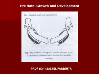
  • 17. The mandibular arch forms the lateral wall of the stomadeum. It gives a bud at its dorsal end which is called as maxillary process. It grows ventro-medially, cranial to the main part of the arch, which is now called as the mandibular process. The mandibular processes grow towards each other and fuse in midline. They now form the lower border of the stomadeum i.e. the lower lip and the lower jaw. Meckel’s Cartilage It is derived from the 1st branchial arch around the 41st – 45th day of intra-uterine life. It extends from the cartilagenous otic capsule to the midline or symphysis and provides a template for guiding the growth of the mandible. A major part of Meckel’s cartilage disappears during growth and the remaining part develops into – mental ossicles, incus and malleus, spine of sphenoid bone, anterior ligament of malleus and spheno-mandibular ligament. The first structure to develop in the primordium of the lower jaw is the mandibular division of the trigeminal nerve. This is followed by mesenchymal condensation forming the first branchial arch. Neurotrophic factors produced by the nerve induce osteogenesis in the ossification centers.
  • 18. A single ossification center for each half of mandible arises in 6th week of intra-uterine life in the region of the bifurcation of the inferior alveolar nerve into mental and incisive branches. The ossifying membrane is located lateral to the Meckel’s cartilage. From this primary center, ossification spreads below and around the inferior alveolar nerve and its incisive and branch and upwards to form a trough for accommodating the developing tooth buds. Spread of the intramembranous ossification dorsally and ventrally forms the body and ramus of the mandible. As ossification continues, the Meckel’s cartilage becomes surrounded and invaded by bone. Ossification stops at the site that later becomes mandibular lingula from where the Meckel;s cartilage continues into the middle ear and develops into the auditory ossicles [malleus and incus].
  • 19.
  • 20. Endochondral Bone Formation It is seen only in 3 areas of mandible – condylar process, coronoid process and mental region. • Condylar Process – At about 5th week of intra-uterine life, an area of mesenchymal condensation can be seen above the ventral part of the developing mandible. This develops into a cone shaped cartilage by about 10th week and starts ossification by 14th week. It then migrates inferiorly and fuses with the mandibular ramus by about 4 months. Much of the cone shaped cartilage is replaced by bone by the middlw of fetal life, but its upper end persists into adulthood acting both as a growth cartilage and an articular cartilage. • Coronoid Process – Secondary accessory cartilages appear in the region of the coronoid process by about the 10-14th week of intra-uterine life. This cartilage is believed to grow as a response to the developing temporalis muscle. The coronoid accessory cartilage becomes incorporated into the expanding intra-membranous bone of the ramus and disappears before birth.
  • 21. • Mental Region – In the mental region, on either side of the symphysis, one or two small cartilages appear and ossify in the 7th month of intra- uterine life to form variable numbers of mental ossicles in the fibrous tissues of the symphysis. These ossicles become incorporated into the intra-membranous bone when the symphysis ossifies completely during the first year of post-natal life.