SlideShare ist ein Scribd-Unternehmen logo
1 von 13
Downloaden Sie, um offline zu lesen
Mechanism of Inactivation of γ‑Aminobutyric Acid Aminotransferase
by (1S,3S)‑3-Amino-4-difluoromethylene-1-cyclopentanoic Acid
(CPP-115)
Hyunbeom Lee,†,⊥
Emma H. Doud,†,‡
Rui Wu,§
Ruslan Sanishvili,∥
Jose I. Juncosa,†,¶
Dali Liu,§
Neil L. Kelleher,†,‡
and Richard B. Silverman*,†,‡
†
Department of Chemistry, Chemistry of Life Processes Institute, and Center for Molecular Innovation and Drug Discovery,
Northwestern University, 2145 Sheridan Road, Evanston, Illinois 60208-3113, United States
‡
Department of Molecular Biosciences, Northwestern University, Evanston, Illinois 60208, United States
§
Department of Chemistry and Biochemistry, Loyola University Chicago, 1068 W. Sheridan Road, Chicago, Illinois 60660, United
States
∥
X-ray Science Division, Argonne National Laboratory, Argonne, Illinois 60439, United States
*S Supporting Information
ABSTRACT: γ-Aminobutyric acid aminotransferase (GABA-AT) is a
pyridoxal 5′-phosphate (PLP)-dependent enzyme that degrades GABA,
the principal inhibitory neurotransmitter in mammalian cells. When the
concentration of GABA falls below a threshold level, convulsions can
occur. Inhibition of GABA-AT raises GABA levels in the brain, which can
terminate seizures as well as have potential therapeutic applications in
treating other neurological disorders, including drug addiction. Among
the analogues that we previously developed, (1S,3S)-3-amino-4-difluoro-
methylene-1-cyclopentanoic acid (CPP-115) showed 187 times greater
potency than that of vigabatrin, a known inactivator of GABA-AT and
approved drug (Sabril) for the treatment of infantile spasms and
refractory adult epilepsy. Recently, CPP-115 was shown to have no
adverse effects in a Phase I clinical trial. Here we report a novel
inactivation mechanism for CPP-115, a mechanism-based inactivator that undergoes GABA-AT-catalyzed hydrolysis of the
difluoromethylene group to a carboxylic acid with concomitant loss of two fluoride ions and coenzyme conversion to
pyridoxamine 5′-phosphate (PMP). The partition ratio for CPP-115 with GABA-AT is about 2000, releasing cyclopentanone-
2,4-dicarboxylate (22) and two other precursors of this compound (20 and 21). Time-dependent inactivation occurs by a
conformational change induced by the formation of the aldimine of 4-aminocyclopentane-1,3-dicarboxylic acid and PMP (20),
which disrupts an electrostatic interaction between Glu270 and Arg445 to form an electrostatic interaction between Arg445 and
the newly formed carboxylate produced by hydrolysis of the difluoromethylene group in CPP-115, resulting in a noncovalent,
tightly bound complex. This represents a novel mechanism for inactivation of GABA-AT and a new approach for the design of
mechanism-based inactivators in general.
■ INTRODUCTION
γ-Aminobutyric acid (GABA) is a major inhibitory neuro-
transmitter in the mammalian central nervous system.1
When
GABA concentrations in the brain fall below a threshold level,
convulsions occur.2
It has also been found that Alzheimer’s
disease,3
Huntington’s chorea,4
Parkinson’s disease,5,6
and drug
addiction7,8
are related to diminished GABA levels in the brain.
GABA is produced by the α-decarboxylation of the excitatory
neurotransmitter L-glutamic acid, catalyzed by the pyridoxal 5′-
phosphate (PLP)-dependent enzyme glutamic acid decarbox-
ylase (GAD, Scheme 1). GABA is metabolized by γ-
aminobutyric acid aminotransferase (GABA-AT), also a PLP-
dependent enzyme, to succinic semialdehyde, which is further
oxidized to succinic acid by succinic semialdehyde dehydrogen-
ase (SSDH). Degradation of GABA to succinic semialdehyde
converts the PLP coenzyme to pyridoxamine 5′-phosphate
(PMP); α-ketoglutarate is used in a second step to return PMP
to PLP with concomitant formation of L-glutamic acid.
Therefore, the concentrations of the inhibitory (GABA) and
excitatory (L-Glu) neurotransmitters are regulated by the
appropriate balance between GAD and GABA-AT.9
When GABA is injected directly into the brain of a
convulsing animal, the seizures are terminated.11
However,
because GABA does not cross the blood−brain barrier,
peripheral GABA administration is not an effective approach
Received: December 2, 2014
Published: January 23, 2015
Article
pubs.acs.org/JACS
© 2015 American Chemical Society 2628 DOI: 10.1021/ja512299n
J. Am. Chem. Soc. 2015, 137, 2628−2640
for the treatment of epilepsy. An alternative approach to
increase GABA levels in the brain is by inhibition of GABA-AT,
which blocks the degradation of GABA.12
A mechanism-based
inactivator, an unreactive compound that initially acts as a
substrate for the target enzyme and is converted to a species
that causes inactivation of that enzyme, generally by covalent
attachment, could increase GABA levels for an extended period
of time.13
The design of these types of inhibitors requires
insight into the mechanism of the target enzyme so that the
catalytic chemistry can be utilized in the activation of the
inactivator; the mechanism of the first half of the “ping-pong”
reaction catalyzed by GABA-AT is shown in Scheme 2.
Incubation of GABA with GABA-AT leads to the conversion
of a lysine-PLP aldimine (1) to a GABA-PLP aldimine (2).
Abstraction of the γ-proton by Lys329 and tautomerization
leads to a PMP aldimine (3); enzyme-catalyzed hydrolysis of
this intermediate leads to succinic semialdehyde and PMP.14
4-Amino-5-hexenoic acid (4, γ-vinyl-GABA; vigabatrin) is a
mechanism-based inactivator of GABA-AT15
that is an FDA-
approved drug (Sabril) to treat infantile spasms and refractory
partial seizures. However, because of a serious side effect,
namely, retinal damage and blindness, a drug with more
potency and less toxicity was desired. Mechanistic studies
performed by Nanavati and Silverman16
(Scheme 3) demon-
strated that vigabatrin inactivates GABA-AT via two mecha-
nisms, a Michael addition reaction and an enamine reaction, in
a ratio of 70:30. In the Michael addition pathway (pathway a),
vigabatrin acts like substrate GABA and undergoes aldimine
formation with PLP, deprotonation, and tautomerization to a
Michael acceptor (5), which undergoes Michael addition by
Lys329, the lysine residue to which PLP is bound in the native
enzyme,17
giving, after protonation, covalent adduct 6. Enzyme-
catalyzed hydrolysis of 5 gives α,β-unsaturated ketone 7, which
may be a possible cause for side effects of the drug. In the
enamine pathway (pathway b), an enamine intermediate (8),
formed by tautomerization through the alkene, attacks the
enzyme-bound PLP to inactivate GABA-AT (9), also by
formation of a covalent adduct. Hydrolysis of 8 gives a weak
electrophile, which is unlikely to proceed through further
reactions in the active site.
To avoid the potentially toxic intermediate (7), computer
modeling based on the crystal structures of GABA-AT18
and
the vigabatrin−PLP complex in GABA-AT17
was carried out to
determine how to prevent the Michael addition pathway. The
model predicted that, in the energy-minimized structure, the
alkene double bond pointed away from the nucleophilic
Lys329; for Michael addition to occur, that bond had to rotate,
allowing the alkene to be aligned with the side chain amine of
Lys329. A conformationally rigid vigabatrin analogue was
prepared (10) to prevent Michael addition, and it was found
that it inactivated GABA-AT exclusively by the enamine
pathway;19
however, it was a weak inactivator. The second
approach to prevent formation of reactive product 7 was to
synthesize a vigabatrin analogue that would give rapid Michael
addition so that the equivalent of intermediate 5 would not be
sufficiently stable to allow the enzyme-catalyzed hydrolysis
leading to the equivalent of 7.20
That approach required the
synthesis of a compound with the alkene pointing toward the
side chain amine of Lys329 (11). The obtained compound
inactivated GABA-AT, but only in the absence of the
electrophilic trapping agent 2-mercaptoethanol. Therefore, an
undesirable reactive intermediate (the corresponding ketoacid
of 11) was formed prior to inactivation. To accelerate the
inactivation rate, two electron-withdrawing fluorine atoms were
Scheme 1. GABA Metabolism10
Scheme 2. First Catalytic Reaction of GABA-AT
Journal of the American Chemical Society Article
DOI: 10.1021/ja512299n
J. Am. Chem. Soc. 2015, 137, 2628−2640
2629
attached to the alkene of 11, and the obtained compound (12)
inactivated GABA-AT, even in the presence of 2-mercaptoe-
thanol. Further studies with 12 (now called CPP-115) showed
that it was at least 187 times more potent an inactivator of
GABA-AT than vigabatrin, was not active in the Cerep panel of
111 pharmacological targets (in vitro assays, carried out
commercially by Cerep, on CPP-115 that cover a broad
range of targets, including receptors, ion channels, enzymes,
transporters, and second messengers), does not bind to three
human GABA transporters GAT1, GAT2, and GAT3, or to
GABAA, GABAB, or GABAC receptors, does not bind to the
hERG channel, does not inhibit or induce cytochrome P450s, is
not metabolized by hepatocytes, is rapidly and completely
orally absorbed and eliminated, has no adverse effect on
respiration, produces no mutations or chromosomal aberra-
tions, and has a considerably lower retinal toxicity liability.21
It
also is 300−600 times more potent than vigabatrin in reducing
cocaine-induced synaptic dopamine levels and in blocking
expression of cocaine-induced conditioned place preference in
freely moving rats. Recently, it was shown to have no adverse
effects in a Phase I clinical trial. Because of the clinical
importance of this new drug candidate, we studied its
inactivation mechanism to determine if it indeed favors the
Michael addition pathway for which it was designed. We have
determined that CPP-115 inactivates GABA-AT via an
unexpected inactivation mechanism, namely, enzyme-catalyzed
conversion of the difluoromethenyl group to a carboxylate,
which causes a conformational change in the enzyme, to give a
tightly bound complex. This inactivation mechanism presents a
novel approach to inactivate GABA-AT and other related
enzyme targets using similar catalytic mechanisms.
■ RESULTS
Inhibition of GABA-AT by Increasing Equivalents of
CPP-115. The turnover number for inhibition was determined
by incubating GABA-AT with various equivalents of CPP-115.
Figure 1 shows the remaining enzyme activity versus time for
0−4 equiv of CPP-115. To determine the turnover number, the
data points at 24 h were replotted as a pseudo-first-order
reaction using the remaining enzyme activity versus equivalents.
The turnover number was found to be 1.3 ± 0.3 per active site.
However, as shown in the replot, nonpseudo-first-order
inactivation kinetics was observed, possibly because of product
inhibition.22
Inactivation of GABA-AT by CPP-115 and Dialysis of
the Inactivated Enzyme. To test if the mechanism involves
irreversible and/or reversible inhibition, the time-dependent
reactivation of GABA-AT was studied. First, the enzyme was
treated with an excess of inactivator; then, the mixture was
dialyzed against bulk buffer. Aliquots at different time intervals
were collected and assayed. Return of enzyme activity was
observed, which displayed both time- and concentration-
dependence (see Supporting Information, Figures S1 and
S2). The data also show that the amount of activity of the
inhibited GABA-AT that returned was dependent on the
amount of CPP-115 used in the incubation and the incubation
time. In Figure S1 (Supporting Information), up to 90% and
60% of enzyme activity was recovered when GABA-AT was
inactivated with 40 and 80 equiv of CPP-115, respectively, for 3
h. However, when GABA-AT was incubated for 24 h, even with
10 equiv of CPP-115, only 50% of the activity returned (Figure
S2, Supporting Information). In the presence of 1000 equiv
(approximately 1 mM) of CPP-115 in the incubation mixture,
Scheme 3. Inactivation Mechanism of GABA-AT by Vigabatrin
Journal of the American Chemical Society Article
DOI: 10.1021/ja512299n
J. Am. Chem. Soc. 2015, 137, 2628−2640
2630
the activity of the enzyme returned to about 8%; with 2000
equiv of CPP-115, the activity return was only 1%, even after 50
h of dialysis.
Fluoride Ion Release during Inactivation of GABA-AT
by 2 mM CPP-115. Upon incubation of GABA-AT with CPP-
115, fluoride ions were continually released, even after the
enzyme’s activity had fallen to almost zero (Figure S3,
Supporting Information). After normalization of the values
with the controls, it was found that 66 ± 3 equiv of fluoride
ions were released slowly over a period of 27 h. However, when
the same experiment was repeated with PLP supplemented in
the mixture, there were 127 ± 5 equiv of fluoride ions released
over time. Because α-ketoglutarate is essential to regenerate
PLP from PMP, if α-ketoglutarate is omitted, the amount of
fluoride ions released per single turnover of inhibition can be
determined. The continual release of fluoride ions was not
observed when there was no α-ketoglutarate present, and only
4.3 ± 0.5 equiv of fluoride ions were detected per enzyme
dimer (Figure S4, Supporting Information). However, when α-
ketoglutarate was added to the inactive enzyme, even without
supplementation with PLP, the fluoride ion concentration
began to increase. A control experiment with no enzyme in the
incubation mixture was performed, which confirmed that no
fluoride ions were released from CPP-115 in the absence of the
enzyme, even if PLP is added.
Inactivation of [3
H]PLP-Reconstituted GABA-AT by
CPP-115. To determine the fate of the coenzyme by
inactivation with CPP-115, apo-GABA-AT was reconstituted
with [3
H]PLP. The maximum recovered activity was 71.4% of
the original, and the activity recovery plateaued after 5 h of the
reactivation process (Table 1), indicating that 29.6% of the
enzyme remained inactive.
[3
H]PLP-GABA-AT was inactivated with CPP-115, and the
released radioactive compounds were analyzed using HPLC
(Figure S5, Supporting Information). The experiment was
performed with two controls: CPP-115 was omitted in a
negative control (all radioactivity should be labeled PLP), and
GABA replaced CPP-115 with α-ketoglutarate omitted in a
positive control (all radioactivity should be PMP). The negative
control released most of its radioactivity as PLP (but none as
PMP), whereas a GABA-incubated control released both PMP
and PLP (Figure S6, Supporting Information). Given that the
positive control with GABA should only produce PMP from
active enzyme, the PLP present in this sample represents that
released from the inactive portion of GABA-AT. After the
background radioactivity from the control experiments was
subtracted from the CPP-115 experiment, CPP-115-inactivated
[3
H]PLP-reconstituted GABA aminotransferase was found to
release 100% of its cofactor as [3
H]PMP (Figure S7,
Supporting Information). The small radioactive peak at 33
min was confirmed to be degraded PLP present in all of the
samples.
Mass Spectrometric Analysis of Metabolites from
Inactivation of GABA-AT with CPP-115. Metabolites
released during inactivation were determined by LC/MS/MS.
After inactivation of GABA-AT by CPP-115 followed by
denaturation and filtration, three metabolites were identified
from the sample solution that were not present in the control
samples: [m/z] 127.0389, 171.0291, and 401.0745 (A, B, and C
in Figure S8, Supporting Information, respectively), corre-
sponding to 22, 21, and 20, respectively, in Scheme 5 (see
Discussion). These parent ions were selected for fragmentation
using normalized collision energies. Fragmentation data for m/
z 127.0389 (Supporting Information Figure S9) confirmed the
structure of 3-oxo-1-cyclopentanecarboxylic acid (22). MS/MS
for m/z 171.0291 and 401.0745 (Supporting Information
Figures S10 and S11, respectively) confirmed the structures of
21 and 20.
Intact Mass Spectrometry of GABA-AT. Since covalent
modification of GABA-AT was not detected using middle down
proteomics (see Figures S12−S14 in Supporting Information),
we analyzed the intact GABA-AT protein to see if any
detectable mass shifts were present. The intact mass data
showed multiple peaks, indicating that the GABA-AT purified
from pig brain was a mixture of GABA-AT species with
different N-termini. Three samples were tested using LC/MS/
MS: free enzyme (negative control), vigabatrin-inactivated
GABA-AT (positive control), and CPP-115-inactivated GABA-
AT (Figure S15, Supporting Information). Vigabatrin-inacti-
vated GABA-AT showed an added mass of 122 Da from the
mass of the native enzyme, which matches the covalent adduct
proposed previously.15
However, CPP-115-inactivated GABA-
AT showed no significant peaks corresponding to any added
mass. To stabilize any potential imine adducts throughout the
LC/MS/MS process, the samples were reduced with sodium
borohydride as described previously for crystallography studies.
Reduction resulted in stabilization of the PLP cofactor on the
enzyme, with an added mass of 236 from the original peak (M).
As expected, the peaks of vigabatrin-inactivated GABA-AT
Figure 1. (Top) Time-dependent inactivation of GABA-AT by 0−4.0
equiv of CPP-115. (Bottom) Determination of the turnover number
for the inactivation of GABA-AT by CPP-115. The data points at 24 h
are replotted as enzyme activity vs equiv of CPP-115.
Table 1. Return of GABA-AT Activity after apo-GABA-AT
Was Incubated with [3
H]PLP
2 h 3 h 4 h 5 h original
dA (mL−1
min−1
) 0.630 0.748 1.504 1.522 2.124
Journal of the American Chemical Society Article
DOI: 10.1021/ja512299n
J. Am. Chem. Soc. 2015, 137, 2628−2640
2631
(Figure S16, Supporting Information) were no different from
those in Figure S15 since the vigabatrin covalent adduct is
stable.15
Interestingly, for CPP-115-inactivated GABA-AT,
reduction had no effect on the resulting data and showed no
added mass on the protein.
X-ray Crystallography of Native and CPP-115-Inacti-
vated GABA-AT. To understand how time-dependent
inactivation of GABA-AT by CPP-115 could occur without
covalent modification of the protein or cofactor, CPP-115-
inactivated and dialyzed GABA-AT were crystallized. The
crystal structures of native GABA-AT from pig brain and
inactivated enzyme were obtained at 1.63 Å and 2.19 Å
resolution, respectively. The crystal structure for the native pig
brain enzyme was very similar to that reported from pig liver by
Storici et al.18
The crystal structures of the native enzyme and
the inactivated enzyme were compared to analyze the difference
in overall structure (Figure 2) and in the active site (Figure 3).
The active site of the inactivated GABA-AT was investigated to
understand the ligand−enzyme interactions (Figure 4); the
omit map supports the ligand interpretation (see Supporting
Information, Figure S17).
■ DISCUSSION
At the start of this investigation, we considered a variety of
likely mechanisms for the inactivation of GABA-AT by CPP-
115 (Schemes 4−8)23,24
and then considered experiments to
differentiate them. All of these inactivation mechanisms are
initiated by the same steps shown in Scheme 2 for substrate
turnover or the two inactivation mechanisms for vigabatrin
shown in Scheme 3. In mechanism 1 (Scheme 4), following γ-
proton abstraction of 13, tautomerization leads to an α,β-
unsaturated intermediate (14), which is attacked by the active
site lysine residue or another base to covalently modify the
enzyme. No fluorine is released, the enzyme is inactivated, and
hydrolysis of 15 gives 16 with release of PMP as the cofactor.
If intermediate 15 in Scheme 4 (where X is either the active
site lysine residue or OH from attack by water) eliminates one
fluoride ion (Scheme 5), the inactivator can form another
reactive α,β-unsaturated imine (17), which can be attacked by a
water molecule to release the other fluoride ion, forming 18.
Depending on what X is, this may result in inactivation or
turnover. If X is the active-site lysine residue (15a, pathway a,
Scheme 5), the intermediate is a stabilized amide; hydrolysis
generates covalent adduct 19 with the release of PMP.
However, if X is a hydroxyl group (15b, pathway b, Scheme
5) formed by water molecule attack on reactive intermediate 14
(Scheme 4), stable dicarboxylate intermediate 20 will form;
hydrolysis gives metabolite 21 and PMP, and subsequent
decarboxylation of 21 gives 22.
The third potential mechanism involves allylic tautomeriza-
tion of external aldimine 13 to form 23 (Scheme 6). Because
23 also is a reactive electrophile, it may undergo Michael
addition to form adduct 24, which could be hydrolyzed to give
25 and release PMP.
In the fourth potential mechanism (Scheme 7), intermediate
23 (Scheme 6) can proceed through three different pathways.
Pathway a generates an enamine (26), similar to that formed
during vigabatrin inactivation; this species might undergo
enamine attack on the Lys329-bound PLP to form covalent
adduct 27, which could hydrolyze to 28. In pathway b, one
fluoride ion is released by nucleophilic substitution (29) before
undergoing transimination to form enamine adduct 30, which
might undergo hydrolysis to 31. Pathway c from 30 involves
elimination of a second fluoride ion (32) followed by hydrolysis
to give 33.
The final potential mechanism involves fluoride ion
elimination from enamine 26 to generate reactive Michael
acceptor 34 (Scheme 8). Hydrolysis of imine 34 (pathway a)
gives 35 and PLP. Attack by an active site nucleophile (either
Lys329 or water; pathway b) gives either covalent adduct 36 or
noncovalent adduct 37, respectively. Elimination of another
fluoride ion forms 38 or 40, each of which can be subsequently
hydrolyzed to covalent adduct 39 or metabolite 41; further
hydrolysis of 39 would also give 41. In either case, this
mechanism releases the cofactor as PLP. All of these possible
mechanisms can be differentiated by determination of the
number of fluoride ions released, the fate of the cofactor, and
the final metabolites or adducts formed; these possibilities are
summarized in Table 2.
Figure 2. Ribbon diagram of the superimposed native GABA-AT
(yellow) and GABA-AT (cyan) bound to CPP-115.
Figure 3. Superimposition of the crystal structures of native GABA-AT
(pink) and CPP-115-inactivated GABA-AT (green).
Journal of the American Chemical Society Article
DOI: 10.1021/ja512299n
J. Am. Chem. Soc. 2015, 137, 2628−2640
2632
The partition ratio, the number of inactivator turnovers per
inactivation event, was investigated by determination of the
number of molecules of CPP-115 that were required to cause
complete enzyme inactivation. Time-dependent inhibition was
observed for 1−4 equiv of CPP-115, although pseudo-first-
order kinetics was not observed (Figure 1, top). A replot of
these data with extrapolation of the first few data points gave a
turnover number of 1.3 ± 0.3 per active site (Figure 1, bottom).
The nonpseudo-first-order inactivation kinetics displayed
indicated possible product inhibition and suggested possibly
two concurrent mechanisms occurring at different rates, maybe
one reversible and one irreversible.
To test whether there was a reversible component to the
inactivation, the titration experiment was repeated, first with 0−
80 equiv of CPP-115 (Figure S1, Supporting Information),
then 0−2000 equiv of CPP-115 (Figure S2, Supporting
Information), followed by dialysis. Aliquots removed during
dialysis exhibited time-dependent reactivation of GABA-AT
over an extended period of time; with increasing equiv of
inactivator, less activity returned with maximal return by 24 h
(maximal return of activity occurred sooner with less
inactivator). It required about 2000 equiv of CPP-115 before
the enzyme reactivation was diminished to an insignificant level,
supporting a reversible and irreversible component to the
inactivation.
During inactivation of GABA-AT by 2 mM CPP-115, it was
found that 66 ± 3 equiv of fluoride ions were released over a 26
h time period; however, enzyme activity was lost within
minutes (Figure S3, Supporting Information). This result
further supports the notion of two different mechanisms being
involved, one reversible and one irreversible, but it does not
resolve the question of how many fluoride ions are released per
molecule. A single-turnover experiment was carried out to
determine the number of fluoride ions per molecule released
with each turnover. This experiment is possible because GABA-
AT converts substrate to product with concomitant conversion
of its PLP to PMP. The reaction cycle cannot restart until α-
ketoglutarate binds and converts the PMP back to PLP with
concomitant formation of L-glutamic acid. By leaving out the α-
ketoglutarate, only one inhibition turnover is possible. Under
those conditions, 4.3 ± 0.5 equiv of fluoride ions was detected
(Figure S4, Supporting Information); because GABA-AT is a
homodimer, two molecules of CPP-115 can be turned over per
enzyme dimer. Therefore, 4 equiv of fluoride ion indicates that
both fluoride ions in CPP-115 are lost per enzyme turnover,
and mechanisms 1, 3, and 4a, which release no fluoride ions
Figure 4. Stereoview of GABA bound by the CPP-115 adduct. The 2Fo−Fc electron-density map is shown as light gray mesh at 1.1σ level around the
CPP-115 adduct.
Scheme 4. First Potential Mechanism of Inactivation of GABA-AT by CPP-115
Journal of the American Chemical Society Article
DOI: 10.1021/ja512299n
J. Am. Chem. Soc. 2015, 137, 2628−2640
2633
during inactivation, and mechanism 4b, which releases one
fluoride ion, can be excluded (Table 2).
The cofactor structure after inactivation by CPP-115 was
determined using GABA-AT containing [3
H]PLP (Figure S5,
Supporting Information). With the aid of two control
experiments (Figure S6, Supporting Information), it was
found that all of the cofactor was converted to PMP after
inactivation (Figure S7, Supporting Information). Therefore,
mechanism 4a, which does not release any cofactor and
mechanisms 4b, 4c, 5a, and 5b, which require the release of
PLP as a cofactor, can be excluded (Table 2).
Of the mechanisms considered, only 2a and 2b remain
plausible (Scheme 5). Mechanism 2b should produce
compounds 20−22. High-resolution LC/MS identified parent
ions with m/z 401.0745, 171.0291, and 127.0389 (Figure S8,
Supporting Information), which corresponds to 20, 21, and 22,
respectively. MS/MS fragmentation produced daughter ions
(Figures S9−S11, Supporting Information) consistent with
these three products and with mechanism 2b. This mechanism
appears to be responsible for the reversible component of the
overall inactivation mechanism; mechanism 2a would be
expected to account for the irreversible component, so it was
imperative to demonstrate covalent attachment to the enzyme
(19, Scheme 5) for substantiation.
Mass spectrometry of the intact mass of CPP-115-inactivated
GABA-AT was carried out using LC/MS/MS on native enzyme
as the negative control and vigabatrin-inactivated GABA-AT as
the positive control (Figure S15, Supporting Information).
Compared with native enzyme, vigabatrin-inactivated GABA-
AT had an added m/z peak of 122 Da, which corresponds to
the expected added mass of the covalent adduct previously
proposed.15
CPP-115-inactivated GABA-AT showed no sig-
nificant peaks corresponding to added mass. In case the
potential covalent adduct with CPP-115 was an imine, the
inactivated enzyme was reduced with sodium borohydride prior
to LC/MS/MS (Figure S16, Supporting Information). Again,
no added mass was detected with the CPP-115 inactivated
enzyme, but the vigabatrin-inactivated enzyme had the expected
added mass. These results were corroborated by results from
peptide proteomics (Supporting Information, Figures S13 and
S14). Therefore, CPP-115 appears to be inactivating GABA-AT
without covalent modification.
The solution to this dilemma came from the X-ray crystal
structure of CPP-115-inactivated pig brain GABA-AT, which
revealed that the inactivator was tightly bound to the protein
noncovalently as 20 (Figures 3 and 4). The inactivated species
binds tightly (stable to dialysis) because of its covalent
attachment to the cofactor and by two strong electrostatic
interactions between the guanidinium groups of Arg192 and
Arg445 and each of the two carboxylate groups of 20. This
unexpected phenomenon is the first time that Arg445 has been
observed interacting directly with a ligand and to be involved in
the inactivation of GABA-AT. On the basis of crystallography
of GABA analogues, it is believed that GABA binds in the active
site by Schiff base formation with PLP and an electrostatic
interaction between the carboxylate of GABA and the
guanidinium group of Arg192. Arg445 is sequestered from
the active site by an electrostatic interaction with Glu270.25
It
has been proposed that this second salt bridge only
disassociates during the second half of catalysis, the
regeneration of PLP from PMP, to aid in the binding of the
second carboxylate of α-ketoglutarate. A network of hydrogen
bonds in the active site of GABA-AT connects Glu270 and
Arg445 to O3′ of the cofactor through the side chain amide of
Gln301. Markova et al. proposed that this network may be
affected when the cofactor is in its PMP form, which weakens
the interaction between Glu270 and Arg445.26
To verify these
hypotheses, many research groups have attempted to
cocrystallize GABA-AT with ligands such as α-KG or glutamate
that may directly show the involvement of this second arginine,
but they have not yet been successful.25−27
Our crystal structure of GABA-AT inactivated by CPP-115
(Figure 3) clearly shows for the first time that the salt bridge
between Arg445 and Glu270 has been broken, and Glu270 is
rotated away from its original position to accommodate a
second guanidinium−carboxylate electrostatic interaction be-
tween Arg445 and the newly formed carboxylate group on 20
Scheme 5. Second Potential Mechanisms of Inactivation of
GABA-AT by CPP-115
Scheme 6. Third Potential Mechanism of Inactivation of GABA-AT by CPP-115
Journal of the American Chemical Society Article
DOI: 10.1021/ja512299n
J. Am. Chem. Soc. 2015, 137, 2628−2640
2634
from enzyme-catalyzed hydrolysis of the difluoromethenyl
group. The two guanidinium−carboxylate interactions, the
hydrophobic interactions of the pyridine ring, and the
interactions of the phosphate group contribute to the
stabilization of the metabolite for tight binding. Therefore,
CPP-115 appears to be, to the best of our knowledge, the first
example of a mechanism-based inactivator that leads to a
conformational change, forming a tightly bound inhibitor−
enzyme complex with a newly exposed residue. Apparently, this
conformational change occurs only about once with 2000
molecules of inactivator; the remaining molecules of 20−22 are
slowly released as products.
The α,β-unsaturated intermediate (14, Scheme 4) generated
during turnover is highly electrophilic, which should have been
attacked by the nucleophilic Lys329 residue and form a
covalent adduct, as it does with vigabatrin. Molecular modeling
studies of the CPP-115−PLP aldimine were performed to
analyze the effect of tautomerization during the inactivation of
GABA-AT by CPP-115 (Figure 5). This model demonstrates
that the PMP-imine form (orange) of the aldimine generated
during enzyme-catalyzed tautomerization shifts the difluor-
omethenyl group (fluorines are in green) away from Lys329,
thereby moving it too far for Michael addition by this lysine
residue. Because of the lack of 1,5 steric interactions (both
carbons opposite the carboxylate group are sp2
-hybridized and
thus planar), the difluoromethenyl group is able to release
strain by moving closer to the carboxylate. As GABA-AT
normally catalyzes hydrolysis of the imine formed by
tautomerization of GABA, the catalytic machinery is in place
for water attack of the Michael acceptor instead. This is
Scheme 7. Fourth Potential Mechanism of Inactivation of GABA-AT by CPP-115
Scheme 8. Fifth Potential Mechanism of Inactivation of GABA-AT by CPP-115
Journal of the American Chemical Society Article
DOI: 10.1021/ja512299n
J. Am. Chem. Soc. 2015, 137, 2628−2640
2635
consistent with the obtained crystal structure and explains why
the inactivation mechanism did not follow that of vigabatrin
and form a covalent adduct.
On the basis of the evidence from fluoride release, cofactor
release, metabolites formed, proteomics, and X-ray crystallog-
raphy, the most consistent mechanism is shown in Scheme 9.
■ CONCLUSIONS
Although CPP-115 was rationally designed to inactivate GABA-
AT via a covalent Michael addition mechanism, the results
described here indicate that it inactivates GABA-AT by
mechanism-based formation of a metabolite that induces a
conformational change and forms a tightly bound complex with
the enzyme (Scheme 9). The requirement of about 2000 equiv
of CPP-115 for complete inactivation of GABA-AT reflects the
number of molecules of product that are released prior to
interception by the enzyme in the correct state and
conformation that lead to inactivation. Proteomic studies
could not reveal a covalent adduct. The crystal structure of
CPP-115-inactivated GABA-AT revealed, for the first time, that
the Glu270-Arg445 salt bridge in the active site can be
disrupted, leading to the formation of a new binding pocket for
the inactivator. Our molecular modeling studies indicate a
movement of the difluoromethenyl group upon tautomeriza-
tion, which shifts that group too far from Lys329 for
nucleophilic attack. This leads to enzyme-catalyzed hydrolysis
of the PMP-aldimine of CPP-115 and either release of products
(2000 times) or inducement of a conformational change to give
a tightly bound PMP complex stabilized by the exposure of
Arg445 and formation of an electrostatic interaction with the
newly formed carboxylate of the inactivator. Once metabolite
20 is tightly bound to the enzyme, it is released from the active
site very slowly, which accounts for the extended period of time
that fluoride ions are released relative to the rate of inhibition of
the enzyme. With our results revealing the possibility of
additional binding with Arg445, future inactivators may be
designed so that the newly formed carboxylate group more
closely mimics the second carboxylate group of α-ketoglutarate
to encourage less turnover prior to conformational change-
induced exposure of Arg445.
■ EXPERIMENTAL PROCEDURES
Analytical Methods. GABA-AT assays were recorded on a
Synergy H1 hybrid multimode microplate reader (Biotek, USA) with
transparent 96-well plates (Greiner bio-one, Monroe, NC). Measure-
ments of pH were performed on a Fisher Scientific AP71 pH/mV/°C
meter with a pH/ATC electrode. Determinations of fluoride ion
concentration were performed on the same meter with a Thermo
Scientific 9609BN combination fluoride electrode. Large-scale dialyses
were performed with Thermo Scientific Slide-A-Lyzer dialysis cassettes
(molecular weight cutoff of 10 kDa) unless otherwise specified. Small-
scale dialyses were performed with EMD Chemicals D-Tube Mini
dialyzer (molecular weight cutoff of 12−14 kDa). Radioactivity was
determined with a Packard TRI-CARB 2100TR liquid scintillation
analyzer using PerkinElmer ULTIMA GOLD scintillation fluid. For
centrifugations, a DuPont Sorvall RC 5B Plus centrifuge was used with
either an SLA-3000 or an SA600 rotor. Eppendorf Minispin plus tubes
were used for microcentrifugation. An IEC clinical centrifuge was used
for the Penefsky spin method. HPLC analysis was performed with
Beckman 125P pumps and a Beckman 166 detector. All of the runs
were monitored at 254 nm. The HPLC columns used were Alltech
C18 analytical and Econosil 10 μm columns. Enzyme purification was
carried out on a GE Healthcare Life Sciences ÄKTA FPLC system.
The column media used were CM Sepharose Fast Flow for weak
cationic exchange mode, DEAE Sepharose Fast Flow for weak anionic
exchange mode, and hydroxylapatite (HA) for mixed mode. A
prepacked Hiprep Sephacryl S-200 high-resolution column was used
for size-exclusion mode. Electrophoresis was carried out on a Bio-Rad
Mini-Protean Tetra Cell using a VWR AccuPower Model 300 power
supply. LC/MS/MS was performed on Thermo Fisher Q Exactive
mass spectrometer. Mass spectrometry of intact mass of GABA-AT
was performed on a Velos Orbitrap Elite (ThermoFisher). Crystallo-
graphic data were collected on beamline 23 ID-B and 23 ID-D of GM/
CA of the Advanced Photon Source (APS) using X-rays of 0.99 Å
wavelength and a Rayonix (formerly MAR-USA) 4 × 4 tiled CCD
detector. All data were indexed, integrated, and scaled with HKL2000.
Data processing statistics are given in Table S1 in Supporting
Information.
Reagents. All reagents and materials were purchased from Sigma-
Aldrich Co. except the following: Centrifugal filters (molecular weight
cutoff value of 10 kDa and 30 kDa) were purchased form EMD
Millipore; Dowex 50 and sodium dodecyl sulfate were purchased from
Bio-Rad; [3
H]sodium borohydride was purchased from American
Radiolabeled Chemicals, Inc.; pig brains were a generous gift from the
Park Packing Co. (Chicago, IL); all of the buffers and solvents used for
FPLC analyses were filtered through GE Healthcare 0.45 μm nylon
membranes. GABase (Pseudomonas fluorescens) and succinic semi-
aldehyde were purchased from Sigma-Aldrich.
Enzyme and Assays. GABA-AT (2.65 mg/mL, specific activity
2.1 unit/mg) was purified from pig brain by the procedure described
previously.28
Succinic semialdehyde dehydrogenase (SSDH) was
obtained from GABAse, a commercially available mixture of SSDH
and GABA-AT, using a known procedure.24
GABA-AT activity was
Table 2. Expected Differences in the Various Inactivation
Mechanisms
Journal of the American Chemical Society Article
DOI: 10.1021/ja512299n
J. Am. Chem. Soc. 2015, 137, 2628−2640
2636
assayed using a published method.29
The final assay solution consisted
of 11 mM GABA, 1.1 mM NADP+
, 5.3 mM α-KG, 2 mM β-
mercaptoethanol, and excess SSDH in 50 mM potassium pyrophos-
phate buffer, pH 8.5. The change in UV absorbance at a wavelength of
340 nm at 25 °C caused by the conversion of NADP+
to NADPH is
proportional to the GABA-AT activity. All enzyme assays were
recorded with a PerkinElmer Lambda 10 UV/vis spectrophotometer.
Syntheses of (1S,3S)-3-Amino-4-difluoromethylene-1-cyclo-
pentanoic Acid (CPP-115). This compound was synthesized
according to the procedure published by Pan et al.24
Inhibition of GABA-AT by Various Equivalents of CPP-115.
GABA-AT (2 μg) was incubated with CPP-115 (0, 0.2, 0.4, 0.6, 0.8,
1.0, 1.2, 1.4, 1.6, 1.8, and 4 equiv) at 25 °C in 50 mM potassium
pyrophosphate buffer solution, containing 2 mM α-ketoglutarate and 1
mM β-mercaptoethanol. Aliquots (2 μL) were withdrawn at timed
intervals and were added immediately to the assay solution (96 μL)
containing excess SSDH (2 μL). The reaction rates were monitored at
340 nm.
Inactivation of GABA-AT by CPP-115 and Dialysis of the
Inactivated Enzyme. GABA-AT (60 μL; final concentration 1 μM)
was preincubated for 3 h with various amounts of CPP-115 (0, 1, 1.8,
4, 40, and 80 equiv) in 50 mM pyrophosphate buffer (pH 8.5)
containing 0.625 mM α-ketoglutarate, in a total volume of 160 μL at
25 °C. In another experiment, it was preincubated for 24 h with
various amounts of CPP-115 (0, 1, 2, 4, 40, 80, 100, 1000, and 2000
equiv). After preincubation, the enzyme solutions were transferred to
D-Tube Mini dialyzers and exhaustively dialyzed against buffer (50
mM pyrophosphate buffer containing 0.1 mM α-ketoglutarate and 0.1
mM PLP, pH 8.5) at 4 °C. The dialysis buffer was exchanged every 4
h, three times. The enzyme activity remaining in each of the solutions
was assayed per time interval.
Analysis of Fluoride Ion Release during Inactivation of
GABA-AT by CPP-115. GABA-AT (150 μL) was incubated in 100
mM potassium pyrophosphate buffer at pH 8.5, containing 2 mM
CPP-115 and 2.5 mM α-ketoglutarate in a total volume of 510 μL. A
control lacking GABA-AT was also incubated. The incubation mixture
was protected from light and was carried out at room temperature.
The experiment was repeated with 2.5 mM PLP supplemented in the
incubation mixture.
To construct a standard curve, the relative potential (mV) of
sodium fluoride solutions in the range of 1 × 10⁻⁶
to 5 × 10⁻⁵
M in
total ionic strength adjustment buffer (TISAB; made from 5.8 g of
sodium chloride and 5.7 mL of acetic acid in 100 mL of water, adjusted
to pH 5.2) was measured. The measured potential was plotted against
the concentration of sodium fluoride. At different incubation time (1,
2, 5, 7, 17, 24, 27 h), each aliquot was removed from the incubation
samples and mixed with TISAB to measure their relative potentials.
Using the formula obtained from the standard curve, the mV readings
were converted to concentration of fluoride ions in the samples. The
readings from the control sample were subtracted from the inactivated
sample, and the concentration was divided by the concentration of
GABA-AT to obtain the equivalents of fluoride ion released per
inactivation event.
Analysis of Fluoride Ion Release during Inactivation of
GABA-AT by CPP-115 without α-Ketoglutarate. The similar
experiment was run as above but without α-ketoglutarate to test the
amount of fluoride released during a one-turnover event. GABA-AT
(200 μL) was incubated in 100 mM potassium pyrophosphate buffer
at pH 8.5, containing 2 mM CPP-115 in a total volume of 510 μL.
When there is no α-ketoglutarate in the mixture to regenerate PLP, the
reaction stops at one turnover per active site.
Radioactive Labeling of [7-3
H]-PLP with Tritiated NaBH4.
Thirty drops of 1 M NaOH were added to 1.8 mL of 0.5 M PLP and
cooled to 0 °C in an ice bath. The mixture of sodium borohydride (5.8
mg, 0.15 mmol) and [3
H]-sodium borohydride (25 mCi) in 450 μL of
0.1 M NaOH was added to the PLP solution and stirred for 1 h at 0
Figure 5. Molecular docking studies of CPP-115-PLP aldimine in PLP-form (cyan) vs CPP-115-PMP-form (orange) in the active site of GABA-AT.
Journal of the American Chemical Society Article
DOI: 10.1021/ja512299n
J. Am. Chem. Soc. 2015, 137, 2628−2640
2637
°C. After being stirred, 120 μL of concd HCl was added to the
solution slowly (became pH 4). To the solution at pH 4, 20 equiv of
ground MnO2 was added, and the mixture was stirred at room
temperature for a total of 2 h. The mixture was brought to pH 8 with 1
M NaOH, and the solution was centrifuged. The supernatants were
collected and were loaded onto a gel filtration column packed with
Bio-Rad AG1-X8 resin (hydroxide form). The mobile phase used was
water and 5 M acetic acid (gradient of 90% water to 0% water),
flowing at 1.5 mL/min for 300 min. Fractions were collected every 10
mL and tested for its UV absorption and radioactivity. The fractions
with desired product were lyophilized. The lyophilized product was
redissolved in 100 μL of 1 mM PLP and loaded onto an HPLC with
an Econosil C18 column (10 mm × 250 mm, 10 μm). The mobile
phase used was 0.1% aqueous TFA with 5% acetonitrile flowing at 0.5
mL/min for 40 min. Under these conditions, PLP eluted at 15 min.
Fractions running with the PLP peak were collected and counted for
radioactivity using liquid scintillation counting. The product was
collected and lyophilized.
Inactivation of [3
H]PLP-Reconstituted GABA-AT by CPP-115.
GABA-AT that had been reconstituted with [3
H]PLP was incubated at
room temperature and protected from light in 100 mM potassium
phosphate buffer containing CPP-115 (2 mM), α-ketoglutarate (3
mM), and β-mercaptoethanol (3 mM), in a total volume of 100 μL at
pH 7.4. A negative control was run under identical conditions as
above, excluding the inactivator. A positive second control was run
with 3 mM GABA in the absence of inactivator and α-ketoglutarate.
The first control should release the cofactor as PLP, and the second
control should release it as PMP. After incubation for 18 h, the activity
of GABA-AT was less than 1% of control, and the solutions were
adjusted to pH 11 with 1 M KOH and incubated for 1 h.
Trifluoroacetic acid (TFA) was added to quench the base and make
the solution 10% v/v TFA. The resulting denatured enzyme solution
was microcentrifuged for 5 min at 10 000 rpm after standing at room
temperature for 10 min. A small amount of white solids was seen at the
bottom of the tube. The supernatants were collected individually. To
wash the pellets, 50 μL of 10% TFA was added to each tube, vortexed,
and microcentrifuged for another 5 min. This process was repeated
three times. The washed pellets were counted for radioactivity. The
supernatant and washes were combined and lyophilized. Cofactor
analysis was carried out by dissolving the solids obtained from
lyophilization with 100 μL of a solution containing 2.5 mM PLP and
2.5 mM PMP as standards and then injecting the samples into the
HPLC with an Econosil C18 column (4.6 mm × 150 mm, 10 μm).
The mobile phase used was 0.1% aqueous TFA flowing at 0.5 mL/min
for 25 min. Then the flow rate was increased to 1 mL/min from 25 to
30 min, and then a solvent gradient into 95% acetonitrile was run over
the next 30 min. Under these conditions, PLP eluted at 15 min and
PMP at 8 min. Fractions were collected every minute, and the
radioactivity was measured by liquid scintillation counting.
Mass Spectrometric Analysis of Metabolites from Inactiva-
tion of GABA-AT with CPP-115. GABA-AT (0.3 nmol) was
incubated in 50 mM ammonium bicarbonate buffer (pH 7.4)
containing 0.1 mM CPP-115, 1 mM α-ketoglutarate, and 0.1 mM
PLP in a total volume of 70 μL at room temperature in the dark for 12
h. A control containing everything except CPP-115 was also incubated.
After 12 h, GABA-AT in the inactivated sample was less than 1% active
vs control. Formic acid (1 μL) was added to each reaction mixture,
and both were centrifuged in a 0.5 mL 30 kDa MWCO centrifuge tube
(Millipore) at 14000g for 4 min or until most of the solution had
passed through. An additional 20 μL of 50 mM ammonium
bicarbonate was added above the filter and centrifuged for 3 min.
The flow-through (20 uL) was injected onto a Luna C18(2) column
(100 A, 2 × 150 mm, 5 μm, Phenomenex). A 60 min gradient (Agilent
1100 HPLC, solvent A = 5% acetonitrile and 0.1% formic acid; solvent
B = 0.1% formic acid in acetonitrile) was run from 2−80% B over 40
min. The LC was directly connected to a Thermo Fisher Q Exactive
mass spectrometer. The top five most abundant ions in negative ion
mode were selected for fragmentation using mass normalized collision
energies. Commercially available 3-oxo-1-cyclopentanecarboxylic acid
(Sigma) was injected as a standard for comparison.
Scheme 9. Mechanism of Inactivation of GABA-AT by CPP-115 Most Consistent with Experimental Evidence
Journal of the American Chemical Society Article
DOI: 10.1021/ja512299n
J. Am. Chem. Soc. 2015, 137, 2628−2640
2638
Intact Mass Spectrometry of GABA-AT. Three reaction samples
were prepared as described previously with only GABA-AT, including
vigabatrin, or CPP-115.30
After incubation with or without inhibitors,
half of each reaction mixture was reduced with 15 mM sodium
cyanoborohydride for 1 h. Both reduced and nonreduced samples
were buffer exchanged into 50 mM ammonium acetate using 30 kDa
MWCO filters (Millipore). Nano-LC/MS/MS runs were done on a 75
μm ID × 10 cm Kinetex C8 (Phenomenex) column connected to an
autosampler (Dionex Ultimate 3000 RSLCnano system) and a Velos
Elite Orbitrap (ThermoFisher) mass spectrometer. Results were
deconvoluted using ProMass Deconvolution software (Thermo
Scientific).
Enzyme Crystallization. Before crystallization, GABA-AT and
CPP-115-inactivated GABA-AT were exchanged into a buffer that
contained 40 mM sodium acetate (pH 5.5). After the initial
crystallization screening and optimization, the proteins were crystal-
lized via the hanging drop method. Hanging drops were prepared by
mixing 1 μL of 12 mg/mL native GABA-AT protein solution and 1 μL
of the reservoir solution, containing 0.1 M ammonium acetate, 0.1 M
bis-Tris (pH 5.5), and 17% w/v PEG 10 000. Crystals appeared within
24 h at 20 °C and grew for 5−6 days before harvesting. Crystals with
good morphology and large sizes were transferred to cryoprotecting
conditions, which contain 20% glycerol in addition to the original
composition of the reservoir solution, before being frozen in liquid
nitrogen. The same method of crystallization was applied for CPP-
115-inactivated GABA-AT; however, before freezing in liquid nitrogen,
the inactivated crystals were transferred to a cryoprotecting solution
that contained 20% glycerol and 2 mM CPP-115, in addition to the
compounds of the reservoir solution. Crystallographic data were
collected on beamlines 23ID-B and 23ID-D of GM/CA@APS of the
Advanced Photon Source (APS) using X-rays of 0.99 Å wavelength
and Rayonix (formerly MAR-USA) 4 × 4 tiled CCD detector with a
300 mm2
sensitive area. All data were indexed, integrated, and scaled
with HKL2000. Data collection and processing statistics are given in
Table S1 (Supporting Information).
Phasing, Model Building, and Refinement. Molecular replace-
ment for the native GABA-AT was carried out using the program
Phaser31
from CCP432
software suite, using the previously reported
coordinates of GABA-AT from pig liver (PDB ID: IOHV) as the
starting search model. The initial Rfree and R factor of the correct
solution were 0.1904 and 0.1934, respectively. The rigid body
refinement was followed by restrained refinement using Refmac5.33
Manual adjustment and modification of the structure based on
electron density maps were performed using the program Coot.34
The
Rfree and R factor values of the final model were 0.1513 and 0.1766,
respectively.
The data sets from the inactivated GABA-AT crystals were
isomorphous to those of native GABA-AT. Therefore, rigid body
refinement could be used directly, placing the previously refined native
GABA-AT model into the asymmetric unit of inactivated GABA-AT
crystals. Model building and refinements of the inactivated structures
were carried out following the same protocol as the native structure.
There were no ligand coordinates included in the refinement until the
refinement converged. Without ligands built in, the Fo−Fc map shows
a well-defined electron density supporting the existence of the bound
ligand.
The structure of the inactivator was made in the program
ChemDraw generating a “Mol” file as the output. This output ligand
structure was then regularized and its chemical restraints were
generated in the program JLigand.35
Using Coot, the inactivator was
manually fit to the residual electron density in the difference (Fo−Fc)
map. The complex structure with the ligand bound was further refined
in Refmac5. The Rfree and R factor for the inactivated structure were
0.1575 and 0.1872, respectively.
Molecular Modeling. All renderings were performed in PyMol.36
Computer simulations were carried out as previously described.37
In
essence, the ligands (including the cofactor) were docked into the
active site of the prepared protein using Autodock 4.2,38
with Lys329
being flexible. The best docked structures were then refined by
molecular mechanics, using GROMACS 4.5.39
The sequence utilized
started with energy minimization, continued with molecular dynamics
(4 ns), and had a final energy minimization step. The final structures
were used for evaluation.
■ ASSOCIATED CONTENT
*S Supporting Information
Reactivation results of GABA-AT after inactivation; fluoride ion
release results; cofactor release results; high resolution mass
spectra; mass fragmentation data of metabolites 20−22; middle
down peptide proteomics results, omit map for inactivated
enzyme, crystallography data collection, processing statistics.
This material is available free of charge via the Internet at
http://pubs.acs.org.
■ AUTHOR INFORMATION
Corresponding Author
*Agman@chem.northwestern.edu
Present Addresses
⊥
Molecular Recognition Research Center, Korea Institute of
Science and Technology (KIST), Seoul, Korea.
¶
Department of Chemistry, Salisbury University, Salisbury, MD
21801.
Notes
The authors declare no competing financial interest.
■ ACKNOWLEDGMENTS
The authors are grateful to the National Institutes of Health for
financial support (grants GM066132 and DA030604 to R.B.S.;
GM067725 to N.L.K.; contracts ACB-12002 and AGM-12006
to R.S. This research used resources of the Advanced Photon
Source, a U.S. Department of Energy (DOE) Office of Science
User Facility operated for the DOE Office of Science by
Argonne National Laboratory under Contract No. DE-AC02-
06CH11357.).
■ REFERENCES
(1) Krnjevic, K. Physiol. Rev. 1974, 54, 418−540.
(2) Karlsson, A.; Fonnum, F.; Malthe-Sørenssen, D. J. Biochem.
Pharmacol. 1974, 23, 3053−3061.
(3) Davies, P. Brain Res. 1979, 171, 319−327.
(4) Perry, T. L.; Hansen, S.; Tischler, B.; Richards, F. M.; Sokol, M.
N. Engl. J. Med. 1973, 289, 395−398.
(5) Maker, H. S.; Weiss, C.; Weissbarth, S.; Silides, D. J.; Whetsell,
W. Ann. Neurol. 1981, 10, 377−383.
(6) Rinne, U. K.; Laaksonen, H.; Riekkinen, P.; Sonninen, V. Eur.
Neurol. 1974, 12, 13−19.
(7) Dewey, S. L.; Morgan, A. E.; Ashby, C. R., Jr.; Horan, B.;
Kushner, S. A.; Logan, J.; Volkow, N. D.; Fowler, J. S.; Gardner, E. L.;
Brodie, J. D. Synapse 1998, 30, 119−129.
(8) Karila, L.; Gorelick, D.; Weinstein, A.; Noble, F.; Benyamina, A.;
Coscas, S.; Blecha, L.; Lowenstein, W.; Martinot, J. L.; Reynaud, M.;
Lepine, J. P. Int. J. Neuropsychopharmacol. 2008, 11, 425−438.
(9) Hearl, W. G.; Churchich, J. E. J. Biol. Chem. 1984, 259, 11459−
11463.
(10) Baxter, C. F.; Roberts, E. J. Biol. Chem. 1958, 233, 1135−1139.
(11) Purpura, D. P.; Girando, M.; Smith, T. A.; Callan, D. A.;
Groundfest, J. J. Neurochem. 1959, 3, 238−268.
(12) Hammond, E. J.; Wilder, B. J. Gen Pharmacol 1985, 16, 441−
447.
(13) (a) Silverman, R. B. Methods Enzymol. 1995, 249, 240−283.
(b) Silverman, R. B. Mechanism-Based Enzyme Inactivation: Chemistry
and Enzymology; CRC Press: Boca Raton, FL, 1988; Vols. I and II.
(14) Silverman, R. B. The organic chemistry of enzyme-catalyzed
reactions; Academic Press: San Diego, CA, 2000.
Journal of the American Chemical Society Article
DOI: 10.1021/ja512299n
J. Am. Chem. Soc. 2015, 137, 2628−2640
2639
(15) Lippert, B.; Metcalf, B. W.; Jung, M. J.; Casara, P. Eur. J.
Biochem. 1977, 74, 441−445.
(16) Nanavati, S. M.; Silverman, R. B. J. Am. Chem. Soc. 1991, 113,
9341−9349.
(17) Storici, P.; De Biase, D.; Bossa, F.; Bruno, S.; Mozzarelli, A.;
Peneff, C.; Silverman, R. B.; Schirmer, T. J. Biol. Chem. 2004, 279 (1),
363−373.
(18) Storici, P.; Capitani, G.; De Biase, D.; Moser, M.; John, R. A.;
Jansonius, J. N.; Schirmer, T. Biochemistry 1999, 38, 8628−8634.
(19) (a) Choi, S.; Storici, P.; Schirmer, T.; Silverman, R. B. J. Am.
Chem. Soc. 2002, 124, 1620−1624. (b) Choi, S.; Silverman, R. B. J.
Med. Chem. 2002, 45, 4531−4539.
(20) Pan, Y.; Qiu, J.; Silverman, R. B. J. Med. Chem. 2003, 46, 5292−
5293.
(21) Silverman, R. B. J. Med. Chem. 2012, 55, 567−575.
(22) (a) Andrić, P.; Meyer, A. S.; Jensen, P. A.; Dam-Johansen, K.
Biotechnol. Adv. 2010, 28 (3), 308−324. (b) Frieden, E.; Walter, C.
Nature 1963, 198 (4883), 834−837.
(23) Pan, Y. Design, synthesis and mechanistic studies of γ-
aminobutyric acid aminotransferase inhibitors. Ph.D. Dissertation,
Northwestern University, Evanston, IL, June 2004.
(24) Silverman, R. B.; Bichler, K. A.; Leon, A. J. J. Am. Chem. Soc.
1996, 118 (6), 1241−1252.
(25) Liu, W.; Peterson, P. E.; Carter, R. J.; Zhou, X.; Langston, J. A.;
Fisher, A. J.; Toney, M. D. Biochemistry 2004, 43 (34), 10896−10905.
(26) Markova, M.; Peneff, C.; Hewlins, M. J. E.; Schirmer, T.; John,
R. A. J. Biol. Chem. 2005, 280 (43), 36409−36416.
(27) Storici, P.; Capitani, G.; Muller, R.; Schirmer, T.; Jansonius, J. N.
J. Mol. Biol. 1999, 285 (1), 297−309.
(28) Silverman, R. B.; Levy, M. A. J. Biol. Chem. 1981, 256, 11565−
11568.
(29) Scott, E. M.; Jakoby, W. B. J. Biol. Chem. 1959, 234, 932−936.
(30) Burke, J. R.; Silverman, R. B. J. Am. Chem. Soc. 1991, 113 (24),
9329−9340.
(31) McCoy, A. J.; Grosse-Kunstleve, R. W.; Adams, P. D.; Winn, M.
D.; Storoni, L. C.; Read, R. J. J. Appl. Crystallogr. 2007, 40, 658.
(32) Collaborative Computational Project, N. Acta Crystallogr., Sect.
D: Biol. Crystallogr. 1994, 50, 760.
(33) Murshudov, G. N.; Vagin, A.; Dodson, J. Acta Crystallogr., Sect.
D: Biol. Crystallogr. 1997, 53, 240.
(34) Emsley, P.; Cowtan, K. Acta Crystallogr., Sect. D: Biol. Crystallogr.
2004, 60, 2126.
(35) Lebedev, A. A.; Young, P.; Isupov, M. N.; Moroz, O. V.; Vagin,
A. A.; Murshudov, G. N. Acta Crystallogr., Sect. D: Biol. Crystallogr.
2012, 68, 431.
(36) The PyMOL Molecular Graphics System, Version 1.5.0.4
Schrödinger, LLC.
(37) Juncosa, J. I.; Groves, A. P.; Xia, G.; Silverman, R. B. Bioorg.
Med. Chem. 2013, 21, 903−911.
(38) Morris, G. M.; Huey, R.; Lindstrom, W.; Sanner, M. F.; Belew,
R. K.; Goodsell, D. S.; Olson, A. J. J. Comput. Chem. 2009, 16, 2785−
2791.
(39) Pronk, S.; Páll, S.; Schulz, R.; Larsson, P.; Bjelkmar, P.;
Apostolov, R.; Shirts, M. R.; Smith, J. C.; Kasson, P. M.; van der Spoel,
D.; Hess, B.; Lindahl, E. Bioinformatics 2013, 29 (7), 845−854.
Journal of the American Chemical Society Article
DOI: 10.1021/ja512299n
J. Am. Chem. Soc. 2015, 137, 2628−2640
2640

Weitere ähnliche Inhalte

Was ist angesagt?

GABA-AT_2
GABA-AT_2GABA-AT_2
GABA-AT_2Rui Wu
 
dipeptide conjugate poster gene kerstanki spring 2016 edits Evans Print
dipeptide conjugate poster gene kerstanki spring 2016 edits Evans Printdipeptide conjugate poster gene kerstanki spring 2016 edits Evans Print
dipeptide conjugate poster gene kerstanki spring 2016 edits Evans PrintGene Kerstanski
 
PRIMARY STRUCTURE OF PEPTIDES
PRIMARY STRUCTURE OF PEPTIDESPRIMARY STRUCTURE OF PEPTIDES
PRIMARY STRUCTURE OF PEPTIDESJyotsnarani Panda
 
تطبيقات الانزيمات فى التصنيع الغذائى
تطبيقات الانزيمات فى التصنيع الغذائىتطبيقات الانزيمات فى التصنيع الغذائى
تطبيقات الانزيمات فى التصنيع الغذائىMohamed Hassanien
 
Protein structure determination
Protein structure determinationProtein structure determination
Protein structure determinationVydehi indraneel
 
Enzymalogy Factors affecting enzyme activity and kinetics
Enzymalogy Factors affecting enzyme activity and kineticsEnzymalogy Factors affecting enzyme activity and kinetics
Enzymalogy Factors affecting enzyme activity and kineticsrohini sane
 
Peptide by KK Sahu sir
Peptide by KK Sahu sirPeptide by KK Sahu sir
Peptide by KK Sahu sirKAUSHAL SAHU
 
L-Glutamate
L-GlutamateL-Glutamate
L-Glutamatesai nath
 
Drugs: From Molecules to Man
Drugs: From Molecules to ManDrugs: From Molecules to Man
Drugs: From Molecules to Manmeducationdotnet
 
Zinc as an essential mineral in type 2 Diabetes.
Zinc as an essential mineral in type 2 Diabetes.Zinc as an essential mineral in type 2 Diabetes.
Zinc as an essential mineral in type 2 Diabetes.Komal Negi
 
Peptides modification creative peptides
Peptides modification creative peptidesPeptides modification creative peptides
Peptides modification creative peptidesCreative Peptides
 
Chemistry of proteins with their clinical applications
Chemistry of proteins with their clinical applicationsChemistry of proteins with their clinical applications
Chemistry of proteins with their clinical applicationsrohini sane
 
Chapter-6 enzymes - Biochemistry
Chapter-6 enzymes - Biochemistry Chapter-6 enzymes - Biochemistry
Chapter-6 enzymes - Biochemistry Areej Abu Hanieh
 
Protein and amino acid metabolism
Protein and amino acid metabolism Protein and amino acid metabolism
Protein and amino acid metabolism Muhammadasif909
 
Solution And Solid Phase Synthesis Publication
Solution  And Solid Phase Synthesis PublicationSolution  And Solid Phase Synthesis Publication
Solution And Solid Phase Synthesis Publicationadotse
 

Was ist angesagt? (20)

GABA-ACs Chem Bio
GABA-ACs Chem BioGABA-ACs Chem Bio
GABA-ACs Chem Bio
 
GABA-AT_2
GABA-AT_2GABA-AT_2
GABA-AT_2
 
Enzymes. classification. isoenzymes
Enzymes. classification. isoenzymesEnzymes. classification. isoenzymes
Enzymes. classification. isoenzymes
 
Anticonvulsants
AnticonvulsantsAnticonvulsants
Anticonvulsants
 
Peptidomimitics
PeptidomimiticsPeptidomimitics
Peptidomimitics
 
dipeptide conjugate poster gene kerstanki spring 2016 edits Evans Print
dipeptide conjugate poster gene kerstanki spring 2016 edits Evans Printdipeptide conjugate poster gene kerstanki spring 2016 edits Evans Print
dipeptide conjugate poster gene kerstanki spring 2016 edits Evans Print
 
PRIMARY STRUCTURE OF PEPTIDES
PRIMARY STRUCTURE OF PEPTIDESPRIMARY STRUCTURE OF PEPTIDES
PRIMARY STRUCTURE OF PEPTIDES
 
تطبيقات الانزيمات فى التصنيع الغذائى
تطبيقات الانزيمات فى التصنيع الغذائىتطبيقات الانزيمات فى التصنيع الغذائى
تطبيقات الانزيمات فى التصنيع الغذائى
 
Protein structure determination
Protein structure determinationProtein structure determination
Protein structure determination
 
Enzymalogy Factors affecting enzyme activity and kinetics
Enzymalogy Factors affecting enzyme activity and kineticsEnzymalogy Factors affecting enzyme activity and kinetics
Enzymalogy Factors affecting enzyme activity and kinetics
 
Peptide by KK Sahu sir
Peptide by KK Sahu sirPeptide by KK Sahu sir
Peptide by KK Sahu sir
 
L-Glutamate
L-GlutamateL-Glutamate
L-Glutamate
 
Drugs: From Molecules to Man
Drugs: From Molecules to ManDrugs: From Molecules to Man
Drugs: From Molecules to Man
 
Zinc as an essential mineral in type 2 Diabetes.
Zinc as an essential mineral in type 2 Diabetes.Zinc as an essential mineral in type 2 Diabetes.
Zinc as an essential mineral in type 2 Diabetes.
 
Peptides modification creative peptides
Peptides modification creative peptidesPeptides modification creative peptides
Peptides modification creative peptides
 
Glutamate seminar
Glutamate seminarGlutamate seminar
Glutamate seminar
 
Chemistry of proteins with their clinical applications
Chemistry of proteins with their clinical applicationsChemistry of proteins with their clinical applications
Chemistry of proteins with their clinical applications
 
Chapter-6 enzymes - Biochemistry
Chapter-6 enzymes - Biochemistry Chapter-6 enzymes - Biochemistry
Chapter-6 enzymes - Biochemistry
 
Protein and amino acid metabolism
Protein and amino acid metabolism Protein and amino acid metabolism
Protein and amino acid metabolism
 
Solution And Solid Phase Synthesis Publication
Solution  And Solid Phase Synthesis PublicationSolution  And Solid Phase Synthesis Publication
Solution And Solid Phase Synthesis Publication
 

Ähnlich wie GABA-AT_1

GABA-AT_3
GABA-AT_3GABA-AT_3
GABA-AT_3Rui Wu
 
Hillman INRC poster-1
Hillman INRC poster-1Hillman INRC poster-1
Hillman INRC poster-1Josh Hillman
 
ppt on neuro transmitters
ppt on neuro transmittersppt on neuro transmitters
ppt on neuro transmittersPratikDas717481
 
G-protein for lecture.ppt
G-protein for lecture.pptG-protein for lecture.ppt
G-protein for lecture.pptsksarje2021
 
2011 O'LearyandLiu et al JBC
2011 O'LearyandLiu et al JBC2011 O'LearyandLiu et al JBC
2011 O'LearyandLiu et al JBCHeather Caballes
 
G-Protein Coupled Receptors and Secondary Messenger Pathways
G-Protein Coupled Receptors and Secondary Messenger PathwaysG-Protein Coupled Receptors and Secondary Messenger Pathways
G-Protein Coupled Receptors and Secondary Messenger PathwaysSaikat Polley
 
Cyclic amp pathway
Cyclic amp pathwayCyclic amp pathway
Cyclic amp pathwayssuser3f154a
 
Cristallo, Tara Murdock Conference Poster
Cristallo, Tara Murdock Conference PosterCristallo, Tara Murdock Conference Poster
Cristallo, Tara Murdock Conference PosterTara Cristallo
 
Investigations of TARP gamma-8 Null Mice
Investigations of TARP gamma-8 Null MiceInvestigations of TARP gamma-8 Null Mice
Investigations of TARP gamma-8 Null MicePatrick Stutz
 
Cell Signalling and Secondary messengers
Cell Signalling and Secondary messengers Cell Signalling and Secondary messengers
Cell Signalling and Secondary messengers VenkatramanAnanthakr
 
The Effect of Curcumin on AMPA Receptors Pharmacokinetics
The Effect of Curcumin on AMPA Receptors PharmacokineticsThe Effect of Curcumin on AMPA Receptors Pharmacokinetics
The Effect of Curcumin on AMPA Receptors PharmacokineticsHasan Arafat
 
Toxicodynamics or pharmacodynamics
Toxicodynamics or pharmacodynamicsToxicodynamics or pharmacodynamics
Toxicodynamics or pharmacodynamicsMuhammad Amir Sohail
 
adenosine monophosphate kinase
adenosine monophosphate kinaseadenosine monophosphate kinase
adenosine monophosphate kinaseBiruk Negussie
 
RECEPTORS and its FAMILIES, Detailed Pharmacology
RECEPTORS and its FAMILIES, Detailed PharmacologyRECEPTORS and its FAMILIES, Detailed Pharmacology
RECEPTORS and its FAMILIES, Detailed PharmacologyAswin Palanisamy
 

Ähnlich wie GABA-AT_1 (20)

GABA-AT_3
GABA-AT_3GABA-AT_3
GABA-AT_3
 
Hillman INRC poster-1
Hillman INRC poster-1Hillman INRC poster-1
Hillman INRC poster-1
 
ppt on neuro transmitters
ppt on neuro transmittersppt on neuro transmitters
ppt on neuro transmitters
 
Penicillins
PenicillinsPenicillins
Penicillins
 
Anticonvulsants
AnticonvulsantsAnticonvulsants
Anticonvulsants
 
G-protein for lecture.ppt
G-protein for lecture.pptG-protein for lecture.ppt
G-protein for lecture.ppt
 
2011 O'LearyandLiu et al JBC
2011 O'LearyandLiu et al JBC2011 O'LearyandLiu et al JBC
2011 O'LearyandLiu et al JBC
 
G-Protein Coupled Receptors and Secondary Messenger Pathways
G-Protein Coupled Receptors and Secondary Messenger PathwaysG-Protein Coupled Receptors and Secondary Messenger Pathways
G-Protein Coupled Receptors and Secondary Messenger Pathways
 
130907 paper study 준섭
130907 paper study 준섭130907 paper study 준섭
130907 paper study 준섭
 
130907 paper study 준섭
130907 paper study 준섭130907 paper study 준섭
130907 paper study 준섭
 
Cyclic amp pathway
Cyclic amp pathwayCyclic amp pathway
Cyclic amp pathway
 
SIGNAL TRANSDUCTION.pptx
SIGNAL TRANSDUCTION.pptxSIGNAL TRANSDUCTION.pptx
SIGNAL TRANSDUCTION.pptx
 
Cristallo, Tara Murdock Conference Poster
Cristallo, Tara Murdock Conference PosterCristallo, Tara Murdock Conference Poster
Cristallo, Tara Murdock Conference Poster
 
Investigations of TARP gamma-8 Null Mice
Investigations of TARP gamma-8 Null MiceInvestigations of TARP gamma-8 Null Mice
Investigations of TARP gamma-8 Null Mice
 
Second Messenger Systems
Second Messenger SystemsSecond Messenger Systems
Second Messenger Systems
 
Cell Signalling and Secondary messengers
Cell Signalling and Secondary messengers Cell Signalling and Secondary messengers
Cell Signalling and Secondary messengers
 
The Effect of Curcumin on AMPA Receptors Pharmacokinetics
The Effect of Curcumin on AMPA Receptors PharmacokineticsThe Effect of Curcumin on AMPA Receptors Pharmacokinetics
The Effect of Curcumin on AMPA Receptors Pharmacokinetics
 
Toxicodynamics or pharmacodynamics
Toxicodynamics or pharmacodynamicsToxicodynamics or pharmacodynamics
Toxicodynamics or pharmacodynamics
 
adenosine monophosphate kinase
adenosine monophosphate kinaseadenosine monophosphate kinase
adenosine monophosphate kinase
 
RECEPTORS and its FAMILIES, Detailed Pharmacology
RECEPTORS and its FAMILIES, Detailed PharmacologyRECEPTORS and its FAMILIES, Detailed Pharmacology
RECEPTORS and its FAMILIES, Detailed Pharmacology
 

Mehr von Rui Wu

SucSase
SucSaseSucSase
SucSaseRui Wu
 
NHase_2
NHase_2NHase_2
NHase_2Rui Wu
 
NHase_1
NHase_1NHase_1
NHase_1Rui Wu
 
pvdq_2
pvdq_2pvdq_2
pvdq_2Rui Wu
 
pvdq_1
pvdq_1pvdq_1
pvdq_1Rui Wu
 
GabR_A
GabR_AGabR_A
GabR_ARui Wu
 

Mehr von Rui Wu (6)

SucSase
SucSaseSucSase
SucSase
 
NHase_2
NHase_2NHase_2
NHase_2
 
NHase_1
NHase_1NHase_1
NHase_1
 
pvdq_2
pvdq_2pvdq_2
pvdq_2
 
pvdq_1
pvdq_1pvdq_1
pvdq_1
 
GabR_A
GabR_AGabR_A
GabR_A
 

GABA-AT_1

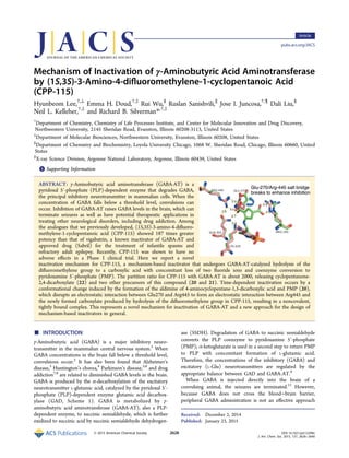
  • 1. Mechanism of Inactivation of γ‑Aminobutyric Acid Aminotransferase by (1S,3S)‑3-Amino-4-difluoromethylene-1-cyclopentanoic Acid (CPP-115) Hyunbeom Lee,†,⊥ Emma H. Doud,†,‡ Rui Wu,§ Ruslan Sanishvili,∥ Jose I. Juncosa,†,¶ Dali Liu,§ Neil L. Kelleher,†,‡ and Richard B. Silverman*,†,‡ † Department of Chemistry, Chemistry of Life Processes Institute, and Center for Molecular Innovation and Drug Discovery, Northwestern University, 2145 Sheridan Road, Evanston, Illinois 60208-3113, United States ‡ Department of Molecular Biosciences, Northwestern University, Evanston, Illinois 60208, United States § Department of Chemistry and Biochemistry, Loyola University Chicago, 1068 W. Sheridan Road, Chicago, Illinois 60660, United States ∥ X-ray Science Division, Argonne National Laboratory, Argonne, Illinois 60439, United States *S Supporting Information ABSTRACT: γ-Aminobutyric acid aminotransferase (GABA-AT) is a pyridoxal 5′-phosphate (PLP)-dependent enzyme that degrades GABA, the principal inhibitory neurotransmitter in mammalian cells. When the concentration of GABA falls below a threshold level, convulsions can occur. Inhibition of GABA-AT raises GABA levels in the brain, which can terminate seizures as well as have potential therapeutic applications in treating other neurological disorders, including drug addiction. Among the analogues that we previously developed, (1S,3S)-3-amino-4-difluoro- methylene-1-cyclopentanoic acid (CPP-115) showed 187 times greater potency than that of vigabatrin, a known inactivator of GABA-AT and approved drug (Sabril) for the treatment of infantile spasms and refractory adult epilepsy. Recently, CPP-115 was shown to have no adverse effects in a Phase I clinical trial. Here we report a novel inactivation mechanism for CPP-115, a mechanism-based inactivator that undergoes GABA-AT-catalyzed hydrolysis of the difluoromethylene group to a carboxylic acid with concomitant loss of two fluoride ions and coenzyme conversion to pyridoxamine 5′-phosphate (PMP). The partition ratio for CPP-115 with GABA-AT is about 2000, releasing cyclopentanone- 2,4-dicarboxylate (22) and two other precursors of this compound (20 and 21). Time-dependent inactivation occurs by a conformational change induced by the formation of the aldimine of 4-aminocyclopentane-1,3-dicarboxylic acid and PMP (20), which disrupts an electrostatic interaction between Glu270 and Arg445 to form an electrostatic interaction between Arg445 and the newly formed carboxylate produced by hydrolysis of the difluoromethylene group in CPP-115, resulting in a noncovalent, tightly bound complex. This represents a novel mechanism for inactivation of GABA-AT and a new approach for the design of mechanism-based inactivators in general. ■ INTRODUCTION γ-Aminobutyric acid (GABA) is a major inhibitory neuro- transmitter in the mammalian central nervous system.1 When GABA concentrations in the brain fall below a threshold level, convulsions occur.2 It has also been found that Alzheimer’s disease,3 Huntington’s chorea,4 Parkinson’s disease,5,6 and drug addiction7,8 are related to diminished GABA levels in the brain. GABA is produced by the α-decarboxylation of the excitatory neurotransmitter L-glutamic acid, catalyzed by the pyridoxal 5′- phosphate (PLP)-dependent enzyme glutamic acid decarbox- ylase (GAD, Scheme 1). GABA is metabolized by γ- aminobutyric acid aminotransferase (GABA-AT), also a PLP- dependent enzyme, to succinic semialdehyde, which is further oxidized to succinic acid by succinic semialdehyde dehydrogen- ase (SSDH). Degradation of GABA to succinic semialdehyde converts the PLP coenzyme to pyridoxamine 5′-phosphate (PMP); α-ketoglutarate is used in a second step to return PMP to PLP with concomitant formation of L-glutamic acid. Therefore, the concentrations of the inhibitory (GABA) and excitatory (L-Glu) neurotransmitters are regulated by the appropriate balance between GAD and GABA-AT.9 When GABA is injected directly into the brain of a convulsing animal, the seizures are terminated.11 However, because GABA does not cross the blood−brain barrier, peripheral GABA administration is not an effective approach Received: December 2, 2014 Published: January 23, 2015 Article pubs.acs.org/JACS © 2015 American Chemical Society 2628 DOI: 10.1021/ja512299n J. Am. Chem. Soc. 2015, 137, 2628−2640
  • 2. for the treatment of epilepsy. An alternative approach to increase GABA levels in the brain is by inhibition of GABA-AT, which blocks the degradation of GABA.12 A mechanism-based inactivator, an unreactive compound that initially acts as a substrate for the target enzyme and is converted to a species that causes inactivation of that enzyme, generally by covalent attachment, could increase GABA levels for an extended period of time.13 The design of these types of inhibitors requires insight into the mechanism of the target enzyme so that the catalytic chemistry can be utilized in the activation of the inactivator; the mechanism of the first half of the “ping-pong” reaction catalyzed by GABA-AT is shown in Scheme 2. Incubation of GABA with GABA-AT leads to the conversion of a lysine-PLP aldimine (1) to a GABA-PLP aldimine (2). Abstraction of the γ-proton by Lys329 and tautomerization leads to a PMP aldimine (3); enzyme-catalyzed hydrolysis of this intermediate leads to succinic semialdehyde and PMP.14 4-Amino-5-hexenoic acid (4, γ-vinyl-GABA; vigabatrin) is a mechanism-based inactivator of GABA-AT15 that is an FDA- approved drug (Sabril) to treat infantile spasms and refractory partial seizures. However, because of a serious side effect, namely, retinal damage and blindness, a drug with more potency and less toxicity was desired. Mechanistic studies performed by Nanavati and Silverman16 (Scheme 3) demon- strated that vigabatrin inactivates GABA-AT via two mecha- nisms, a Michael addition reaction and an enamine reaction, in a ratio of 70:30. In the Michael addition pathway (pathway a), vigabatrin acts like substrate GABA and undergoes aldimine formation with PLP, deprotonation, and tautomerization to a Michael acceptor (5), which undergoes Michael addition by Lys329, the lysine residue to which PLP is bound in the native enzyme,17 giving, after protonation, covalent adduct 6. Enzyme- catalyzed hydrolysis of 5 gives α,β-unsaturated ketone 7, which may be a possible cause for side effects of the drug. In the enamine pathway (pathway b), an enamine intermediate (8), formed by tautomerization through the alkene, attacks the enzyme-bound PLP to inactivate GABA-AT (9), also by formation of a covalent adduct. Hydrolysis of 8 gives a weak electrophile, which is unlikely to proceed through further reactions in the active site. To avoid the potentially toxic intermediate (7), computer modeling based on the crystal structures of GABA-AT18 and the vigabatrin−PLP complex in GABA-AT17 was carried out to determine how to prevent the Michael addition pathway. The model predicted that, in the energy-minimized structure, the alkene double bond pointed away from the nucleophilic Lys329; for Michael addition to occur, that bond had to rotate, allowing the alkene to be aligned with the side chain amine of Lys329. A conformationally rigid vigabatrin analogue was prepared (10) to prevent Michael addition, and it was found that it inactivated GABA-AT exclusively by the enamine pathway;19 however, it was a weak inactivator. The second approach to prevent formation of reactive product 7 was to synthesize a vigabatrin analogue that would give rapid Michael addition so that the equivalent of intermediate 5 would not be sufficiently stable to allow the enzyme-catalyzed hydrolysis leading to the equivalent of 7.20 That approach required the synthesis of a compound with the alkene pointing toward the side chain amine of Lys329 (11). The obtained compound inactivated GABA-AT, but only in the absence of the electrophilic trapping agent 2-mercaptoethanol. Therefore, an undesirable reactive intermediate (the corresponding ketoacid of 11) was formed prior to inactivation. To accelerate the inactivation rate, two electron-withdrawing fluorine atoms were Scheme 1. GABA Metabolism10 Scheme 2. First Catalytic Reaction of GABA-AT Journal of the American Chemical Society Article DOI: 10.1021/ja512299n J. Am. Chem. Soc. 2015, 137, 2628−2640 2629
  • 3. attached to the alkene of 11, and the obtained compound (12) inactivated GABA-AT, even in the presence of 2-mercaptoe- thanol. Further studies with 12 (now called CPP-115) showed that it was at least 187 times more potent an inactivator of GABA-AT than vigabatrin, was not active in the Cerep panel of 111 pharmacological targets (in vitro assays, carried out commercially by Cerep, on CPP-115 that cover a broad range of targets, including receptors, ion channels, enzymes, transporters, and second messengers), does not bind to three human GABA transporters GAT1, GAT2, and GAT3, or to GABAA, GABAB, or GABAC receptors, does not bind to the hERG channel, does not inhibit or induce cytochrome P450s, is not metabolized by hepatocytes, is rapidly and completely orally absorbed and eliminated, has no adverse effect on respiration, produces no mutations or chromosomal aberra- tions, and has a considerably lower retinal toxicity liability.21 It also is 300−600 times more potent than vigabatrin in reducing cocaine-induced synaptic dopamine levels and in blocking expression of cocaine-induced conditioned place preference in freely moving rats. Recently, it was shown to have no adverse effects in a Phase I clinical trial. Because of the clinical importance of this new drug candidate, we studied its inactivation mechanism to determine if it indeed favors the Michael addition pathway for which it was designed. We have determined that CPP-115 inactivates GABA-AT via an unexpected inactivation mechanism, namely, enzyme-catalyzed conversion of the difluoromethenyl group to a carboxylate, which causes a conformational change in the enzyme, to give a tightly bound complex. This inactivation mechanism presents a novel approach to inactivate GABA-AT and other related enzyme targets using similar catalytic mechanisms. ■ RESULTS Inhibition of GABA-AT by Increasing Equivalents of CPP-115. The turnover number for inhibition was determined by incubating GABA-AT with various equivalents of CPP-115. Figure 1 shows the remaining enzyme activity versus time for 0−4 equiv of CPP-115. To determine the turnover number, the data points at 24 h were replotted as a pseudo-first-order reaction using the remaining enzyme activity versus equivalents. The turnover number was found to be 1.3 ± 0.3 per active site. However, as shown in the replot, nonpseudo-first-order inactivation kinetics was observed, possibly because of product inhibition.22 Inactivation of GABA-AT by CPP-115 and Dialysis of the Inactivated Enzyme. To test if the mechanism involves irreversible and/or reversible inhibition, the time-dependent reactivation of GABA-AT was studied. First, the enzyme was treated with an excess of inactivator; then, the mixture was dialyzed against bulk buffer. Aliquots at different time intervals were collected and assayed. Return of enzyme activity was observed, which displayed both time- and concentration- dependence (see Supporting Information, Figures S1 and S2). The data also show that the amount of activity of the inhibited GABA-AT that returned was dependent on the amount of CPP-115 used in the incubation and the incubation time. In Figure S1 (Supporting Information), up to 90% and 60% of enzyme activity was recovered when GABA-AT was inactivated with 40 and 80 equiv of CPP-115, respectively, for 3 h. However, when GABA-AT was incubated for 24 h, even with 10 equiv of CPP-115, only 50% of the activity returned (Figure S2, Supporting Information). In the presence of 1000 equiv (approximately 1 mM) of CPP-115 in the incubation mixture, Scheme 3. Inactivation Mechanism of GABA-AT by Vigabatrin Journal of the American Chemical Society Article DOI: 10.1021/ja512299n J. Am. Chem. Soc. 2015, 137, 2628−2640 2630
  • 4. the activity of the enzyme returned to about 8%; with 2000 equiv of CPP-115, the activity return was only 1%, even after 50 h of dialysis. Fluoride Ion Release during Inactivation of GABA-AT by 2 mM CPP-115. Upon incubation of GABA-AT with CPP- 115, fluoride ions were continually released, even after the enzyme’s activity had fallen to almost zero (Figure S3, Supporting Information). After normalization of the values with the controls, it was found that 66 ± 3 equiv of fluoride ions were released slowly over a period of 27 h. However, when the same experiment was repeated with PLP supplemented in the mixture, there were 127 ± 5 equiv of fluoride ions released over time. Because α-ketoglutarate is essential to regenerate PLP from PMP, if α-ketoglutarate is omitted, the amount of fluoride ions released per single turnover of inhibition can be determined. The continual release of fluoride ions was not observed when there was no α-ketoglutarate present, and only 4.3 ± 0.5 equiv of fluoride ions were detected per enzyme dimer (Figure S4, Supporting Information). However, when α- ketoglutarate was added to the inactive enzyme, even without supplementation with PLP, the fluoride ion concentration began to increase. A control experiment with no enzyme in the incubation mixture was performed, which confirmed that no fluoride ions were released from CPP-115 in the absence of the enzyme, even if PLP is added. Inactivation of [3 H]PLP-Reconstituted GABA-AT by CPP-115. To determine the fate of the coenzyme by inactivation with CPP-115, apo-GABA-AT was reconstituted with [3 H]PLP. The maximum recovered activity was 71.4% of the original, and the activity recovery plateaued after 5 h of the reactivation process (Table 1), indicating that 29.6% of the enzyme remained inactive. [3 H]PLP-GABA-AT was inactivated with CPP-115, and the released radioactive compounds were analyzed using HPLC (Figure S5, Supporting Information). The experiment was performed with two controls: CPP-115 was omitted in a negative control (all radioactivity should be labeled PLP), and GABA replaced CPP-115 with α-ketoglutarate omitted in a positive control (all radioactivity should be PMP). The negative control released most of its radioactivity as PLP (but none as PMP), whereas a GABA-incubated control released both PMP and PLP (Figure S6, Supporting Information). Given that the positive control with GABA should only produce PMP from active enzyme, the PLP present in this sample represents that released from the inactive portion of GABA-AT. After the background radioactivity from the control experiments was subtracted from the CPP-115 experiment, CPP-115-inactivated [3 H]PLP-reconstituted GABA aminotransferase was found to release 100% of its cofactor as [3 H]PMP (Figure S7, Supporting Information). The small radioactive peak at 33 min was confirmed to be degraded PLP present in all of the samples. Mass Spectrometric Analysis of Metabolites from Inactivation of GABA-AT with CPP-115. Metabolites released during inactivation were determined by LC/MS/MS. After inactivation of GABA-AT by CPP-115 followed by denaturation and filtration, three metabolites were identified from the sample solution that were not present in the control samples: [m/z] 127.0389, 171.0291, and 401.0745 (A, B, and C in Figure S8, Supporting Information, respectively), corre- sponding to 22, 21, and 20, respectively, in Scheme 5 (see Discussion). These parent ions were selected for fragmentation using normalized collision energies. Fragmentation data for m/ z 127.0389 (Supporting Information Figure S9) confirmed the structure of 3-oxo-1-cyclopentanecarboxylic acid (22). MS/MS for m/z 171.0291 and 401.0745 (Supporting Information Figures S10 and S11, respectively) confirmed the structures of 21 and 20. Intact Mass Spectrometry of GABA-AT. Since covalent modification of GABA-AT was not detected using middle down proteomics (see Figures S12−S14 in Supporting Information), we analyzed the intact GABA-AT protein to see if any detectable mass shifts were present. The intact mass data showed multiple peaks, indicating that the GABA-AT purified from pig brain was a mixture of GABA-AT species with different N-termini. Three samples were tested using LC/MS/ MS: free enzyme (negative control), vigabatrin-inactivated GABA-AT (positive control), and CPP-115-inactivated GABA- AT (Figure S15, Supporting Information). Vigabatrin-inacti- vated GABA-AT showed an added mass of 122 Da from the mass of the native enzyme, which matches the covalent adduct proposed previously.15 However, CPP-115-inactivated GABA- AT showed no significant peaks corresponding to any added mass. To stabilize any potential imine adducts throughout the LC/MS/MS process, the samples were reduced with sodium borohydride as described previously for crystallography studies. Reduction resulted in stabilization of the PLP cofactor on the enzyme, with an added mass of 236 from the original peak (M). As expected, the peaks of vigabatrin-inactivated GABA-AT Figure 1. (Top) Time-dependent inactivation of GABA-AT by 0−4.0 equiv of CPP-115. (Bottom) Determination of the turnover number for the inactivation of GABA-AT by CPP-115. The data points at 24 h are replotted as enzyme activity vs equiv of CPP-115. Table 1. Return of GABA-AT Activity after apo-GABA-AT Was Incubated with [3 H]PLP 2 h 3 h 4 h 5 h original dA (mL−1 min−1 ) 0.630 0.748 1.504 1.522 2.124 Journal of the American Chemical Society Article DOI: 10.1021/ja512299n J. Am. Chem. Soc. 2015, 137, 2628−2640 2631
  • 5. (Figure S16, Supporting Information) were no different from those in Figure S15 since the vigabatrin covalent adduct is stable.15 Interestingly, for CPP-115-inactivated GABA-AT, reduction had no effect on the resulting data and showed no added mass on the protein. X-ray Crystallography of Native and CPP-115-Inacti- vated GABA-AT. To understand how time-dependent inactivation of GABA-AT by CPP-115 could occur without covalent modification of the protein or cofactor, CPP-115- inactivated and dialyzed GABA-AT were crystallized. The crystal structures of native GABA-AT from pig brain and inactivated enzyme were obtained at 1.63 Å and 2.19 Å resolution, respectively. The crystal structure for the native pig brain enzyme was very similar to that reported from pig liver by Storici et al.18 The crystal structures of the native enzyme and the inactivated enzyme were compared to analyze the difference in overall structure (Figure 2) and in the active site (Figure 3). The active site of the inactivated GABA-AT was investigated to understand the ligand−enzyme interactions (Figure 4); the omit map supports the ligand interpretation (see Supporting Information, Figure S17). ■ DISCUSSION At the start of this investigation, we considered a variety of likely mechanisms for the inactivation of GABA-AT by CPP- 115 (Schemes 4−8)23,24 and then considered experiments to differentiate them. All of these inactivation mechanisms are initiated by the same steps shown in Scheme 2 for substrate turnover or the two inactivation mechanisms for vigabatrin shown in Scheme 3. In mechanism 1 (Scheme 4), following γ- proton abstraction of 13, tautomerization leads to an α,β- unsaturated intermediate (14), which is attacked by the active site lysine residue or another base to covalently modify the enzyme. No fluorine is released, the enzyme is inactivated, and hydrolysis of 15 gives 16 with release of PMP as the cofactor. If intermediate 15 in Scheme 4 (where X is either the active site lysine residue or OH from attack by water) eliminates one fluoride ion (Scheme 5), the inactivator can form another reactive α,β-unsaturated imine (17), which can be attacked by a water molecule to release the other fluoride ion, forming 18. Depending on what X is, this may result in inactivation or turnover. If X is the active-site lysine residue (15a, pathway a, Scheme 5), the intermediate is a stabilized amide; hydrolysis generates covalent adduct 19 with the release of PMP. However, if X is a hydroxyl group (15b, pathway b, Scheme 5) formed by water molecule attack on reactive intermediate 14 (Scheme 4), stable dicarboxylate intermediate 20 will form; hydrolysis gives metabolite 21 and PMP, and subsequent decarboxylation of 21 gives 22. The third potential mechanism involves allylic tautomeriza- tion of external aldimine 13 to form 23 (Scheme 6). Because 23 also is a reactive electrophile, it may undergo Michael addition to form adduct 24, which could be hydrolyzed to give 25 and release PMP. In the fourth potential mechanism (Scheme 7), intermediate 23 (Scheme 6) can proceed through three different pathways. Pathway a generates an enamine (26), similar to that formed during vigabatrin inactivation; this species might undergo enamine attack on the Lys329-bound PLP to form covalent adduct 27, which could hydrolyze to 28. In pathway b, one fluoride ion is released by nucleophilic substitution (29) before undergoing transimination to form enamine adduct 30, which might undergo hydrolysis to 31. Pathway c from 30 involves elimination of a second fluoride ion (32) followed by hydrolysis to give 33. The final potential mechanism involves fluoride ion elimination from enamine 26 to generate reactive Michael acceptor 34 (Scheme 8). Hydrolysis of imine 34 (pathway a) gives 35 and PLP. Attack by an active site nucleophile (either Lys329 or water; pathway b) gives either covalent adduct 36 or noncovalent adduct 37, respectively. Elimination of another fluoride ion forms 38 or 40, each of which can be subsequently hydrolyzed to covalent adduct 39 or metabolite 41; further hydrolysis of 39 would also give 41. In either case, this mechanism releases the cofactor as PLP. All of these possible mechanisms can be differentiated by determination of the number of fluoride ions released, the fate of the cofactor, and the final metabolites or adducts formed; these possibilities are summarized in Table 2. Figure 2. Ribbon diagram of the superimposed native GABA-AT (yellow) and GABA-AT (cyan) bound to CPP-115. Figure 3. Superimposition of the crystal structures of native GABA-AT (pink) and CPP-115-inactivated GABA-AT (green). Journal of the American Chemical Society Article DOI: 10.1021/ja512299n J. Am. Chem. Soc. 2015, 137, 2628−2640 2632
  • 6. The partition ratio, the number of inactivator turnovers per inactivation event, was investigated by determination of the number of molecules of CPP-115 that were required to cause complete enzyme inactivation. Time-dependent inhibition was observed for 1−4 equiv of CPP-115, although pseudo-first- order kinetics was not observed (Figure 1, top). A replot of these data with extrapolation of the first few data points gave a turnover number of 1.3 ± 0.3 per active site (Figure 1, bottom). The nonpseudo-first-order inactivation kinetics displayed indicated possible product inhibition and suggested possibly two concurrent mechanisms occurring at different rates, maybe one reversible and one irreversible. To test whether there was a reversible component to the inactivation, the titration experiment was repeated, first with 0− 80 equiv of CPP-115 (Figure S1, Supporting Information), then 0−2000 equiv of CPP-115 (Figure S2, Supporting Information), followed by dialysis. Aliquots removed during dialysis exhibited time-dependent reactivation of GABA-AT over an extended period of time; with increasing equiv of inactivator, less activity returned with maximal return by 24 h (maximal return of activity occurred sooner with less inactivator). It required about 2000 equiv of CPP-115 before the enzyme reactivation was diminished to an insignificant level, supporting a reversible and irreversible component to the inactivation. During inactivation of GABA-AT by 2 mM CPP-115, it was found that 66 ± 3 equiv of fluoride ions were released over a 26 h time period; however, enzyme activity was lost within minutes (Figure S3, Supporting Information). This result further supports the notion of two different mechanisms being involved, one reversible and one irreversible, but it does not resolve the question of how many fluoride ions are released per molecule. A single-turnover experiment was carried out to determine the number of fluoride ions per molecule released with each turnover. This experiment is possible because GABA- AT converts substrate to product with concomitant conversion of its PLP to PMP. The reaction cycle cannot restart until α- ketoglutarate binds and converts the PMP back to PLP with concomitant formation of L-glutamic acid. By leaving out the α- ketoglutarate, only one inhibition turnover is possible. Under those conditions, 4.3 ± 0.5 equiv of fluoride ions was detected (Figure S4, Supporting Information); because GABA-AT is a homodimer, two molecules of CPP-115 can be turned over per enzyme dimer. Therefore, 4 equiv of fluoride ion indicates that both fluoride ions in CPP-115 are lost per enzyme turnover, and mechanisms 1, 3, and 4a, which release no fluoride ions Figure 4. Stereoview of GABA bound by the CPP-115 adduct. The 2Fo−Fc electron-density map is shown as light gray mesh at 1.1σ level around the CPP-115 adduct. Scheme 4. First Potential Mechanism of Inactivation of GABA-AT by CPP-115 Journal of the American Chemical Society Article DOI: 10.1021/ja512299n J. Am. Chem. Soc. 2015, 137, 2628−2640 2633
  • 7. during inactivation, and mechanism 4b, which releases one fluoride ion, can be excluded (Table 2). The cofactor structure after inactivation by CPP-115 was determined using GABA-AT containing [3 H]PLP (Figure S5, Supporting Information). With the aid of two control experiments (Figure S6, Supporting Information), it was found that all of the cofactor was converted to PMP after inactivation (Figure S7, Supporting Information). Therefore, mechanism 4a, which does not release any cofactor and mechanisms 4b, 4c, 5a, and 5b, which require the release of PLP as a cofactor, can be excluded (Table 2). Of the mechanisms considered, only 2a and 2b remain plausible (Scheme 5). Mechanism 2b should produce compounds 20−22. High-resolution LC/MS identified parent ions with m/z 401.0745, 171.0291, and 127.0389 (Figure S8, Supporting Information), which corresponds to 20, 21, and 22, respectively. MS/MS fragmentation produced daughter ions (Figures S9−S11, Supporting Information) consistent with these three products and with mechanism 2b. This mechanism appears to be responsible for the reversible component of the overall inactivation mechanism; mechanism 2a would be expected to account for the irreversible component, so it was imperative to demonstrate covalent attachment to the enzyme (19, Scheme 5) for substantiation. Mass spectrometry of the intact mass of CPP-115-inactivated GABA-AT was carried out using LC/MS/MS on native enzyme as the negative control and vigabatrin-inactivated GABA-AT as the positive control (Figure S15, Supporting Information). Compared with native enzyme, vigabatrin-inactivated GABA- AT had an added m/z peak of 122 Da, which corresponds to the expected added mass of the covalent adduct previously proposed.15 CPP-115-inactivated GABA-AT showed no sig- nificant peaks corresponding to added mass. In case the potential covalent adduct with CPP-115 was an imine, the inactivated enzyme was reduced with sodium borohydride prior to LC/MS/MS (Figure S16, Supporting Information). Again, no added mass was detected with the CPP-115 inactivated enzyme, but the vigabatrin-inactivated enzyme had the expected added mass. These results were corroborated by results from peptide proteomics (Supporting Information, Figures S13 and S14). Therefore, CPP-115 appears to be inactivating GABA-AT without covalent modification. The solution to this dilemma came from the X-ray crystal structure of CPP-115-inactivated pig brain GABA-AT, which revealed that the inactivator was tightly bound to the protein noncovalently as 20 (Figures 3 and 4). The inactivated species binds tightly (stable to dialysis) because of its covalent attachment to the cofactor and by two strong electrostatic interactions between the guanidinium groups of Arg192 and Arg445 and each of the two carboxylate groups of 20. This unexpected phenomenon is the first time that Arg445 has been observed interacting directly with a ligand and to be involved in the inactivation of GABA-AT. On the basis of crystallography of GABA analogues, it is believed that GABA binds in the active site by Schiff base formation with PLP and an electrostatic interaction between the carboxylate of GABA and the guanidinium group of Arg192. Arg445 is sequestered from the active site by an electrostatic interaction with Glu270.25 It has been proposed that this second salt bridge only disassociates during the second half of catalysis, the regeneration of PLP from PMP, to aid in the binding of the second carboxylate of α-ketoglutarate. A network of hydrogen bonds in the active site of GABA-AT connects Glu270 and Arg445 to O3′ of the cofactor through the side chain amide of Gln301. Markova et al. proposed that this network may be affected when the cofactor is in its PMP form, which weakens the interaction between Glu270 and Arg445.26 To verify these hypotheses, many research groups have attempted to cocrystallize GABA-AT with ligands such as α-KG or glutamate that may directly show the involvement of this second arginine, but they have not yet been successful.25−27 Our crystal structure of GABA-AT inactivated by CPP-115 (Figure 3) clearly shows for the first time that the salt bridge between Arg445 and Glu270 has been broken, and Glu270 is rotated away from its original position to accommodate a second guanidinium−carboxylate electrostatic interaction be- tween Arg445 and the newly formed carboxylate group on 20 Scheme 5. Second Potential Mechanisms of Inactivation of GABA-AT by CPP-115 Scheme 6. Third Potential Mechanism of Inactivation of GABA-AT by CPP-115 Journal of the American Chemical Society Article DOI: 10.1021/ja512299n J. Am. Chem. Soc. 2015, 137, 2628−2640 2634
  • 8. from enzyme-catalyzed hydrolysis of the difluoromethenyl group. The two guanidinium−carboxylate interactions, the hydrophobic interactions of the pyridine ring, and the interactions of the phosphate group contribute to the stabilization of the metabolite for tight binding. Therefore, CPP-115 appears to be, to the best of our knowledge, the first example of a mechanism-based inactivator that leads to a conformational change, forming a tightly bound inhibitor− enzyme complex with a newly exposed residue. Apparently, this conformational change occurs only about once with 2000 molecules of inactivator; the remaining molecules of 20−22 are slowly released as products. The α,β-unsaturated intermediate (14, Scheme 4) generated during turnover is highly electrophilic, which should have been attacked by the nucleophilic Lys329 residue and form a covalent adduct, as it does with vigabatrin. Molecular modeling studies of the CPP-115−PLP aldimine were performed to analyze the effect of tautomerization during the inactivation of GABA-AT by CPP-115 (Figure 5). This model demonstrates that the PMP-imine form (orange) of the aldimine generated during enzyme-catalyzed tautomerization shifts the difluor- omethenyl group (fluorines are in green) away from Lys329, thereby moving it too far for Michael addition by this lysine residue. Because of the lack of 1,5 steric interactions (both carbons opposite the carboxylate group are sp2 -hybridized and thus planar), the difluoromethenyl group is able to release strain by moving closer to the carboxylate. As GABA-AT normally catalyzes hydrolysis of the imine formed by tautomerization of GABA, the catalytic machinery is in place for water attack of the Michael acceptor instead. This is Scheme 7. Fourth Potential Mechanism of Inactivation of GABA-AT by CPP-115 Scheme 8. Fifth Potential Mechanism of Inactivation of GABA-AT by CPP-115 Journal of the American Chemical Society Article DOI: 10.1021/ja512299n J. Am. Chem. Soc. 2015, 137, 2628−2640 2635
  • 9. consistent with the obtained crystal structure and explains why the inactivation mechanism did not follow that of vigabatrin and form a covalent adduct. On the basis of the evidence from fluoride release, cofactor release, metabolites formed, proteomics, and X-ray crystallog- raphy, the most consistent mechanism is shown in Scheme 9. ■ CONCLUSIONS Although CPP-115 was rationally designed to inactivate GABA- AT via a covalent Michael addition mechanism, the results described here indicate that it inactivates GABA-AT by mechanism-based formation of a metabolite that induces a conformational change and forms a tightly bound complex with the enzyme (Scheme 9). The requirement of about 2000 equiv of CPP-115 for complete inactivation of GABA-AT reflects the number of molecules of product that are released prior to interception by the enzyme in the correct state and conformation that lead to inactivation. Proteomic studies could not reveal a covalent adduct. The crystal structure of CPP-115-inactivated GABA-AT revealed, for the first time, that the Glu270-Arg445 salt bridge in the active site can be disrupted, leading to the formation of a new binding pocket for the inactivator. Our molecular modeling studies indicate a movement of the difluoromethenyl group upon tautomeriza- tion, which shifts that group too far from Lys329 for nucleophilic attack. This leads to enzyme-catalyzed hydrolysis of the PMP-aldimine of CPP-115 and either release of products (2000 times) or inducement of a conformational change to give a tightly bound PMP complex stabilized by the exposure of Arg445 and formation of an electrostatic interaction with the newly formed carboxylate of the inactivator. Once metabolite 20 is tightly bound to the enzyme, it is released from the active site very slowly, which accounts for the extended period of time that fluoride ions are released relative to the rate of inhibition of the enzyme. With our results revealing the possibility of additional binding with Arg445, future inactivators may be designed so that the newly formed carboxylate group more closely mimics the second carboxylate group of α-ketoglutarate to encourage less turnover prior to conformational change- induced exposure of Arg445. ■ EXPERIMENTAL PROCEDURES Analytical Methods. GABA-AT assays were recorded on a Synergy H1 hybrid multimode microplate reader (Biotek, USA) with transparent 96-well plates (Greiner bio-one, Monroe, NC). Measure- ments of pH were performed on a Fisher Scientific AP71 pH/mV/°C meter with a pH/ATC electrode. Determinations of fluoride ion concentration were performed on the same meter with a Thermo Scientific 9609BN combination fluoride electrode. Large-scale dialyses were performed with Thermo Scientific Slide-A-Lyzer dialysis cassettes (molecular weight cutoff of 10 kDa) unless otherwise specified. Small- scale dialyses were performed with EMD Chemicals D-Tube Mini dialyzer (molecular weight cutoff of 12−14 kDa). Radioactivity was determined with a Packard TRI-CARB 2100TR liquid scintillation analyzer using PerkinElmer ULTIMA GOLD scintillation fluid. For centrifugations, a DuPont Sorvall RC 5B Plus centrifuge was used with either an SLA-3000 or an SA600 rotor. Eppendorf Minispin plus tubes were used for microcentrifugation. An IEC clinical centrifuge was used for the Penefsky spin method. HPLC analysis was performed with Beckman 125P pumps and a Beckman 166 detector. All of the runs were monitored at 254 nm. The HPLC columns used were Alltech C18 analytical and Econosil 10 μm columns. Enzyme purification was carried out on a GE Healthcare Life Sciences ÄKTA FPLC system. The column media used were CM Sepharose Fast Flow for weak cationic exchange mode, DEAE Sepharose Fast Flow for weak anionic exchange mode, and hydroxylapatite (HA) for mixed mode. A prepacked Hiprep Sephacryl S-200 high-resolution column was used for size-exclusion mode. Electrophoresis was carried out on a Bio-Rad Mini-Protean Tetra Cell using a VWR AccuPower Model 300 power supply. LC/MS/MS was performed on Thermo Fisher Q Exactive mass spectrometer. Mass spectrometry of intact mass of GABA-AT was performed on a Velos Orbitrap Elite (ThermoFisher). Crystallo- graphic data were collected on beamline 23 ID-B and 23 ID-D of GM/ CA of the Advanced Photon Source (APS) using X-rays of 0.99 Å wavelength and a Rayonix (formerly MAR-USA) 4 × 4 tiled CCD detector. All data were indexed, integrated, and scaled with HKL2000. Data processing statistics are given in Table S1 in Supporting Information. Reagents. All reagents and materials were purchased from Sigma- Aldrich Co. except the following: Centrifugal filters (molecular weight cutoff value of 10 kDa and 30 kDa) were purchased form EMD Millipore; Dowex 50 and sodium dodecyl sulfate were purchased from Bio-Rad; [3 H]sodium borohydride was purchased from American Radiolabeled Chemicals, Inc.; pig brains were a generous gift from the Park Packing Co. (Chicago, IL); all of the buffers and solvents used for FPLC analyses were filtered through GE Healthcare 0.45 μm nylon membranes. GABase (Pseudomonas fluorescens) and succinic semi- aldehyde were purchased from Sigma-Aldrich. Enzyme and Assays. GABA-AT (2.65 mg/mL, specific activity 2.1 unit/mg) was purified from pig brain by the procedure described previously.28 Succinic semialdehyde dehydrogenase (SSDH) was obtained from GABAse, a commercially available mixture of SSDH and GABA-AT, using a known procedure.24 GABA-AT activity was Table 2. Expected Differences in the Various Inactivation Mechanisms Journal of the American Chemical Society Article DOI: 10.1021/ja512299n J. Am. Chem. Soc. 2015, 137, 2628−2640 2636
  • 10. assayed using a published method.29 The final assay solution consisted of 11 mM GABA, 1.1 mM NADP+ , 5.3 mM α-KG, 2 mM β- mercaptoethanol, and excess SSDH in 50 mM potassium pyrophos- phate buffer, pH 8.5. The change in UV absorbance at a wavelength of 340 nm at 25 °C caused by the conversion of NADP+ to NADPH is proportional to the GABA-AT activity. All enzyme assays were recorded with a PerkinElmer Lambda 10 UV/vis spectrophotometer. Syntheses of (1S,3S)-3-Amino-4-difluoromethylene-1-cyclo- pentanoic Acid (CPP-115). This compound was synthesized according to the procedure published by Pan et al.24 Inhibition of GABA-AT by Various Equivalents of CPP-115. GABA-AT (2 μg) was incubated with CPP-115 (0, 0.2, 0.4, 0.6, 0.8, 1.0, 1.2, 1.4, 1.6, 1.8, and 4 equiv) at 25 °C in 50 mM potassium pyrophosphate buffer solution, containing 2 mM α-ketoglutarate and 1 mM β-mercaptoethanol. Aliquots (2 μL) were withdrawn at timed intervals and were added immediately to the assay solution (96 μL) containing excess SSDH (2 μL). The reaction rates were monitored at 340 nm. Inactivation of GABA-AT by CPP-115 and Dialysis of the Inactivated Enzyme. GABA-AT (60 μL; final concentration 1 μM) was preincubated for 3 h with various amounts of CPP-115 (0, 1, 1.8, 4, 40, and 80 equiv) in 50 mM pyrophosphate buffer (pH 8.5) containing 0.625 mM α-ketoglutarate, in a total volume of 160 μL at 25 °C. In another experiment, it was preincubated for 24 h with various amounts of CPP-115 (0, 1, 2, 4, 40, 80, 100, 1000, and 2000 equiv). After preincubation, the enzyme solutions were transferred to D-Tube Mini dialyzers and exhaustively dialyzed against buffer (50 mM pyrophosphate buffer containing 0.1 mM α-ketoglutarate and 0.1 mM PLP, pH 8.5) at 4 °C. The dialysis buffer was exchanged every 4 h, three times. The enzyme activity remaining in each of the solutions was assayed per time interval. Analysis of Fluoride Ion Release during Inactivation of GABA-AT by CPP-115. GABA-AT (150 μL) was incubated in 100 mM potassium pyrophosphate buffer at pH 8.5, containing 2 mM CPP-115 and 2.5 mM α-ketoglutarate in a total volume of 510 μL. A control lacking GABA-AT was also incubated. The incubation mixture was protected from light and was carried out at room temperature. The experiment was repeated with 2.5 mM PLP supplemented in the incubation mixture. To construct a standard curve, the relative potential (mV) of sodium fluoride solutions in the range of 1 × 10⁻⁶ to 5 × 10⁻⁵ M in total ionic strength adjustment buffer (TISAB; made from 5.8 g of sodium chloride and 5.7 mL of acetic acid in 100 mL of water, adjusted to pH 5.2) was measured. The measured potential was plotted against the concentration of sodium fluoride. At different incubation time (1, 2, 5, 7, 17, 24, 27 h), each aliquot was removed from the incubation samples and mixed with TISAB to measure their relative potentials. Using the formula obtained from the standard curve, the mV readings were converted to concentration of fluoride ions in the samples. The readings from the control sample were subtracted from the inactivated sample, and the concentration was divided by the concentration of GABA-AT to obtain the equivalents of fluoride ion released per inactivation event. Analysis of Fluoride Ion Release during Inactivation of GABA-AT by CPP-115 without α-Ketoglutarate. The similar experiment was run as above but without α-ketoglutarate to test the amount of fluoride released during a one-turnover event. GABA-AT (200 μL) was incubated in 100 mM potassium pyrophosphate buffer at pH 8.5, containing 2 mM CPP-115 in a total volume of 510 μL. When there is no α-ketoglutarate in the mixture to regenerate PLP, the reaction stops at one turnover per active site. Radioactive Labeling of [7-3 H]-PLP with Tritiated NaBH4. Thirty drops of 1 M NaOH were added to 1.8 mL of 0.5 M PLP and cooled to 0 °C in an ice bath. The mixture of sodium borohydride (5.8 mg, 0.15 mmol) and [3 H]-sodium borohydride (25 mCi) in 450 μL of 0.1 M NaOH was added to the PLP solution and stirred for 1 h at 0 Figure 5. Molecular docking studies of CPP-115-PLP aldimine in PLP-form (cyan) vs CPP-115-PMP-form (orange) in the active site of GABA-AT. Journal of the American Chemical Society Article DOI: 10.1021/ja512299n J. Am. Chem. Soc. 2015, 137, 2628−2640 2637
  • 11. °C. After being stirred, 120 μL of concd HCl was added to the solution slowly (became pH 4). To the solution at pH 4, 20 equiv of ground MnO2 was added, and the mixture was stirred at room temperature for a total of 2 h. The mixture was brought to pH 8 with 1 M NaOH, and the solution was centrifuged. The supernatants were collected and were loaded onto a gel filtration column packed with Bio-Rad AG1-X8 resin (hydroxide form). The mobile phase used was water and 5 M acetic acid (gradient of 90% water to 0% water), flowing at 1.5 mL/min for 300 min. Fractions were collected every 10 mL and tested for its UV absorption and radioactivity. The fractions with desired product were lyophilized. The lyophilized product was redissolved in 100 μL of 1 mM PLP and loaded onto an HPLC with an Econosil C18 column (10 mm × 250 mm, 10 μm). The mobile phase used was 0.1% aqueous TFA with 5% acetonitrile flowing at 0.5 mL/min for 40 min. Under these conditions, PLP eluted at 15 min. Fractions running with the PLP peak were collected and counted for radioactivity using liquid scintillation counting. The product was collected and lyophilized. Inactivation of [3 H]PLP-Reconstituted GABA-AT by CPP-115. GABA-AT that had been reconstituted with [3 H]PLP was incubated at room temperature and protected from light in 100 mM potassium phosphate buffer containing CPP-115 (2 mM), α-ketoglutarate (3 mM), and β-mercaptoethanol (3 mM), in a total volume of 100 μL at pH 7.4. A negative control was run under identical conditions as above, excluding the inactivator. A positive second control was run with 3 mM GABA in the absence of inactivator and α-ketoglutarate. The first control should release the cofactor as PLP, and the second control should release it as PMP. After incubation for 18 h, the activity of GABA-AT was less than 1% of control, and the solutions were adjusted to pH 11 with 1 M KOH and incubated for 1 h. Trifluoroacetic acid (TFA) was added to quench the base and make the solution 10% v/v TFA. The resulting denatured enzyme solution was microcentrifuged for 5 min at 10 000 rpm after standing at room temperature for 10 min. A small amount of white solids was seen at the bottom of the tube. The supernatants were collected individually. To wash the pellets, 50 μL of 10% TFA was added to each tube, vortexed, and microcentrifuged for another 5 min. This process was repeated three times. The washed pellets were counted for radioactivity. The supernatant and washes were combined and lyophilized. Cofactor analysis was carried out by dissolving the solids obtained from lyophilization with 100 μL of a solution containing 2.5 mM PLP and 2.5 mM PMP as standards and then injecting the samples into the HPLC with an Econosil C18 column (4.6 mm × 150 mm, 10 μm). The mobile phase used was 0.1% aqueous TFA flowing at 0.5 mL/min for 25 min. Then the flow rate was increased to 1 mL/min from 25 to 30 min, and then a solvent gradient into 95% acetonitrile was run over the next 30 min. Under these conditions, PLP eluted at 15 min and PMP at 8 min. Fractions were collected every minute, and the radioactivity was measured by liquid scintillation counting. Mass Spectrometric Analysis of Metabolites from Inactiva- tion of GABA-AT with CPP-115. GABA-AT (0.3 nmol) was incubated in 50 mM ammonium bicarbonate buffer (pH 7.4) containing 0.1 mM CPP-115, 1 mM α-ketoglutarate, and 0.1 mM PLP in a total volume of 70 μL at room temperature in the dark for 12 h. A control containing everything except CPP-115 was also incubated. After 12 h, GABA-AT in the inactivated sample was less than 1% active vs control. Formic acid (1 μL) was added to each reaction mixture, and both were centrifuged in a 0.5 mL 30 kDa MWCO centrifuge tube (Millipore) at 14000g for 4 min or until most of the solution had passed through. An additional 20 μL of 50 mM ammonium bicarbonate was added above the filter and centrifuged for 3 min. The flow-through (20 uL) was injected onto a Luna C18(2) column (100 A, 2 × 150 mm, 5 μm, Phenomenex). A 60 min gradient (Agilent 1100 HPLC, solvent A = 5% acetonitrile and 0.1% formic acid; solvent B = 0.1% formic acid in acetonitrile) was run from 2−80% B over 40 min. The LC was directly connected to a Thermo Fisher Q Exactive mass spectrometer. The top five most abundant ions in negative ion mode were selected for fragmentation using mass normalized collision energies. Commercially available 3-oxo-1-cyclopentanecarboxylic acid (Sigma) was injected as a standard for comparison. Scheme 9. Mechanism of Inactivation of GABA-AT by CPP-115 Most Consistent with Experimental Evidence Journal of the American Chemical Society Article DOI: 10.1021/ja512299n J. Am. Chem. Soc. 2015, 137, 2628−2640 2638
  • 12. Intact Mass Spectrometry of GABA-AT. Three reaction samples were prepared as described previously with only GABA-AT, including vigabatrin, or CPP-115.30 After incubation with or without inhibitors, half of each reaction mixture was reduced with 15 mM sodium cyanoborohydride for 1 h. Both reduced and nonreduced samples were buffer exchanged into 50 mM ammonium acetate using 30 kDa MWCO filters (Millipore). Nano-LC/MS/MS runs were done on a 75 μm ID × 10 cm Kinetex C8 (Phenomenex) column connected to an autosampler (Dionex Ultimate 3000 RSLCnano system) and a Velos Elite Orbitrap (ThermoFisher) mass spectrometer. Results were deconvoluted using ProMass Deconvolution software (Thermo Scientific). Enzyme Crystallization. Before crystallization, GABA-AT and CPP-115-inactivated GABA-AT were exchanged into a buffer that contained 40 mM sodium acetate (pH 5.5). After the initial crystallization screening and optimization, the proteins were crystal- lized via the hanging drop method. Hanging drops were prepared by mixing 1 μL of 12 mg/mL native GABA-AT protein solution and 1 μL of the reservoir solution, containing 0.1 M ammonium acetate, 0.1 M bis-Tris (pH 5.5), and 17% w/v PEG 10 000. Crystals appeared within 24 h at 20 °C and grew for 5−6 days before harvesting. Crystals with good morphology and large sizes were transferred to cryoprotecting conditions, which contain 20% glycerol in addition to the original composition of the reservoir solution, before being frozen in liquid nitrogen. The same method of crystallization was applied for CPP- 115-inactivated GABA-AT; however, before freezing in liquid nitrogen, the inactivated crystals were transferred to a cryoprotecting solution that contained 20% glycerol and 2 mM CPP-115, in addition to the compounds of the reservoir solution. Crystallographic data were collected on beamlines 23ID-B and 23ID-D of GM/CA@APS of the Advanced Photon Source (APS) using X-rays of 0.99 Å wavelength and Rayonix (formerly MAR-USA) 4 × 4 tiled CCD detector with a 300 mm2 sensitive area. All data were indexed, integrated, and scaled with HKL2000. Data collection and processing statistics are given in Table S1 (Supporting Information). Phasing, Model Building, and Refinement. Molecular replace- ment for the native GABA-AT was carried out using the program Phaser31 from CCP432 software suite, using the previously reported coordinates of GABA-AT from pig liver (PDB ID: IOHV) as the starting search model. The initial Rfree and R factor of the correct solution were 0.1904 and 0.1934, respectively. The rigid body refinement was followed by restrained refinement using Refmac5.33 Manual adjustment and modification of the structure based on electron density maps were performed using the program Coot.34 The Rfree and R factor values of the final model were 0.1513 and 0.1766, respectively. The data sets from the inactivated GABA-AT crystals were isomorphous to those of native GABA-AT. Therefore, rigid body refinement could be used directly, placing the previously refined native GABA-AT model into the asymmetric unit of inactivated GABA-AT crystals. Model building and refinements of the inactivated structures were carried out following the same protocol as the native structure. There were no ligand coordinates included in the refinement until the refinement converged. Without ligands built in, the Fo−Fc map shows a well-defined electron density supporting the existence of the bound ligand. The structure of the inactivator was made in the program ChemDraw generating a “Mol” file as the output. This output ligand structure was then regularized and its chemical restraints were generated in the program JLigand.35 Using Coot, the inactivator was manually fit to the residual electron density in the difference (Fo−Fc) map. The complex structure with the ligand bound was further refined in Refmac5. The Rfree and R factor for the inactivated structure were 0.1575 and 0.1872, respectively. Molecular Modeling. All renderings were performed in PyMol.36 Computer simulations were carried out as previously described.37 In essence, the ligands (including the cofactor) were docked into the active site of the prepared protein using Autodock 4.2,38 with Lys329 being flexible. The best docked structures were then refined by molecular mechanics, using GROMACS 4.5.39 The sequence utilized started with energy minimization, continued with molecular dynamics (4 ns), and had a final energy minimization step. The final structures were used for evaluation. ■ ASSOCIATED CONTENT *S Supporting Information Reactivation results of GABA-AT after inactivation; fluoride ion release results; cofactor release results; high resolution mass spectra; mass fragmentation data of metabolites 20−22; middle down peptide proteomics results, omit map for inactivated enzyme, crystallography data collection, processing statistics. This material is available free of charge via the Internet at http://pubs.acs.org. ■ AUTHOR INFORMATION Corresponding Author *Agman@chem.northwestern.edu Present Addresses ⊥ Molecular Recognition Research Center, Korea Institute of Science and Technology (KIST), Seoul, Korea. ¶ Department of Chemistry, Salisbury University, Salisbury, MD 21801. Notes The authors declare no competing financial interest. ■ ACKNOWLEDGMENTS The authors are grateful to the National Institutes of Health for financial support (grants GM066132 and DA030604 to R.B.S.; GM067725 to N.L.K.; contracts ACB-12002 and AGM-12006 to R.S. This research used resources of the Advanced Photon Source, a U.S. Department of Energy (DOE) Office of Science User Facility operated for the DOE Office of Science by Argonne National Laboratory under Contract No. DE-AC02- 06CH11357.). ■ REFERENCES (1) Krnjevic, K. Physiol. Rev. 1974, 54, 418−540. (2) Karlsson, A.; Fonnum, F.; Malthe-Sørenssen, D. J. Biochem. Pharmacol. 1974, 23, 3053−3061. (3) Davies, P. Brain Res. 1979, 171, 319−327. (4) Perry, T. L.; Hansen, S.; Tischler, B.; Richards, F. M.; Sokol, M. N. Engl. J. Med. 1973, 289, 395−398. (5) Maker, H. S.; Weiss, C.; Weissbarth, S.; Silides, D. J.; Whetsell, W. Ann. Neurol. 1981, 10, 377−383. (6) Rinne, U. K.; Laaksonen, H.; Riekkinen, P.; Sonninen, V. Eur. Neurol. 1974, 12, 13−19. (7) Dewey, S. L.; Morgan, A. E.; Ashby, C. R., Jr.; Horan, B.; Kushner, S. A.; Logan, J.; Volkow, N. D.; Fowler, J. S.; Gardner, E. L.; Brodie, J. D. Synapse 1998, 30, 119−129. (8) Karila, L.; Gorelick, D.; Weinstein, A.; Noble, F.; Benyamina, A.; Coscas, S.; Blecha, L.; Lowenstein, W.; Martinot, J. L.; Reynaud, M.; Lepine, J. P. Int. J. Neuropsychopharmacol. 2008, 11, 425−438. (9) Hearl, W. G.; Churchich, J. E. J. Biol. Chem. 1984, 259, 11459− 11463. (10) Baxter, C. F.; Roberts, E. J. Biol. Chem. 1958, 233, 1135−1139. (11) Purpura, D. P.; Girando, M.; Smith, T. A.; Callan, D. A.; Groundfest, J. J. Neurochem. 1959, 3, 238−268. (12) Hammond, E. J.; Wilder, B. J. Gen Pharmacol 1985, 16, 441− 447. (13) (a) Silverman, R. B. Methods Enzymol. 1995, 249, 240−283. (b) Silverman, R. B. Mechanism-Based Enzyme Inactivation: Chemistry and Enzymology; CRC Press: Boca Raton, FL, 1988; Vols. I and II. (14) Silverman, R. B. The organic chemistry of enzyme-catalyzed reactions; Academic Press: San Diego, CA, 2000. Journal of the American Chemical Society Article DOI: 10.1021/ja512299n J. Am. Chem. Soc. 2015, 137, 2628−2640 2639
  • 13. (15) Lippert, B.; Metcalf, B. W.; Jung, M. J.; Casara, P. Eur. J. Biochem. 1977, 74, 441−445. (16) Nanavati, S. M.; Silverman, R. B. J. Am. Chem. Soc. 1991, 113, 9341−9349. (17) Storici, P.; De Biase, D.; Bossa, F.; Bruno, S.; Mozzarelli, A.; Peneff, C.; Silverman, R. B.; Schirmer, T. J. Biol. Chem. 2004, 279 (1), 363−373. (18) Storici, P.; Capitani, G.; De Biase, D.; Moser, M.; John, R. A.; Jansonius, J. N.; Schirmer, T. Biochemistry 1999, 38, 8628−8634. (19) (a) Choi, S.; Storici, P.; Schirmer, T.; Silverman, R. B. J. Am. Chem. Soc. 2002, 124, 1620−1624. (b) Choi, S.; Silverman, R. B. J. Med. Chem. 2002, 45, 4531−4539. (20) Pan, Y.; Qiu, J.; Silverman, R. B. J. Med. Chem. 2003, 46, 5292− 5293. (21) Silverman, R. B. J. Med. Chem. 2012, 55, 567−575. (22) (a) Andrić, P.; Meyer, A. S.; Jensen, P. A.; Dam-Johansen, K. Biotechnol. Adv. 2010, 28 (3), 308−324. (b) Frieden, E.; Walter, C. Nature 1963, 198 (4883), 834−837. (23) Pan, Y. Design, synthesis and mechanistic studies of γ- aminobutyric acid aminotransferase inhibitors. Ph.D. Dissertation, Northwestern University, Evanston, IL, June 2004. (24) Silverman, R. B.; Bichler, K. A.; Leon, A. J. J. Am. Chem. Soc. 1996, 118 (6), 1241−1252. (25) Liu, W.; Peterson, P. E.; Carter, R. J.; Zhou, X.; Langston, J. A.; Fisher, A. J.; Toney, M. D. Biochemistry 2004, 43 (34), 10896−10905. (26) Markova, M.; Peneff, C.; Hewlins, M. J. E.; Schirmer, T.; John, R. A. J. Biol. Chem. 2005, 280 (43), 36409−36416. (27) Storici, P.; Capitani, G.; Muller, R.; Schirmer, T.; Jansonius, J. N. J. Mol. Biol. 1999, 285 (1), 297−309. (28) Silverman, R. B.; Levy, M. A. J. Biol. Chem. 1981, 256, 11565− 11568. (29) Scott, E. M.; Jakoby, W. B. J. Biol. Chem. 1959, 234, 932−936. (30) Burke, J. R.; Silverman, R. B. J. Am. Chem. Soc. 1991, 113 (24), 9329−9340. (31) McCoy, A. J.; Grosse-Kunstleve, R. W.; Adams, P. D.; Winn, M. D.; Storoni, L. C.; Read, R. J. J. Appl. Crystallogr. 2007, 40, 658. (32) Collaborative Computational Project, N. Acta Crystallogr., Sect. D: Biol. Crystallogr. 1994, 50, 760. (33) Murshudov, G. N.; Vagin, A.; Dodson, J. Acta Crystallogr., Sect. D: Biol. Crystallogr. 1997, 53, 240. (34) Emsley, P.; Cowtan, K. Acta Crystallogr., Sect. D: Biol. Crystallogr. 2004, 60, 2126. (35) Lebedev, A. A.; Young, P.; Isupov, M. N.; Moroz, O. V.; Vagin, A. A.; Murshudov, G. N. Acta Crystallogr., Sect. D: Biol. Crystallogr. 2012, 68, 431. (36) The PyMOL Molecular Graphics System, Version 1.5.0.4 Schrödinger, LLC. (37) Juncosa, J. I.; Groves, A. P.; Xia, G.; Silverman, R. B. Bioorg. Med. Chem. 2013, 21, 903−911. (38) Morris, G. M.; Huey, R.; Lindstrom, W.; Sanner, M. F.; Belew, R. K.; Goodsell, D. S.; Olson, A. J. J. Comput. Chem. 2009, 16, 2785− 2791. (39) Pronk, S.; Páll, S.; Schulz, R.; Larsson, P.; Bjelkmar, P.; Apostolov, R.; Shirts, M. R.; Smith, J. C.; Kasson, P. M.; van der Spoel, D.; Hess, B.; Lindahl, E. Bioinformatics 2013, 29 (7), 845−854. Journal of the American Chemical Society Article DOI: 10.1021/ja512299n J. Am. Chem. Soc. 2015, 137, 2628−2640 2640