
Empfohlen
Weitere ähnliche Inhalte
Was ist angesagt?
Was ist angesagt? (20)
Ähnlich wie Cerebral Malaria
Ähnlich wie Cerebral Malaria (20)
Mehr von Randolph Tulsie
Kürzlich hochgeladen
Kürzlich hochgeladen (20)
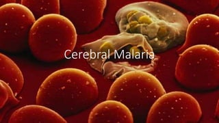
Cerebral Malaria
- 2. Introduction • Malaria remains one of the most prevalent infectious diseases in the world. • The World Health Organization (WHO) reports that 50% of the world’s population living in 109 countries are still at risk of malaria • Cerebral malaria is the most severe pathology caused by the malaria parasite, Plasmodium falciparum. • There are four species of human malaria, but Plasmodium falciparum causes nearly all the deaths and neurological complications. • Coma is a characteristic and ominous feature of falciparum malaria and, despite treatment, is associated with death rates of ~20% among adults and 15% among children
- 3. Pathophysiology • P falciparum is transmitted by female Anopheles mosquitoes. In humans, although the parasite undergoes development in the liver, it is the erythrocytic cycle that is responsible for disease. • The histopathological hallmark of cerebral malaria is engorgement of cerebral capillaries and venules with parasitised red blood cells (PRBCs) and non-paratised RBCs (NPRBCs). • This is due to sequestration, cyto adherence and rosetting.
- 4. Pathophysiology • Sequestration –accumulation of red cells containing mature forms of the parasite (trophozoites and meronts) in the microvasculature of organs (most commonly the brain) secondary to cyto-adherence and rosetting • Cyto-adherence- a specific interaction between PRBCs and the vascular endothelium mediated by plasmodium derived proteins on the surface of PRBCs and modified erythrocyte cell wall proteins on endothelial cells. • The adhesion of the PRBCs reduces the microvascular blood flow, which may explain organ and tissue dysfunction such as coma. • Rosetting - The adherence of NPRBCs to PRBCs results in agglutination within microvasculature also contributing to ischemia and organ damage.
- 6. Clinical Manifestations • Cerebral Malaria can occur in less than two weeks after a mosquito bite and may develop after 2 to 7 d of fever. • Can begin as AMS or delirium and progress to unrousable coma • Seizures, usually generalized and often repeated, occur in ~10% of adults and up to 50% of children with cerebral malaria. • On direct ophthalmoscopy retinal haemorrhages are found in about 15% of patients • Signs of multi-organ failure include jaundice, metabolic acidosis and acute pulmonary edema. • Hypoglycemia is common along with anemia. • Rarely, patients with severe malaria have disseminated intravascular coagulation
- 7. Diagnosis • The diagnosis of malaria rests on the demonstration of asexual forms of the parasite in stained peripheral-blood smears. • Giemsa at pH 7.2 is the preferred stain • Both Thick and Thin Blood Smears should be taken and fixed appropriately • THICK FILM = advantage of concentrating the parasites (by 40- to 100-fold compared with a thin blood film) and thus increasing diagnostic sensitivity. • THIN SMEAR = Determine level of parasitemia (quantify as # of infected RBC per 1000 RBCs) • Rapid, simple, sensitive, and specific antibody-based diagnostic stick or card tests that detect P. falciparum–specific Malaria
- 8. Lab Findings • Anemia = usually normochromic, normocytic • WBC = elevated in cerebral malaria; with reactive lymphocytosis and eosinophilia in the weeks after the acute infection. • ESR/CRP elevated • Severe infections may be accompanied by prolonged prothrombin and partial thromboplastin times and by more severe thrombocytopenia. • Hypoglycemia, and metabolic acidosis may be present with electrolyte imbalance. • the mean cerebrospinal fluid (CSF) Findings • opening pressure at lumbar puncture is ~160 mm; • usually the CSF content is normal • or there is a slight elevation of total protein level (<1.0 g/L [<100 mg/dL]) and cell count (<20/μL).
- 9. Management : AntiMalarial Agents • Artesunate is the drug of choice for all patients with severe malaria everywhere. • Artesunate (2.4 mg/kg stat IV followed by 2.4 mg/kg at 12 and 24 h and then daily if necessary) OR • Artemether (3.2 mg/kg stat IM followed by 1.6 mg/ kg qd) • Quinine dihydrochloride (20 mg of salt/kg infused over 4 h, followed by 10 mg of salt/kg infused over 2–8 h q8hr) • The administration of quinidine must be closely monitored if dysrhythmias and hypotension are to be avoided. • If total plasma levels exceed 8 μg/mL or the QTc interval exceeds 0.6 s or the QRS complex widens by more than 25% of baseline, then infusion rates should be slowed or infusion stopped temporarily.
- 10. Management: Supportive Care • Severe falciparum malaria constitutes a medical emergency requir- ing intensive nursing care and careful management. • RBS Monitoring q 4hrly ; All patients should receive a continuous infusion of dextrose, and blood concentrations ideally should be maintained above 4 mmol/L or 70mg/dl. • Blood Transfusions for severe anemia (HCT <20%) • Patients who develop spontaneous bleeding should be given fresh blood and IV vitamin K. • Convulsions should be treated with IV or rectal benzodiazepines and, if necessary, respiratory support. • Monitor BUN and Cr and assess Fluid status daily to prevent overload. • Patients who develop AKI or metabolic acidosis may require dialysis.
- 11. Follow-on treatment • Following initial parenteral treatment for atleast 24hrs, once the patient can tolerate oral therapy, it is essential to continue and complete treatment with an effective oral antimalarial using a full course of an effective ACT • artemether plus lumefantrine • artesunate (plus clindamycin or doxycycline) • quinine (plus clindamycin or doxycycline).
- 12. References • Harrison’s Principles of Internal Medicine 19th Ed. • Medscape • WHO’s Guidelines for the treatment of malaria, 2nd Ed.