
Empfohlen
Weitere ähnliche Inhalte
Was ist angesagt?
Was ist angesagt? (20)
Andere mochten auch
Andere mochten auch (10)
Ähnlich wie Medicinal chemistry Basics
Ähnlich wie Medicinal chemistry Basics (20)
Mehr von Rahul Patil PhD
Mehr von Rahul Patil PhD (7)
Kürzlich hochgeladen
Kürzlich hochgeladen (20)
Medicinal chemistry Basics
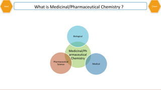
- 1. Med Medicinal/Ph armaceutical Chemistry Biological Medical Pharmaceutical Science What is Medicinal/Pharmaceutical Chemistry ? Chem
- 2. HISTORY OF MEDICINAL CHEMISTRY Medicinal chemistry received formal recognition in academic pharmacy in 1932 Ehrlich’s “Side chain theory” and chemotherapy and Fischer’s lock-and key theory Birth of Modern Med Chem 1800 Synthesis of Urea 1828 started Organic medicinal Chemistry Emperor Frederick II issued the Magna Carta of pharmacy in 1240 1500 BC Egyptian papyrus ebers 700 drugs originated from animal/plants/minerals 2000 BC Materia Medica 250 vegetables drug And 120 mineral drugs Med Chem
- 3. What is DrugMed Chem No drug is totally safe. Drugs vary in the side effects they might have. The dose level of a compound determines whether it will act a medicine or as a poison. The therapeutic index is a measure of a drugs beneficial effect at a low dose versus its harmful effects at higher dose. A high therapeutic index indicates a large safety margin between beneficial and toxic doses The principle of selective toxicity means that useful drugs show toxicity against foreign or abnormal cells but not against normal host cell why, where, how drug act ? Drug : Any substance which generate biological response. Good or Bad drugs
- 4. Drug TargetMed Chem Why should chemicals, some of which have remarkably simple structures, have such an important effect on such a complicated and large structure as a human being ? Cytoplasm Nucleus Nuclear membrane Cell membrane
- 5. Chemical Structure of Cell wallMed Chem Glycoprotein Lipid bilayer phosphatidylcholine, phosphatidylethanolamine, phosphatidylserine, and phosphatidylinositol
- 6. Chemical Structure of Cell wallMed Chem Glycoprotein O-aglycon N-aglycon Glycophorin
- 7. Drug targets at the molecular levelMed Chem Proteins : Enzymes Receptors Transport proteins Nucleic Acids : DNA RNA Biological 10% Receptor Agonist 12% Receptor Antagonist 24% Ion Channel Modulator 8% Enzyme Inhibitor 38% Misc 8%
- 8. Drug targets at the molecular levelMed Chem The equilibrium of a drug being bound and unbound to its target.
- 9. Intermolecular bonding forcesMed Chem Electrostatic or ionic bonds Hydrogen bonds Van der Waals interactions Dipole-Dipole and ion-Dipole interactions Repulsive interactions The role of water and hydrophobic interactions Electrostatic or ionic bonds Hydrogen bondsCovalent bonds Van der Waals interactions 1.5–2.2 Å1.0–1.5 Å
- 10. Med ChemElectrostatic or ionic bonds An ionic or electrostatic bond is the strongest of the intermolecular bonds Closer the charged atoms stronger the bond Strength of bond depend on environment Most important initial interaction as the drug enters the binding site
- 11. Hydrogen bondsMed Chem (X, Y = oxygen or nitrogen; HBD = hydrogen bond donor, HBA = hydrogen bond acceptor) For H bond require an electron-rich hetero- atom (With lone pair) and an electron-deficient hydrogen The hydrogen bond (5 to 30 kJ/mole) is stronger than a van der Waals interaction, but weaker than covalent or ionic bonds. hydrogen bond donor (HBD) Has covalently bonded electro +Ve H hydrogen bond acceptor (HBA) provide electro –Ve atom hydrogen bond flip-flop
- 13. Hydrogen bondsMed Chem Orbital overlap in a hydrogen bond. Weak form of sigma bonding Bond angel 130-180 moderated bond strength 90 bond angel week bond Optimum orientation is where bond angel is 180 where it is strongest bond
- 14. Hydrogen bond donor (HBD)/acceptor (HBA)Med Chem R-F Fluorine is highly electronegative atom With 3 lone pair of electron…. But still weak HBA ?????
- 15. Van der Waals interactionsMed Chem
- 18. Repulsive interactionsMed Chem Induced dipole interaction between an alkyl ammonium ion and an aromatic ring. Nicotinic acetylcholine receptor tryptophan residue
- 19. Repulsive interactionsMed Chem Important to keep molecule at specific distance. If molecules come too close there molecular orbitals start to overlap and this results in repulsion. Same group try to repel each other…For example, two charged groups of identical charge are repelled.
- 20. The role of water and hydrophobic interactionsMed Chem Ritonavir
- 21. The role of water Hydrophobic interactions.Med Chem
- 22. Pharmacokinetic issues and medicinesMed Chem Pharmacodynamics is the study of how a drug binds to its target binding site and produces a pharmacological effect The study of how a drug is absorbed, distributed, metabolized, and excreted (known as ADME in the pharmaceutical industry) is called pharmacokinetics. ‘what the body does to the drug’ ‘what the drug does to the body’
- 23. Classification of drugsMed Chem Pharmacological effect analgesics, antipsychotics, antihypertensive, anti-asthmatics, and antibiotics Chemical structure penicillin's, barbiturates, opiates, steroids, and catecholamine's ephalosporins, sulphonamides, opioids, and glucocorticoids target system target molecule anticholinesterases are drugs which act by inhibiting the enzyme acetylcholinesterase neurotransmitter
- 24. Naming of drugs and medicinesMed Chem Ro31-8959, ABT-538, and MK-639 were compounds prepared by Roche, Abbott, and Merck Promising anti-HIV drugs and were named saquinavir, ritonavir , and indinavir In market brand name (proprietary) Fortovase ®, Norvir ® and Crixivan Different formulation has different brand name For generic (non-proprietary) Recommended International Nonproprietary Name (rINN), which is usually identical To the name Of The drug.
- 25. Protein Structure and function of proteinsMed Chem The 20 common amino acids found in humans
- 26. Primary Structure of proteinsMed Chem Met-encephalin (one of the body’s own painkillers) The primary structure is the order in which the individual amino acids making up the protein are linked together through peptide bonds
- 27. The secondary structure of proteinsMed Chem The secondary structure of proteins consists of regions of ordered structure adopted by the protein chain. The -helix The -pleated sheet (antiparallel arrangement)
- 28. The secondary structure of proteinsMed Chem Hydrogen bonding in antiparallel and parallel ß-sheets (the arrows are pointing to the C –terminal end of the chain) The -turn showing hydrogen bonding between the first and third peptide bond.
- 29. The tertiary structure of proteinsMed Chem Human cyclindependent kinase 2 (CDK2), where cylinders represent -helices and arrows represent ß-sheets.
- 30. The tertiary structure of proteinsMed Chem Tertiary structure formation as a result of intramolecular interactions. With the exception of disulphide bonds, the bonding interactions involved in tertiary structure are the same as the intermolecular bonds
- 31. The tertiary structure of proteinsMed Chem Covalent bonds-disulphides links Ionic or electrostatic bonds Hydrogen bonds Van der Waals and hydrophobic interactions
- 32. The quartarnary structure of proteinsMed Chem Only proteins that are made up of a number of protein subunits have quaternary structure.
Hinweis der Redaktion
- Medicinal chemistry is a chemistry-based discipline, also involving aspects of biological, medical and pharmaceutical sciences. It is concerned with the invention, discovery, design, identification and Preparation of biologically active compounds (drug), the study of their metabolism, the interpretation of their mode of action at the molecular level and the construction of structure-activity relationships
- Emperor Frederick II issued the Magna Carta of pharmacy in 1240 which legally separated the professions of pharmacy and medicine The Wöhler synthesis is the conversion of ammonium cyanate into urea.[1] This chemical reaction was discovered in 1828 by Friedrich Wöhler in an attempt to synthesize ammonium cyanate Side-chain theory (German, Seitenkettentheorie) is a theory proposed by Paul Ehrlich (1854–1915) to explain the immune response in living cells. Ehrlich theorized from very early in his career that chemical structure could be used to explain why the immune response occurred in reaction to infection. He believed that toxins and antitoxins were chemical substances at a time when very little was known about their nature Ehrlich supposed that living cells have side-chains in the same way dyes have side-chains which are related to their coloring properties. These side chains can link with a particular toxin, just as Emil Fischer said enzymes must bind to their receptors "like a key in a lock."
- Admittedly, some come quite close to the ideal. Penicillin, for example, has been one of the safest and most effective antibacterial agents ever discovered. Morphine is one such example. It is an excellent analgesic, yet there are serious side effects, such as tolerance, respiratory depression, and addiction. It can even kill if taken in excess. Many drugs are effective because they are toxic to ‘problem cells’, but not normal cells. For example, antibacterial, antifungal, and antiprotozoal drugs are useful in medicine when they show a selective toxicity to microbial cells, rather than mammalian cells.
- Drugs may be mere chemicals, but they are entering a world of chemical reactions with which they interact. Therefore, there should be nothing odd in the fact that they can have an effetct.. The surprising thing might be that they can have such specific effects. This is more a result of where they act in the body—the drug targets. As life is made up of cells, then quite clearly drugs must act on cells. nucleus . This acts as the ‘control centre’ The DNA—which acts as the blueprint for the construction of all the cell’s proteins. Suffice it to say that different drugs act on molecular targets at different locations in the cell.
- Some proteins lie attached to the inner or the outer surface of the membrane. Others are embedded in the membrane with part of their structure exposed to one surface or both. Portions of protein that are embedded in the cell membrane have a large number of hydrophobic amino acids, whereas those portions that stick out from the Surface have a large number of hydrophilic amino acids
- Some proteins lie attached to the inner or the outer surface of the membrane. Others are embedded in the membrane with part of their structure exposed to one surface or both. Portions of protein that are embedded in the cell membrane have a large number of hydrophobic amino acids, whereas those portions that stick out from the Surface have a large number of hydrophilic amino acids Glycoproteins are also formed in the cytosol, but their functions and the pathways producing these modifications in this compartment are less well understood A Glycophorin is a sialoglycoprotein of the membrane of a red blood cell. It is a membrane-spanning protein and carries sugar molecules. It is heavily glycosylated (60%). Glycophorins are rich in sialic acid, which gives the red blood cells a very hydrophilic-charged coat. This enables them to circulate without adhering to other cells or vessel walls
- We shall now move to the molecular level, because it is here that we can truly appreciate how drugs work. The main molecular targets for drugs are proteins These are large molecules ( macromolecules ) that have molecular weights measured in the order of several thousand atomic mass units. They are much bigger than the typical drug, which has a molecular weight in the order of a few hundred atomic mass units. Th e interaction of a drug with a macromolecular target involves a process Known as binding. There is usually a specific area of the macromolecule where this takes place, known as the binding site
- Typically, this takes the form of a hollow or canyon on the surface of the macromolecule allowing the drug to sink into the body of the larger molecule. Some drugs react with the binding site and become permanently attached via a covalent bond that has a bond strength of 200–400 kJ mol . However, most drugs interact through weaker forms of interaction known as intermolecular bonds . These include electrostatic or ionic bonds, hydrogen bonds, van der Waals interactions, dipole–dipole interactions, and hydrophobic interactions. It is also possible for these interactions to take place within a molecule, in which case they are called intramolecular bonds Th e study of how drugs interact with their targets through binding interactions and produce a pharmacological eff ect is known as pharmacodynamics
- There are several types of intermolecular bonding interactions, Which differ in their bond strengths. The number and types of these interactions depend on the structure of the drug and the functional groups that are present. Thus, each drug may use one or more of the following interactions, but not necessarily all of them.
- An ionic or electrostatic bond is the strongest of the intermolecular bonds (20–40 kJ mol ) and takes place between groups that have opposite charges, such as a carboxylate ion and an aminium ion. The strength of the interaction is inversely proportional to the distance between the two charged atoms ts. Usually, the binding sites of macromolecules are more hydrophobic in nature than the surface and so this enhances the eff ect of an ionic interaction. The drop off in ionic bonding strength with separation is less than in other intermolecular interactions, so if an Ionic interaction is possible, it is likely to be the most important initial Interaction as the drug enters the binding site.
- A hydrogen bond can vary substantially in strength and normally takes place between an electron-rich hetero- atom and an electron-deficient hydrogen ( Fig. 1.6 ). The electron-rich heteroatom has to have a lone pair of electrons And is usually oxygen or nitrogen. The hydrogen bond (5 to 30 kJ/mole) is stronger than a van der Waals interaction, but weaker than covalent or ionic bonds. The electron-deficient hydrogen is usually linked by a covalent bond to an electronegative atom, such as oxygen or nitrogen Some functional groups can act both as hydrogen bond donors and hydrogen bond acceptors (e.g. OH, NH ). When such a group is present in a binding site, it is possible that it might bind to one ligand as a hydrogen bond donor and to another as a hydrogen bond acceptor. Th is characteristic is given the term hydrogen bond flip-flop
- 35% ammonia Melting point−57.5 °C (−71.5 °F; 215.7 K)Boiling point37.7 °C (99.9 °F; 310.8 K Nitrogen and oxygen are the most common atoms involved as hydrogen bond acceptors in biological systems.
- Hydrogen bonds have been viewed as a weak form of electrostatic interaction because the heteroatom is slightly negative and the hydrogen is slightly positive. However, there is more to hydrogen bonding than an attraction between partial charges. Unlike other intermolecular interactions, an interaction of orbitals takes place Between the two molecules The orbital containing the lone pair of electrons on heteroatom (Y) interacts with the atomic orbitals normally involved in the covalent Bond between X and H. This results in a weak form of sigma (s) bonding and has an important directional consequence that is not evident in electrostatic bonds. The optimum orientation is where the X–H bond points directly to the lone pair on Y such that the angle formed between X, H, and Y is 180°. This is observed in very strong hydrogen bonds. However, the angle can vary between 130° and 180° for moderately strong hydrogen bonds, and can be as low as 90° for weak hydrogen bonds. Th e lone pair orbital of Y also has a directional property depending on its hybridization. For example, the nitrogen of a pyridine ring is sp hybridized and so the lone pair points directly away from the ring and in the same plane
- Any feature that affects the electron density of the hydrogen bond acceptor is likely to affect its ability to act as a hydrogen bond acceptor; the greater the electron density of the heteroatom, the greater its strength as a hydrogen bond acceptor. For example, the oxygen of a negatively charged carboxylate ion is a stronger hydrogen bond acceptor than the oxygen of the uncharged carboxylic acid Phosphate ions can also act as good hydrogen bond acceptors. Most hydrogen bond acceptors present in drugs and binding sites are neutral functional groups, such as ethers, alcohols, phenols, amides, amines, and ketones. These groups will form moderately strong hydrogen bonds. It has been proposed that the pi (p) systems present in alkynes and aromatic rings are regions of high electron density and can act as hydrogen bond acceptors. However, the electron density in these systems is diff use and so the hydrogen bonding interaction is much weaker than those involving oxygen or nitrogen. As a result, aromatic rings and alkynes are only likely to be significant hydrogen bond acceptors if they interact with a strong hydrogen bond donor, such as an alkylammonium ion (NHR ). More subtle effects can influence whether an atom is a good hydrogen bond acceptor or not. For example, the nitrogen atom of an aliphatic tertiary amine is a better hydrogen bond acceptor than the nitrogen of an amide or an aniline Similarly, the ability of a carbonyl group to act as a hydrogen bond acceptor varies depending on the functional group involved. Good hydrogen bond donors contain an electron deficient proton linked to oxygen or nitrogen. The more electron-deficient The proton, the better it will act as a hydrogen bond donor. For example, a proton attached to a positively charged nitrogen atom Acts as a stronger hydrogen bond donor than the proton of a primary or secondary amine. Hydrogen bonds (16 to 60 kj per mol) are 10 time weaker than covalent bone. HB distance 1.5–2.2 Å compared with 1.0–1.5 Å for Covalent bond. The strength of a hydrogen bond depends on how strong the hydrogen bond acceptor and the hydrogen bond donor are. A good hydrogen bond acceptor has to be electronegative and have a lone pair of electrons. Nitrogen and oxygen are the most common atoms involved as hydrogen bond acceptors in biological systems. Nitrogen has one lone pair of electrons and can act as an acceptor for one hydrogen bond; oxygen has two lone pairs of electrons and can act as an acceptor for two hydrogen bonds. Several drugs and macromolecular targets contain a Sulphur atom, which is also electronegative. However, sulphur is a weak hydrogen bond acceptor because its lone pairs are in third-shell orbitals that are larger and more diffuse. This means that the orbitals concerned interact less efficiently with the small 1s orbitals of hydrogen atoms. Fluorine, which is present in several drugs. High electronegative with 3 lone pairs, but weak HBA because of its clings on tightly to its lone pairs of electrons. This is in contrast to fluoride ions which are very strong hydrogen bond acceptors.
- Van der Waals interactions are very weak interactions that are typically 2–4 kJ mol in strength. They involve interactions between hydrophobic regions of different molecules, such as aliphatic substituents or the overall carbon skeleton. The electronic distribution in neutral, non-polar regions is never totally even or symmetrical, and there are always transient areas of high and low electron Densities leading to temporary dipoles. The dipoles in one molecule can induce dipoles in a neighboring molecule, leading to weak interactions Between the two molecules ( The strength of these interactions falls off rapidly the further the two molecules are apart, decreasing to the seventh power of the separation. Therefore, the drug has to be close to the target binding site before the interactions become important. Van der Waals interactions are Also referred to as London forces . Although the interactions are individu-ally weak, there may be many such interactions between a drug and its target, and so the overall contribution of van der Waals interactions is oft en crucial to binding. The London dispersion force is the weakest intermolecular force. The Londondispersion force is a temporary attractive force that results when the electrons in two adjacent atoms occupy positions that make the atoms form temporary dipoles. Thisforce is sometimes called an induced dipole-induced dipole attraction.
- Many molecules have a permanent dipole moment resulting from the different electronegativity's of the atoms and functional groups present. For example, a ketone has a dipole moment due to the different electronegativity's of the carbon and oxygen making up the carbonyl bond. The binding site also contains functional groups, so it is inevitable that it too will have various local dipole moments. The strength of dipole–dipole interactions reduces with the cube of the distance between the two dipoles. This means that dipole–dipole interactions fall away more quickly with distance than electrostatic interactions, but less quickly than van der Waals interactions. Interactions involving an induced dipole moment have been proposed. Th ere is evidence that an aromatic ring can interact with an ionic group such as a quaternary Ammonium ion. Such an interaction is feasible if the positive charge of the quaternary ammonium group distorts the p electron cloud of the aromatic ring to produce a dipole moment where the face of the aromatic ring is electron-rich and the edges are electron-deficient ( Fig. 1.17 ). Th is is also called a cation-pi interaction . An important neurotransmitter called acetylcholine forms this type of interaction with its binding site (section 22.5).
- An ion–dipole interaction is where a charged or ionic group in one molecule interacts with a dipole in a second molecule ( Fig. 1.16 ). Th is is stronger than a dipole– dipole interaction and falls off less rapidly with separation (decreasing relative to the square of The separation).
- Interactions involving an induced dipole moment have been proposed. Th ere is evidence that an aromatic ring can interact with an ionic group such as a quaternary Ammonium ion. Such an interaction is feasible if the positive charge of the quaternary ammonium group distorts the p electron cloud of the aromatic ring to produce a dipole moment where the face of the aromatic ring is electron-rich and the edges are electron-deficient ( Fig. 1.17 ). Th is is also called a cation-pi interaction . An important neurotransmitter called acetylcholine forms this type of interaction with its binding site (section 22.5). Cationic Acetylcholine binding to a tryptophan residue of the nicotinamide acetylcholine receptor via a cation–π effect. Acetylcholine receptors are therapeutic targets for a large host of neurological disorders, including Parkinson's disease, Alzheimer's disease, schizophrenia, depression and autism. The nAChR is especially important in binding nicotine in the brain, and plays a key role in nicotine addiction. Nicotine has a similar pharmacophore to acetylcholine, especially when protonated. Strong evidence supports cation-π interactions being central to the ability of nictotine to selectivity activate brain receptors without affecting muscle activity
- So far we have concentrated on attractive forces, which increase in strength the closer the molecules approach each othe
- A crucial feature that is often overlooked when consider-ing the interaction of a drug with its target is the role of water. Th e macromolecular targets in the body exist in an aqueous environment and the drug has to travel through that environment in order to reach its target; therefore, both the drug and the macromolecule are solvated with water molecules before they meet each other. The water molecules surrounding the drug and the target binding site have to be stripped away befor the interactions described above can take place. Th is requires energy and if the energy required to desolvate both the drug and the binding site is greater than the stabilization energy gained by the binding interactions, then the drug may be ineff ective. In certain cases, it has even proved benefi cial to remove a polar binding group from a drug in order to lower its energy of desolvation. For example, this was carried out during the development of the anti- viral drug ritonavir (
- Sometimes polar groups are added to a drug to increase its water solubility. If this is the case, it is important that such groups Are positioned in such a way that they protrude from the binding site when the drug binds; in other words, they are solvent- accessible or solvent exposed. In This way, the water that solvates this highly polar group does not have to be stripped Away and there is no energy penalty when the drug binds to its target It is not possible for water to solvate the non-polar or hydrophobic regions of a drug or its target binding site. Instead, the surrounding water molecules form strongerthan-usual interactions with each other, resulting in a more ordered layer of water next to the non-polar surface. Th is represents a negative entropy due to the increase in order. When the hydrophobic region of a drug interacts with a hydrophobic region of a binding site, these water molecules are freed and become less ordered ( Th is leads to an increase in entropy and a gain in binding energy. * Th e interactions involved are small at 0.1–0.2 kJ mol -1 for each Å 2 of hydrophobic surface, but overall they can be substantial. Sometimes, a hydrophobic region in the drug may not be suffi ciently close to a hydrophobi
- However, a drug capable of binding to a particular target is not necessarily going to be useful as a clinical agent or Medicine. orally administered drug….. stomach acids….digestive enzymes in the intestine. Absorb from gut into the blood supply And survive In liver where enzymes try to Destroy it (drug metabolism). It has to be distributed round the Body and not Get mopped up By fat tissue. It should not be excreted too rapidly or else frequent doses will be required To maintain activity. However, It should Not be excreted too slowly or its effects could linger on longer than required. Pharmacokinetics has sometimes been described as ‘what the body does to the drug’ as opposed to pharmacodynamics—‘what the drug does to the body’.
- proprietary, brand, or trade name, which only the company can use. Fortovase ®, Norvir ® and Crixivan Th e proprietary names are also specifi c for the preparation or formulation of the drug. For example, Fortovase ® ® (or Fortovase ) is a preparation containing 200 mg of saquinavir in a gel-fi lled, beige-coloured capsule. If the formulation is changed, then a diff erent name is used. For example, Roche sell a diff erent preparation of saquinavir called Invirase TM which consists of a brown/green capsule contain- ing 200 mg of saquinavir as the mesylate salt.
- Th e vast majority of drugs used in medicine are targeted to proteins, such as receptors, enzymes, and transport proteins. Th erefore, it is important to understand protein structure in order to understand drug action on proteins
- The peptide bond in proteins is planar in nature as a result of the resonance structure shown in Fig. 2.3 . This gives the peptide bond a significant double bond character, which prevents rotation. As a result, bond rotation in the Protein backbone is only possible for the bonds on either side of each peptide bond. This has an important consequence for protein tertiary structure. There are two possible conformations for the peptide bond ( Fig. 2.4 ). Th e trans conformation is the one that is normally present in proteins as the cis conformation leads to a steric clash between the residues. However, the cis conformation is possible for peptide bonds next to a proline residue.
- Th e -helix results from coiling of the protein chain such that the peptide bonds making up the backbone are able to form hydrogen bonds between each other. These hydrogen bonds are directed along the axis of the helix, as shown The side chains of the component amino acids stick out at right angles from the helix, thus minimizing steric interactions and further stabilizing the structure. Th e ß-pleated sheet is a layering of protein chains one on top of another, as shown in Fig.. Here, too, the structure is held together by hydrogen bonds between the peptide chains. The side chains are situated at right angles to the sheets—once again to reduce steric interactions. The chains in ß-sheets can run in opposite directions (antiparallel) or in the same direction (parallel)
- A ß-turn allows the polypeptide chain to turn abruptly and go in the opposite direction. This is important in allowing the protein to adopt a more globular compact shape. A hydrogen bonding interaction between the fi rst and third peptide bond of the turn is important in stabilizing the turn. ). Less abrupt changes in the direction of the polypeptide chain can also take place Through longer loops, which are less regular in their structure, but often rigid and well defined.
- The tertiary structure is the overall three-dimensional shape of a protein. Structural proteins are quite ordered in shape, whereas globular proteins, such as enzymes and receptors (Chapters 3 and 4), fold up to form more complex structures. The tertiary structure of enzymes and receptors is crucial to their function and also to their interaction with drugs; therefore, it is important to appreciate the forces that control tertiary structure. Globular proteins oft en contain regions of ordered secondary structure, the extent of which varies from protein To protein. For example, cyclin-dependent kinase 2 (a protein that catalyses phosphorylation reactions) has several regions of a-helices and ß-pleated sheets whereas the digestive enzyme chymotrypsin has very little secondary structure. Nevertheless, the protein chains in both cyclin-dependent kinase 2 and chymotrypsin fold up to form a complex, but distinctive, globular shape. How does this come about? he answer lies in the fact that a protein is not just a bland length of string. It contains a range of different chemical functional groups along its length— not only peptide links, but also the side chains of each amino acid. These can interact with each other such that there is either an attractive interaction or a repulsive interaction. Thus, the protein will twist and turn to minimize the unfavourable interactions and maximize the favourable ones until the most stable shape or conformation is found—the tertiary structure
- With the exception of disulphide bonds, the bonding interactions involved in tertiary structure are the same as the intermolecular bonds described in section
- Cysteine has a residue containing a thiol group capable of forming a covalent bond in the protein tertiary structure. When two such residues are close together, a covalent disulphide bond can be formed as a result of oxidation. A covalent bridge is thus formed between two diff erent parts of the protein chain ( Fig. 2.11 ). It should be noted hat the two cysteine residues involved in this bond for- mation may be far apart from each other in the primary structure of the protein, but are brought close together as a result of protein folding. An ionic bond or salt bridge can be formed between the carboxylate ion of an acidic residue, such as aspartic acid or glutamic acid, and the aminium ion of a basic residue, such as lysine, arginine, or histidine ( Fig. 2.12 ). Th is is the strongest of the intramolecular bonds. Hydrogen bonds can be viewed as a weak form of ionic interaction as they involve interactions between atoms having partial charges. Th ey can be formed between a large number of amino acid side chains, such as serine, threonine, aspartic acid, glutamic acid, glutamine, lysine, arginine, histidine, tryptophan, tyrosine, and asparagine. Two examples are shown in Fig. 2.13 . Th e amino acids alanine, valine, leucine, isoleucine, phenylalanine, and proline all have hydrophobic side chains capable of interacting with each other by van der Waals interactions.
- For example, haemoglobin is made up of four protein molecules—two identical alpha subunits and two identical beta subunits As this must inevitably involve interactions between the exterior surfaces of proteins, ionic bonding can be more important to quaternary structure than it is to tertiary structure. Nevertheless, hydrophobic and van der Waals interactions have a role to play. It is not possible for a protein to fold up such that all its hydrophobic groups are placed towards the centre. Some of these groups may be stranded on the surface. If they form a small hydrophobic area on the Protein surface, there is a distinct advantage for two protein molecules to form a dimer such That the two hydrophobic areas face each other rather than be exposed to an aqueous Environment. It is also possible for protein molecule to be interlock…