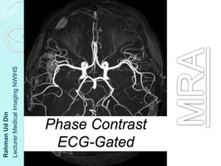
Phase Contrast and ECG-Gated MRA
•Als PPTX, PDF herunterladen•
0 gefällt mir•368 views

Phase contrast MRI uses changes in the phase of tissue magnetization from moving protons to create angiographic images and measure flow velocity. ECG-gated MRI acquires images during systole and diastole, subtracting the former to highlight arteries based on their faster flow during cardiac contraction. Typical velocity encoding values are 20-30 cm/s for veins, 40-60 cm/s for higher arterial flow, and over 60 cm/s to determine flow direction and velocity.

Empfohlen
Weitere ähnliche Inhalte
Was ist angesagt?
Was ist angesagt? (20)
Ähnlich wie Phase Contrast and ECG-Gated MRA
Ähnlich wie Phase Contrast and ECG-Gated MRA (20)
Mehr von Rahman Ud Din
Mehr von Rahman Ud Din (11)
Kürzlich hochgeladen
Kürzlich hochgeladen (20)
Phase Contrast and ECG-Gated MRA
- 2. Learning Outcomes Phase Contrast MRA VENC (Velocity Encoding) ECG-Gated FSE MRA
- 3. Phase Contrast MRA PC-MRA uses change in the phase of TM of the blood and form an image The selective phase shift of the moving p+ is produced GRE of a given strength is applied to both stationery as well as moving p+ causing phase shift in both but at different rates Initial RF excitation pulse brings all p+ in phase Second GRE of same amplitude and duration but of opposite polarity is applied
- 5. In stationery p+ reversal of phase shift occur of exact amount Canceling the effect of original phase shift Resulting into no net phase shift Hence, flowing p+ changed their position the phase shift will not be corrected This shift is directly proportional to the change in location or distance the p+ travel b/w application of first and second gradients The phase shift is used by PC-MRA to create angiographic image and to measure flow velocity
- 7. Velocity encoding gradients are applied in one or all three directions to acquire quantitative information Velocity encoding technique compensates for projected flow velocity within the vessel by controlling the amplitude or strength of the bipolar gradient If velocity encoding (VENC) is selected lower than the velocity within the vessel- aliasing occurs Aliasing results in low signal intensity in the centre of the vessel and Better delineation of the vessel wall
- 9. Typical values of the VENC are ◦ 20-30 cm/s for venous flow ◦ 40-60 cm/s for higher velocity with some aliasing ◦ 60-80 cm/s to determine velocity and flow direction PC-MRA provides information about flow direction If flow is encoded from superior to inferior Head will appears bright and feet appears black PC-MRA are in 2D or 3D acquisition 2D used in routine practice bcoz of acceptable acquisition time of 1-3 minutes
- 10. Phase Contrast MRA Typical Venc values for different flow measurement are listed below: Aorta: 150 cm/s MPA: 120 cm/s Aqueduct: 8-12 cm/s
- 11. ECG-Gated FSE MRA In this technique, images of the vessels are acquired in systole and in diastole On diastole image for both artery and vein are both bright On systole artery flow will appear black due to its fast movement while veins will appear bright due to it slow flow The systole images are subtracted from diastole images resulted bright blood in arteriography
- 12. Advantages are; ◦ Relative short-time, sensitive to slow flow, acquire coronal plane ◦ Not possible in patient with arrhythmia Used in peripheral arteries and aorta FBI (Toshiba), NATIVE SPACE (s), TRANCE & flow