Reverse Total Shoulder Replacement: Anatomy, Rehabilitation, and Clinical Implications
•Als PPTX, PDF herunterladen•
18 gefällt mir•3,770 views

A review of the reverse total shoulder replacement surgery and it's clinical implications for both physical rehabilitation and functional anatomy. Objectives: Understand basic anatomy of the shoulder complex and its implications for shoulder replacement Understand indications for shoulder replacement Understand differences between standard and reverse total shoulder replacements Understand precautions following rTSA Understand important concepts in rehabilitation following rTSA

Empfohlen
Empfohlen
Weitere ähnliche Inhalte
Was ist angesagt?
Was ist angesagt? (20)
Ähnlich wie Reverse Total Shoulder Replacement: Anatomy, Rehabilitation, and Clinical Implications
Ähnlich wie Reverse Total Shoulder Replacement: Anatomy, Rehabilitation, and Clinical Implications (20)
Kürzlich hochgeladen
Kürzlich hochgeladen (20)
Reverse Total Shoulder Replacement: Anatomy, Rehabilitation, and Clinical Implications
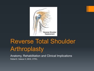
- 1. Reverse Total Shoulder Arthroplasty Anatomy, Rehabilitation and Clinical Implications Rafael E. Salazar II, MHS, OTR/L
- 2. Objectives Understand basic anatomy of the shoulder complex and its implications for shoulder replacement Understand indications for shoulder replacement Understand differences between standard and reverse total shoulder replacements Understand precautions following rTSA Understand important concepts in rehabilitation following rTSA
- 4. Anatomy of the Shoulder Complex 4 main components: Glenohumeral (GH) joint Acromioclavicular (AC) joint Sternoclavicular joint Scapulothoracic joint (gliding mechanism) (Peat, 1986) (Terry & Chopp, 2000)
- 5. Sternoclavicular Joint & Acromioclavicular Joint SC Joint Functions: Acts as only bony attachment of the upper extremity (UE) to the axial Skeleton Allows for elevation, depression, protraction and retraction of clavicle Axial rotation with shoulder elevation (Terry & Chopp, 2000) AC Joint Functions: Contributes to total arm movement as well as transmitting force between clavicle and acromion (Peat, 1986) Elevation/Depression in relation to scapular movement Axial rotation with elevation of the shoulder (Peat, 1986) (Terry & Chopp, 2000)
- 6. Scapulothoracic Mechanism Has NO bony attachments to the axial skeleton other than through AC and SC joints (Culham & Pete, 1993) Held in place through axioscapular muscles to include: Trapezius, serratus anterior, rhomboid major/minor, levator scapulae Function: Allows increased shoulder mov’ts Approx. 1 degree of scapulothoracic elevation for every 2 degrees of GH elevation (Terry & Chopp, 2000)
- 8. Glenohumeral Joint Synovial Ball and Socket Joint Large head of humerus on smaller, shallow glenoid fossa About 25-30% of humeral head is in contact with glenoid Relies on both static and dynamic stabilizing forces to provide stability (Terry & Chopp, 2000)
- 9. Static Stabilizers of the GH Joint Articular Surface Glenoid articular cartilage is thicker at periphery Glenoid Labrum Dense fibrous tissue; extends articular surfaces, increasing stability Also acts as an anchor point for capsuloligamentus structures Joint Capsule Approximately twice the surface area of humeral head Reciprocally tighten and loosen with rotation of the arm to limit translation Ligaments Coracohumeral ligament Glenohumeral ligaments (Terry & Chopp, 2000)
- 10. Dynamic Stabilizers of the GH Joint Rotator Cuff Muscles: Subscapularis Infraspiantus Supraspinatus Teres Minor All of the tendons of the RC muscles blend intricately with the fibrous capsule (Peat, 1986) Contraction of the Rotator Cuff results in concavity- compression Centers the humeral head; important stabilizing mechanism (Terry & Chopp, 2000)
- 11. Other Shoulder Muscles Deltoid Muscle Functions to assist in GH flexion and abduction and extension Biceps 2 Heads: long head and short head Long Head is located between supraspinatus and subscapularis and functions as a humeral head depressor during abduction (Terry & Chopp, 2000)
- 12. Indications for TSA: Total Shoulder Replacement Advanced Glenohumeral pathology (degenerative changes) caused by: Osteoarthritis (OA) Rheumatoid Arthritis (RA) Osteonecrosis Fractures of the Humeral Head RC tear arthropathy May be treated with hemiarthroplasty (Boudreau, S., Boudreau, E., Higgins, & Wilcox, 2007)
- 13. TSA vs rTSA Total Shoulder Arthroplasty Used to treat degenerative changes of the GH joint when the RC is intact or repairable Reverse Total Shoulder Arthroplasty Developed in 1980s in Europe, approved by FDA in 2004 Used to treat GH arthritis when it is associated with irreparable RC damage, complex fractures, or revisions of standard TSA with deficient RC tendons (Brigham and Women’s Hospital, Inc. Department of Rehabilitation services, 2011)
- 14. Indications for Reverse Total Shoulder Replacement Additional Indications for Reverse Total Shoulder Replacement: Proximal humeral fracture with malunion/nonunion Post traumatic arthritis Deficient Rotator Cuff Rotator Cuff Tear Arthropathy Severe humeral head collapse following massive RC tear Revision of previosuly failed conventional TSA (Drake, O’Conner, & Edwards, 2010)
- 15. Rotator Cuff Tear Arthropathy 3 Critical Features: Rotator Cuff Insufficiency Superior Migration of the humeral head Degenerative Changes of the GH joint (Nam et al., 2012) End stage glenohumeral arthritis Caused by a high riding humerus following a RC tear (Drake, O’Conner, & Edwards, 2010)
- 16. Massive and Irreparable RC Tear
- 17. Contraindications for rTSA 1. Deltoid Function is required for active elevation following a rTSA Absence of severe impairment of the deltoid is a contraindication 2. Isolated Supraspinatus Tear (SST) Isolated SST will not produce an imbalanced shoulder Can be treated by standard TSA 3. Massive irreparable RC tear without arthritis and full or nearly full active elevation Likely has a balanced shoulder Nonoperative Modalities indicated NSAID and Corticosteroid Injections If continued pain, imaging to determine presence of Long head of Biceps tendon Tenotomy may decrease pain and restore function (Drake, O’Conner, & Edwards, 2010)
- 18. Precautions Rehabilitation Following rTSA 12 wks postoperatively No movement in extension, adduction, & internal rotation No extension beyond neutral (Brigham and Women’s Hospital, Inc. Department of Rehabilitation services, 2011)
- 19. Important Concepts: Rehabilitation of rTSA cont. 1. rTSA Design alters the center of rotation medially and inferiorly Enhances the torque produced by deltoid as well as the line of pull/action of the deltoid 2. Deltoid Function Stability and mobility of the shoulder is now dependent upon the deltoid 3. Function Goal is to maximize overall upper extremity function while respecting tissue constraints 4. ROM Normal/full AROM is not expected following rTSA (Brigham and Women’s Hospital, Inc. Department of Rehabilitation services, 2011)
- 20. Joint Protection Higher risk of shoulder dislocation following rTSA than standard TSA Dislocations occur with operative arm in IR, adduction, and extension This positions allows prosthesis to dislocate anteriorly and inferiorly Limit functional activities such as tucking in a shirt or reaching behind one’s hip At least 12 weeks postoperatively (Boudreau, S., Boudreau, E., Higgins, & Wilcox, 2007)
- 21. Deltoid Function Enhancing deltoid function is the most important rehabilitation concept in the strengthening phase of postoperative rehabilitation. Stability and mobility of shoulder is dependent on deltoid and periscapular musculature Must assist patients to learn recruiting strategies to make deltoid primary shoulder mover (Boudreau, S., Boudreau, E., Higgins, & Wilcox, 2007)
- 22. ROM and Functional Expectations Expectation for functional and ROM gains should be set on a case-by-case basis ROM gains will be dependent on: Underlying pathology The status of external rotators The extent to which the deltoid and periscapular musculature can be rehabilitated Guideline: Functional active elevation of at least 105 degrees should be expected 80-120 degrees of elevation 30 degrees of ER (Boudreau, S., Boudreau, E., Higgins, & Wilcox, 2007)
- 23. Rehabilitation Protocol Phase I Immediate postsurgical/joint protection 4-6 weeks post-operatively Active elbow, wrist, digit ROM PROM to Shoulder Submaximal deltoid isometrics Phase II Active ROM and early strengthening Approx. 6-12 weeks AAROM/AROM gentle strengthening Avoid poor/inappropriate motor patterns Periscapular and deltoid strengthening should progress to isotonic between 6-8 wks Phase III Moderate strengthening 12+ weeks postoperatively Initiate when patient demonstrates appropriate PROM/AAROM/AROM while demonstrating appropriate shoulder mechanics Goal is to advance strengthening and increase functional independence Phase IV Independent and progressive home exercise program Return to light household work and leisure activities Initiated once patient is d/c’d from rehabilitation services(Boudreau, S., Boudreau, E., Higgins, & Wilcox, 2007) (Brigham and Women’s Hospital, Inc. Department of Rehabilitation services, 2011)
- 24. Summary Popularity of rTSA is growing rapidly in the U.S.A. To date, optimal postoperative rehab plan/protocol has not been established Minimal research regarding long-term outcomes of patients following rTSA Use of rTSA to treat RC tear arthropathy is clinically sound Changes shoulder mechanics and enhances deltoid function in absence of RC Postoperative treatment of clients with rTSA is different than those following a standard TSA Physician, therapist, and client should work together to develop a postoperative rehab plan (client centered care) Further research is required in regards to long-term results of rTSA and an optimal postoperative rehabilitation plan
- 25. References Brigham and Women's Hospital, Inc. Department of Rehabilitation Services. (2011). Reverse Total Shoulder Arthroplasty Protocol. Brigham and Women's Hospital, Inc. Department of Rehabilitation Services. (2007). Total Shoulder Arthroplasty/Hemiarthroplasty Protocol. Boudreau, S., Boudreau, E., Higgins, L., & Wilcox, R. (2007). Rehabilitation Following Reverse Total Shoulder Arthroplasty. Ther Journal of Orthopaedic & Sports Physical Therapy, 734-743. Retrieved August 30, 2015, from www.jospt.org Culham, E., & Peat, M. (1993). Functional Anatomy of the Shoulder Complex. Journal of Orthopaedic & Sports Physical Therapy, 18(1), 342-350. Retrieved August 30, 2015, from www.jospt.org Drake, G., O’Connor, D., & Edwards, T. (2010). Indications for Reverse Total Shoulder Arthroplasty in Rotator Cuff Disease.Clinical Orthopaedics and Related Research, 468(6), 1526-1533. doi: http://dx.doi.org/10.1007/s11999-009-1188-9 Nam, D., Maak, T., Raphael, B., Kepler, C., Cross, M., & Warren, R. (2012). Rotator Cuff Tear Arthropathy: Evaluation, Diagnosis, and Treatment. The Journal of Bone and Joint Surgery (American), 94(6). http://dx.doi.org/10.2106/JBJS.K.00746 Peat, M. (1986). Functional Anatomy of the Shoulder COmplex. Journal of the American Physical Therapy Association, 66(12), 1855-1865. Retrieved August 30, 2015, from http://ptjournal.apta.org/content/66/12/1855 Terry, G., & Chopp, T. (2000). Functional Anatomy of the Shoulder. Journal of Athletic Training, 35(3), 348-255. Retrieved September 29, 2015, from www.journalofathletictraining.org Wilcox, R., Arslanian, L., & Millett, P. (2005). Rehabilitation Following Total Shoulder Arthroplasty. Journal of Orthopaedic & Sports Physical Therapy, 35(12), 821-836. Retrieved August 30, 2015, from www.jospt.org Images: https://www.google.com/imghp?hl=en&tab=wi&ei=H9rwVe- VKIe1ggSczqA4&ved=0CBYQqi4oAQ https://www.youtube.com/watch?v=l7h2FJnSXyw
- 26. Questions?
Hinweis der Redaktion
- https://www.youtube.com/watch?v=l7h2FJnSXyw <iframe width="560" height="315" src="https://www.youtube.com/embed/l7h2FJnSXyw" frameborder="0" allowfullscreen></iframe>