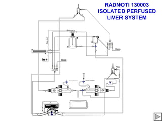
16. Flow Path:
Perfusate is selected
either from the primary
reservoir or the
secondary reservoir
17. Flow Path:
Perfusate is selected
either from the primary
reservoir or the
secondary reservoir
using the three way
stopcock located at the
outlet of the secondary
reservoir.
18. Flow Path:
Perfusate is selected
either from the primary
reservoir or the
secondary reservoir
using the three way
stopcock located at the
outlet of the secondary
reservoir.
19. Flow Path:
Perfusate is selected
either from the primary
reservoir or the
secondary reservoir
using the three way
stopcock located at the
outlet of the secondary
reservoir. The perfusate
then travels to the
peristaltic pump
22. Flow Path:
and up to the bubble trap
compliance chamber Tip: on initial priming or
filling of the system you will
most likely need to close
the compliance port
stopcock
23. Flow Path:
and up to the bubble trap
compliance chamber Tip: on initial priming or
filling of the system you will
most likely need to close
the compliance port
stopcock
and open the bubble trap
vent stopcock
24. Flow Path:
and up to the bubble trap
compliance chamber Tip: on initial priming or
filling of the system you will
most likely need to close
the compliance port
stopcock
and open the bubble trap
vent stopcock
so that the trap will have the
opportunity to fill
25. Flow Path:
and up to the bubble trap
compliance chamber When running in constant
pressure mode, perfusate
flow from the pump that is
greater than the flow rate of
the organ , will exit the
bubble trap via the
compliance port.
26. Flow Path:
and up to the bubble trap
compliance chamber Generally you can leave this
port open after the system
has been primed.
27. Flow Path:
and up to the bubble trap
compliance chamber Generally you can leave this
port open after the system
has been primed.
Over flow exiting the
compliance port is directed
to the overflow manifold.
28. Flow Path:
and up to the bubble trap
compliance chamber
The overflow manifold
will allow you to select
where the overflowing
perfusate is to be
directed.
29. Flow Path:
and up to the bubble trap
compliance chamber
Valve 1 directs flow
back to the primary
reservoir.
30. Flow Path:
and up to the bubble trap
compliance chamber
Valve 1 directs flow
back to the primary
reservoir.
Valve 2 directs flow
back to the secondary
reservoir.
31. Flow Path:
and up to the bubble trap
compliance chamber
Valve 1 directs flow
back to the primary
reservoir.
Valve 2 directs flow
back to the secondary
reservoir.
Valve 3 directs flow Out
to Waste.
32. Flow Path:
and up to the bubble trap
compliance chamber
the out flow of the bubble
trap then flows to the
inflow manifold.
33. Flow Path:
and up to the bubble trap
compliance chamber
the out flow of the bubble
trap then flows to the
inflow manifold.
Tip: When
initially priming
or flushing the
system, the
inflow and
outflow cannulae
will have to be
coupled. This
can be done by
using a small
section of tygon
tubing and
pushing the
cannulae tips in
either side.
34. Flow Path:
The perfusate then
passes through the
cannulated organ (or
tygon coupler when
priming or flushing) and
into the outflow manifold.
35. Flow Path:
Perfusate then flows out
to through the flow meter
( if so equipped) to the
outflow bubble trap.
36. Flow Path:
Perfusate then flows out
to through the flow meter
( if so equipped) to the
outflow bubble trap.
NOTE:
The vent
stopcock and
the out flow
stopcock of the
bubble trap
should be in the
closed position.
37. Flow Path:
Perfusate then flows out
to through the flow meter
( if so equipped) to the
outflow bubble trap.
The perfusate then is
drawn be the second
peristaltic pump head
and pushed to the three
way outflow manifold.
38. Flow Path:
At this point the
perfusate can be directed
to :
1. The primary reservoir
39. Flow Path:
At this point the
perfusate can be directed
to :
1. The primary reservoir
2. The secondary
reservoir
40. Flow Path:
At this point the
perfusate can be directed
to :
1. The primary reservoir
2. The secondary
reservoir
3. Waste
Sink or
collection flask
43. Flow Path:
Example:
If you are flushing the
system close valve 1 and
2
Leaving the valve open to
waste will direct the flow
out to waste.
44. Flow Path:
ALERT!
SPECIAL NOTE:
when flushing the
organ you will
want to protect
components that
may be sensitive to
the initial effluent
by diverting flow
around the
component.
45. Flow Path:
SPECIAL NOTE:
when flushing the
organ you will
want to protect
components that
may be sensitive to
the initial effluent
by diverting flow
around the
component.
46. Flow Path:
SPECIAL NOTE:
In this diagram the
flow meter would
need to be
protected.
47. Flow Path:
SPECIAL NOTE:
In this diagram the
flow meter would
need to be
protected. This is
done by using the
three way
stopcock on the
outflow manifold to
bypass the flow
meter.
48. Flow Path:
SPECIAL NOTE:
In this diagram the
flow meter would
need to be
protected. This is
done by using the
three way
stopcock on the
outflow manifold to
bypass the flow
meter.
51. Flow Path:
Example:
If the organ is flushed
and you wish to have a
recirculating path...
In this case back to the
primary reservoir...
52. Flow Path:
Example:
If the organ is flushed
and you wish to have a
recirculating path...
In this case back to the
primary reservoir…
Close valve 3 (waste)
53. Flow Path:
Example:
If the organ is flushed
and you wish to have a
recirculating path...
In this case back to the
primary reservoir…
Close valve 3 (waste)
Close valve 2
54. Flow Path:
Example:
If the organ is flushed
and you wish to have a
recirculating path...
In this case back to the
primary reservoir…
Close valve 3 (waste)
Close valve 2.
Open valve 1
55. Flow Path:
Example:
If the organ is flushed
and you wish to have a
recirculating path...
Or
To recirculate back to the
secondary reservoir…
56. Flow Path:
Example:
If the organ is flushed
and you wish to have a
recirculating path...
Or
to recirculate back to the
secondary reservoir…
Close valve 1
57. Flow Path:
Example:
If the organ is flushed
and you wish to have a
recirculating path...
Or
to recirculate back to the
secondary reservoir…
Close valve 1
Open valve 2
58. Flow Path:
Example:
If the organ is flushed
and you wish to have a
recirculating path...
Or
to recirculate back to the
secondary reservoir…
Close valve 1
Open valve 2
Close valve 3 (waste)
61. INITIAL START UP
Make sure reservoir
♦Fill the primary selection stopcock is
in the off position
reservoir with buffer
prior to filling.
62. INITIAL START UP
WARNING: ALWAYS MAKE SURE
THERE IS AN OPENING TO
♦Fill the primary ATMOSPHERE ON THE PRIMARY
reservoir with buffer RESERVOIR. IF FOR ANY REASON
GAS PREASURE IS ALLOWED TO
♦Turn on and adjust BUILD IN THE RESERVOIR RISK OF
gas from tank SERIOUS INJURY OR FATALITY MAY
OCCUR.
From tank
63. INITIAL START UP
♦Fill the primary Note:
reservoir with buffer Adjust gas so that
♦Turn on and adjust a gentle stream of
gas from tank bubbles flows from
the gas dispersion
frit in the primary
reservoir. This will
oxygenate the
buffer and serve as
a visual
representation that
gas is flowing
From tank through the
Membrane
Oxygenator.
64. INITIAL START UP
♦Fill the primary
reservoir with buffer
♦Turn on and adjust
gas from tank
♦Open compliance
port stopcock.
65. INITIAL START UP
♦Fill the primary
reservoir with buffer
♦Turn on and adjust
gas from tank
♦Open compliance
port stopcock.
♦Close valve 1 and 2
on three way out
flow manifold.
66. INITIAL START UP
♦Fill the primary
reservoir with buffer
♦Turn on and adjust
gas from tank
♦Open compliance
port stopcock.
♦Close valve 1 and 2
on three way out
flow manifold.
♦Open valve 3
(waste) on three way
out flow manifold
67. INITIAL START UP
♦Fill the primary
reservoir with buffer
♦Turn on and adjust
gas from tank
♦Open compliance
port stopcock.
♦Close valve 1 and 2
on three way out
flow manifold.
♦Open valve 3
(waste) on three way
out flow manifold
♦Close valve 2 and 3
on three-way
overflow manifold
68. INITIAL START UP
♦Fill the primary
reservoir with buffer
♦Turn on and adjust
gas from tank
♦Open compliance
port stopcock.
♦Close valve 1 and 2
on three way out
flow manifold.
♦Open valve 3
(waste) on three way
out flow manifold
♦Close valve 2 and 3
on three-way
overflow manifold
♦Open valve 1
(primary reservoir
return) on three-way
overflow manifold.
70. INITIAL START UP
♦Close the
stopcocks for the
inflow and outflow
pressure
transducers.
♦Direct the outflow
manifold three way
stopcock to pass
through the
manifold.
71. INITIAL START UP
♦Close the
stopcocks for the
inflow and outflow
pressure
transducers.
♦Direct the outflow
manifold three way
stopcock to pass
through the
manifold.
♦Close the drain and
relief ports
stopcocks on the out
flow bubble trap.
72. INITIAL START UP
♦Close the
stopcocks for the
inflow and outflow
pressure
transducers.
♦Direct the outflow
manifold three way
stopcock to pass
through the
manifold.
♦Close the drain and
relief ports
stopcocks on the out
flow bubble trap.
♦Couple the Inflow
and Outflow
cannulae.
73. INITIAL START UP
♦Close the
stopcocks for the
inflow and outflow
pressure
transducers.
♦Direct the outflow
manifold three way
stopcock to pass
through the
manifold.
♦Close the drain and
relief ports
stopcocks on the out
flow bubble trap.
♦Couple the Inflow
and Outflow
cannulae.
♦Turn on peristaltic
pump and turn
reservoir selection
three way stopcock
to feed from primary
ON
reservoir.
74. INITIAL START UP
The system will now
circulate buffer
driven by the
peristaltic pump.
It will take several
moments to purge
air from the lines.
You will most likely
have to open and
close the vent and
overflow ports in the
bubble trap
compliance chamber
temporarily to build
up some perfusate.
Once it
approximately two
thirds full, return the
valves to their
previous position.
ON
75. INITIAL START UP
Once the system is
primed, Turn off the
peristaltic pump.
Close stopcocks at
the inflow and
outflow manifold.
This will trap the
buffer in the lines
and keep the system
primed.
77. Signal Generation
Now is a good time
to calibrate the
pressure
transducers and ion
selective electrodes
(if so equipped.)
78. Signal Generation
For the pressure
Pressure
Transducers
transducers typically
they will have to be
filled with fluid and
purged of bubbles.
79. Signal Generation Pressure Transducers
For the pressure
transducers typically
they will have to be
filled with fluid and
purged of bubbles.
Set the three-way
stopcock at the
outflow pressure
transducer so that
flow is accepted
from the outflow
manifold and the
purge port.
80. Signal Generation Pressure Transducers
For the pressure
transducers typically
they will have to be
filled with fluid and
purged of bubbles.
Set the three-way
stopcock at the
outflow pressure
transducer so that
flow is accepted
from the outflow
Vent port
manifold and the
stopcock
purge port.
(not shown)
Fill a disposable
syringe with buffer.
Open the transducer
purge port (one way
stopcock not
shown.) and gently
fill the pressure
transducer dome
causing air to be
purged.
82. Signal Generation Pressure Transducers
The pressure
transducers can be
calibrated to your
data acquisition at
this time.
Set the three-way
stopcock controlling
flow to the
transducer to the
closed position.
83. Signal Generation Pressure Transducers
The pressure
transducers can be
calibrated to your
data acquisition at
this time.
Set the three-way
stopcock controlling
flow to the
transducer to the
closed position.
Open the purge
stopcock (not
shown) on the
transducer.
84. Signal Generation Pressure Transducers
The pressure
transducers can be
calibrated to your
data acquisition at
this time.
Set the three-way
stopcock controlling
flow to the
transducer to the
closed position.
Open the purge
stopcock (not
shown) on the
transducer.
This will be your
zero pressure
calibration point.
85. Signal Generation Pressure Transducer
Return the
stopcocks to their
previous position
(accepting flow from
the outflow
manifold) and
setting the purge
port stopcock (not
shown) to the closed
position.
This will be you're
high pressure
calibration point.
86. Signal Generation Pressure Transducer
Note:
Return the The pressure head is
stopcocks to their determined by the
previous position elevation of the
(accepting flow from bubble trap
the outflow compliance
manifold) and chamber. The
setting the purge distance from the
port stopcock (not chamber to the
shown) to the closed pressure transducer
position. can be calculated to
a known pressure.
This will be you're
high pressure Distance in mm
calibration point. divided by 13.6 = mm
of mercury
perfusion pressure
should be 10-15 mm
Hg (15-25cm above
the liver.)
87. Signal Generation Pressure Transducer
Note:
Repeat the The pressure head is
procedure for the determined by the
inflow manifold elevation of the
pressure transducer. bubble trap
compliance
chamber. The
distance from the
chamber to the
pressure transducer
can be calculated to
a known pressure.
Distance in mm
divided by 13.6 = mm
of mercury.
89. Signal Generation pH Electrodes
The pH electrodes are
plugged directly from
the mili volt adapter to
the data acquisition
interface.
Ideally a pH electrode
will output a voltage of
0mV in a pH 7 buffer.
This can very by +/-
50mV based on the
individual pH
electrode.
The Nernst equation
tells us that a pH
buffer 4 should be
160mV greater (more
positive) than the
reading in a pH 7.
The reading in a pH10
should be -160mV less
(more negative than
that of a reading in a
pH7 buffer.
The 160 comes from
being 3(7-4)*59(Nernst
value at 20
degrees)=168mV for
100% slope and 160 is
slightly less than 100%
theoretical.
91. Signal Generation O2 Electrodes
Prior to calibration
of the oxygen
electrode, the
electrode should be
examined to insure
that the electrode
membrane is intact
and the interior
chamber is full of
buffer. If the
electrode requires
maintenance
please refer to the
manufacturers
instructions.
92. Signal Generation O2 electrodes
To obtain zero
oxygen reading, the
physiological
buffer, placed in a
vented calibration
container should be
gassed for at least
10 to 30 minutes
with pure nitrogen
at a rate of 3-6
bubbles per/sec to
maintain a constant
temperature and
gas saturation.
93. Signal Generation O2 Electrodes
To obtain zero
oxygen reading, the
physiological
buffer, placed in a
vented calibration
container should be
gassed for at least
10 to 30 minutes
with pure nitrogen
at a rate of 3-6
bubbles per/sec to
maintain a constant
temperature and
gas saturation.
The electrode is
then inserted into
the calibration
chamber and
monitored until the
reading is stabile.
Once the reading
has stabilized the
reading can bet set
to zero using the
amplifier gain and
offset adjustments.
The acquisition
software can use
this as the low
point calibration.
94. Signal Generation O2 Electrodes
The electrode is then
removed and inserted
into aerated container
of buffer. In this case
either the inflow or out
flow manifold with
perfusate being
pumped through. After
stabilizing the reading
would be adjusted
using the amplifier
gain and offset based
on the gas mixture
(room air 21% oxygen,
gas cylinder 100%
oxygen, gas cylinder
95% oxygen etc…)
This can be used as
your high point
calibration in the
datacquisiton
software.
95. Signal Generation O2 Electrodes
It is recommended
that the procedure
is repeated three
times in order to
insure the readings
are stable and
reproducible.
97. Signal Generation Temperature
Temperature probes
are preset and will not
require calibration. It
is recommended that
they be verified
periodically measuring
a known temperature
such as your heater
circulator bath and the
result compared to the
read out of the bath.
99. Signal Generation Flow Meter
The Flow Meter is
preset and will not
require calibration. It
is recommended that
it be verified
periodically measuring
a known flow and the
read out verified.
100. Liver Preparation
1. The animal is anesthetized and placed on its back; the anesthetic used may be a general
anesthetic such as isoflurane or phenobarbital, depending upon the protocol requirements. Test
for the depth of anesthesia via toe pinch, eye reflex, etc.
2. For best positioning, the limbs are retracted and secured with tape or string.
3. The abdomen is wiped with 70% alcohol; the abdomen can be shaved, although this is not
necessary.
4. A midline incision is made by lifting the skin with forceps and cutting the tissue. The abdomen
is cut with the blunt end of blunt/sharp scissors from above the bladder to just below the
diaphragm (rib cage). Care must be taken not to cut the abdomen or internal organs.
5. The incision is extended into horizontally flaps on both left and right to expose the liver and
intestines. The internal organ should be handled gingerly, especially the liver which is soft and
easily damaged.
101. Liver Preparation
6. The intestines are carefully moved to the left side of the animal, exposing the liver and surrounding
vasculature.
7. The vena cava, portal vein, mesenteric veins and arteries and bile duct are located.
8. Using a curved needle, non-cutting preferred and 00 or smaller silk suture, a suture is passed beneath
the portal vein, near the liver and past any branches. A second suture is passed beneath the vens cava
distal to this first suture(~5-10 mm). Note that the cannula will be inserted between the two sutures,
moved forward towards the liver and its tip secured by the suture closest to the liver. The distal suture
will be used to occlude portal vein blood flow.
9 A suture is passed beneath the vena cava, above the right renal vein. A suture is placed beneath the
mesenteric vein.
10. A suture is passed beneath the bile duct, the bile cannula is inserted and the cannula secured via
suture. (bile duct cannula is PE 10 tubing with cuff).
11. The appropriate sized portal vein cannula is selected by comparing the cannula tip to vein diameter.
The cannula is then placed on the end of the perfusion line and the line flushed by opening the stopcock
to clear air bubbles. The stopcock to the line is then closed. The pump should be cycling to permit
perfusion to occur as soon as the stopcock is opened.
102. Liver Preparation
12. Heparin (1000 units) is injected into the tail vein or into the vena cava, below the renal vein.
13. Note that at this point the experimenter must work efficiently so that the liver is not ischemic; total
time without blood flow to the liver should be less than 2 minutes from steps 14-17.
14. The distal portal vein suture is tied to occlude blow flow, the vessel then nicked to permit insertion of
the cannula, the cannula tip slid into the vein past the first suture and the cannula secured using the first
suture. Blood will back flush into the cannula.
15. The stopcock is opened to allow a small amount of fluid (<5ml) into the liver. The liver should not be
over expanded.
16. The vena cava is immediately cut below the suture loop to permit blood to drain out.
17. The stopcock is then re-opened and the liver perfused from the reservoir. A successful perfusion will
have the liver eventually blanching to an even beige color, without spotches or mottling.
18. The mesenteric veins are tied off with the previously placed suture.
19. The chest cavity is opened midline using scissors and the heart exposed. The atrial- thoracic vena
cava cannula is inserted through a cut placed in the right atria into the thoracic vena cava and secured
with suture. The cannula should fully dilated the vena cava to reduce backpressure.
103. Liver Preparation
20. The vena cava suture below the liver is secured and the stopcock to the atrial-thoracic vena cava
cannula is opened. This then forces fluid to exit from the liver into the atrial-thoracic vena cava cannula.
21. The lines to the bile duct, portal vein and vena cava are held in position
The liver is removed from the donor by supporting the diaphragm at the midline with forceps and
carefully cutting the diaphragm around the rib cage, first on side and then the other. Any attachment of
the liver to surrounding tissue are carefully removed.
22. The three lines are supported and the liver is then transferred to the organ chamber.
23. As indicators of a successful perfusion, liver flow should be at least 4 ml/minute/gram liver (for
perfusates without blood or other oxygen carriers) and bile flow 1-2 micro liters minute.
104. Post Experimental Clean Up
Glassware Maintenance & Post Experimental Cleanup
Post Experimental Cleanup
After the experiment has been completed, the experimenter should take care to scrupulously clean the
equipment. It is important to remember that the solutions that can sustain the heart and muscle will also provide
excellent media for bacteria. The cleaning procedures will be dependent upon the types of chemicals and
biological materials that are being used, the types of measurements that are being made and what substances
can interfere with those measurements and the frequency of the use of the equipment and number of operators
involved. Non-phosphate soaps are preferred, since insoluble phosphates can form from calcium and
magnesium in physiological salt solutions. Note that bactericidal soaps may contain iodine or other materials
which can affect isolated tissues and cells. Cleaning supplies and equipment, such as brushes, should be used
only for cleaning this glassware and not used for other lab cleaning procedures. Questions and procedures
noted here should be adjusted in accordance with your licensed procedures and the recommendations of your
safety personnel.
105. Post Experimental Cleanup
Shared equipment is the most difficult to maintain properly. In order to maintain equipment properly, it is generally
best (1) to assign the maintenance or the oversight of the equipment to one individual, who will monitor
equipment and maintain cleaning supplies (2) to have written protocols posted with the equipment (3) to have a
logbook where cleaning dates, as well as notification of problems, suggestions, etc., can be recorded.
Often overlooked as a source of contamination is the water circulator supply. This should be kept clean and the
bath rinsed and solution changed to reduce precipitate build up. Covering equipment to reduce air borne
contamination from microbes and spores is useful. Note that when baths are used intermittently, the lack of
frequent cleaning and the lack of solutions rinsing out bacteria that are deposited in the tubing may result in a
contamination problem when the system is finally used. A convenient rule of thumb for testing for contamination
in preparations that you have found reliable is that two consecutive experimental failures that are not explained
by an obviously damaged sample, poor surgical or dissection techniques or solution problems may be caused by
bath contamination.
Glassware
Much of the Radnoti apparatus is borosilicate glass, which can be cleaned with a wide range of soaps, ethyl
alcohol, dilute HCl or HNO3 (0.1 M) or other solvents. Extensive flushing with distilled, deionized water to remove
all traces of the cleaning agents and salts is recommended. Large glassware, such as reservoirs or assemblies
can be flushed in place, but care must be taken to thoroughly clean aerators, stopcocks and associated parts.
Aerators should be blown dry using gas or air at the final water rinse. If acid is used, the runoff water should not
be more acidic than the normal water pH. As with the use of any chemicals, proper protective gear and training
are essential to reduce personnel hazards and experimental and environmental contamination. Heated acid or
chromic acid is generally not recommended due to personnel hazards and possible heavy metal contamination of
the system.
If very lipohilic substances (prostaglandins, ionophores, certain dyes, etc.) are used, rinses with ethyl alcohol or
the most appropriate organic solvent can be used first, but this will necessitate thorough cleaning afterward to
remove any traces of the organic solvent.
106. Post Experimental Cleanup
Use of toxins, biohazardous materials and radiochemicals can present considerable complications to a
generalized cleaning procedure. Having an apparatus and a contained area dedicated to these procedures
reduces problems. Diluted bleach can be used on glassware, but must be rinsed extensively. The use of
disposable tubing and stopcocks will assist in cleanup, as will scheduling a run of these procedures, rather
than intermittent experiments, if non-dedicated equipment must be used. Glassware can be sterilized but all
fixtures, such as aerators, stopcocks caps, etc., should be removed prior to sterilization.
The glass aerators can be cleaned with water, or dilute acid if clogged. The use of water or gas under high
pressure can result in damage to the glassware and personnel and therefore is not recommended. After a
general soap and water rinse to remove soluble materials, cleaning with 0.1M HCl or 0.1 M HNO3 for several
hours or overnight, followed by an extensive water rinse, will usually remove most contaminants. If this does
not work, 1 M acid can be tried. Because the glass frit filaments are thin, high concentrations of acids, or
especially alkalis, can destroy them and are not recommended.
Non-glass items
Initial cleaning of non-glass items should be with aqueous soap solutions. Depending upon the chemical
resistance of the materials, the use of other solvents, cleaning procedures or sterilization may be possible.
Areas and items to be especially well cleaned are the aerator, tubing, syringe ports, cannulae, pressure
transducer fittings, septa, balloon and other catheters and electrodes (oxygen, pacing, ion selective, etc.).
Tubing should be inspected at the pump head for wear. Note that the interior of tubing can gradually be
roughened during use and the abraded areas will form sites for bacterial growth. Tubing should be a high
grade with low plasticizer leaching. Note that silicone tubing is very permeant to gases, so it should not be
generally used to transport gassed solutions.