
Empfohlen
Weitere ähnliche Inhalte
Ähnlich wie middle ear.pptx
Ähnlich wie middle ear.pptx (20)
Kürzlich hochgeladen
Kürzlich hochgeladen (20)
middle ear.pptx
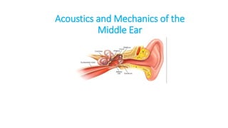
- 1. Acoustics and Mechanics of the Middle Ear
- 2. • Sound- form of energy produced by a vibrating object. • A sound wave consists of compression and rarefaction of molecules of the medium (air, liquid or solid) in which it travels. • Frequency. It is the number of cycles per second. The unit of frequency is Hertz (Hz) . • A sound of 1000 Hz means 1000 cycles per second. • Pure tone. A single frequency sound is called a pure tone, e.g. a sound of 250, 500 or 1000 Hz
- 3. • Complex sound. Sound with more than one frequency is called a complex sound. Human voice is a complex sound. • Pitch. It is a subjective sensation produced by frequency of sound. Higher the frequency, greater is the pitch. • Overtones. A complex sound has a fundamental frequency, i.e. the lowest frequency at which a source vibrates. All fre- quencies above that tone are called the overtones. • Overtones determines quality or the timbre of sound.
- 4. Intensity - strength of sound which determines its loudness. - measured in decibels Whisper = 30 dB Normal conversation =60 dB Shout =90dB Discomfort of the ear =120dB Pain in the ear =130dB
- 5. • Decibel (dB). It is 1/10th of a bel • represents a logarithmic ratio between two sounds, the sound being described and the reference sound. • In audiology, sound is measured as sound pressure level (SPL) • It is compared with the reference sound which has an SPL of 0.0002 dynes/cm2 or 20 μPa (micropascals),
- 6. • Roughly corresponds to the threshold of hear- ing in normal subjects at 1000 Hz. • Sound in dB =10 log Power of S1 /Power of S0 , S0= reference sound • If a sound has an SPL of 1000, i.e. (10^3) times the reference sound, it is expressed as 20 × 3 = 60 dB.
- 7. • Masking- It is a phenomenon to produce inaudibility of one sound by the presentation of another. • Masking of non-test ear is essential in all bone conduction tests, • But for air conduction tests, it is required only when difference of hearing between two ears exceeds 40 dB.
- 8. MIDDLE EAR It together with the eustachian tube, aditus, antrum and mastoid air cells is called middle ear cleft .
- 9. • It is lined by mucous membrane and filled with air. Divided into: (i) mesotympanum ( opposite the pars tensa), (ii) epitympanum or the attic ( above the pars tensa ) (iii) hypotympanum (below the level of pars tensa)
- 10. Middle ear is like a six-sided box with a roof, a floor, medial, lateral, anterior and posterior walls
- 11. • Roof is formed by a thin plate of bone called tegmen tympani. • It separates tympanic cavity from the middle cranial fossa. • The floor is also a thin plate of bone, which separates tym- panic cavity from the jugular bulb
- 12. • The anterior wall has a thin plate of bone, which separates the cavity from internal carotid artery. • Two openings- -the lower one for the eustachian tube -the upper one for the canal of tensor tympani muscle.
- 13. • The posterior wall lies close to the mastoid air cells. • It presents a bony projection called pyramid - attatch tendon of the stapedius muscle • Aditus, opening through which attic communicates with antrum, . • Facial nerve runs in the posterior wall just behind the pyramid.
- 14. • Facial recess or the posterior sinus is a depression in the posterior wall lateral to the pyramid. Surgically, facial recess gives direct access through this into middle ear without disturbing posterior canal wall .
- 15. • Medial wall is formed by the labyrinth. • It presents a bulge called promontory which is due to the basal coil of cochlea • oval window into which is fixed the footplate of stapes.
- 16. • Round window or the fenestra cochleae which is covered by the secondary tympanic membrane. • Above the oval window is the canal for facial nerve. • The tendon of tensor tympani takes a turn here to get attachment to the neck of malleus. • The lateral wall is formed largely by the tympanic membrane
- 17. OSSICLES OF THE MIDDLE EAR Three ossicles in the middle ear—the malleus, incus and stapes . Footplate of stapes is held in the oval window by annular ligament. The ossicles conduct sound energy from the tympanic membrane to the oval window and then to the inner ear fluid.
- 18. Sound Transmission in normal Ear • Acoustic signals transmitted from external environment to fluid-filled inner ear • The transmission of sound power at an air-fluid interface depends on relative impedances of air and fluid . • ln the case of inner ear, only about 0.1% of the intensity of an incident sound wave is transmitted to the fluid, • This is equivalent to a 30 dB loss.
- 19. • The middle ear couples sound signals from ear canal to the cochlea • Primarily through action of tympanic membrane and the ossicular chain.
- 20. Key transformer within the middle ear • It is ratio of the tympanic membrane area (ATM) to area of the stapes footplate (AFP). • Another transformer is the ossicular lever: this is the lever action due to differing lengths of the maleus and long process of incus • The theoretical (ideal) middle ear gain is 28 dB, whereas the actual (measured) middle-ear gain is only about 20 dB.
- 21. • The Middle ear acts as a transformer to increase sound pressure at footplate relative to that at tympanic membrane , at the expense of a decrease in stapes volume velocity relative to the tympanic membrane volume velocity • The major transformer mechanism is the ratio of the tympanic membrane area to the stapes footplate area (the area ratio)
- 22. • Tympanic has an area that is 20 times larger than the footplate • The sound pressure applied to the inner ear by the stapes footplate should be 20 times or 26 dB , than at the tympanic membrane . • Ossicular lever: the lever action due to different lengths of the malleus and incus arms around the axis of rotation of the ossicles .
- 23. • The malleus and incus lever arms , the ratio is i.e 1.3, it causes , only a small 2 dB increase in sound pressure applied by the stapes to the inner ear • Thus, if these transformers acted ideally, then the theoretical middle- ear sound pressure gain is about 28 dB • 26 dB area ratio+ 2 dB ossicular lever
- 24. Actual middle-ear gain is frequency dependent, and is only about 20 dB at best (around 1,000 Hz). The theoretical middle-ear gain, which is approximately 28 dB, is independent of frequency.
- 25. Ossicular coupling • Tympano-ossicular system transforms sound pressure in the ear canal to sound pressure at the oval window. • Acoustic coupling • Motion of the tympanic membrane in response to ear canal sound creates sound pressure in the middle ear cavity • In the normal ear, the magnitude of this acoustically-coupled window pressure difference is small, on the order of 60 dB less than ossicular coupling • Hence, ossicular coupling dominates normal middle-ear function
- 26. • The effective stimulus to the inner ears is a difference in sound pressure between the oval and round windows. • The middle ear maximizes this window pressure difference via 2 mechanism 1- tympano-ossicular system increases the sound pressure at the oval window of the inner ear -At the same time, the intact tympanic membrane reduces the sound pressure by 10 to 20 dB compared to the sound pressure in the ear canal
- 27. 2 - Presence of middle-ear air outside the round window permits the window to move freely when the inner ear is stimulated by motion of the footplate
- 28. ACOUSTICS AND MECHANICS OF DISEASED MIDDLE EARS • Ossicular Interruption With an Intact Tympanic Membrane • Loss of the Tympanic Membrane, Malleus, and lncus • Tympanic Membrane Perforation • Middle Ear Effusion • Tympanic Membrane Atelectasis • Third Window Lesions of the Inner Ear
- 29. Tympanic Membrane Perforation • Perforations of the tympanic membrane cause a conductive hearing loss that can range from negligible to 50 dB . • The primary mechanism of conductive loss due to a perforation is a reduction in ossicular coupling caused by a loss in the sound-pressure difference across the tympanic membrane.
- 30. References • Glasscock-Shambaugh Surgery of the ear-6th Edition • Dhingra’s Disease of Ear, Nose and Throat and HNS- 6th Edition THANK YOU