Comparative study of respiratory organs
•
0 gefällt mir•1,116 views

Comparative study of respiratory organs

Empfohlen
Empfohlen
Weitere ähnliche Inhalte
Was ist angesagt?
Was ist angesagt? (20)
Ähnlich wie Comparative study of respiratory organs
Ähnlich wie Comparative study of respiratory organs (20)
Mehr von Prabhakar Pawar
Mehr von Prabhakar Pawar (18)
Kürzlich hochgeladen
Kürzlich hochgeladen (20)
Comparative study of respiratory organs
- 1. 2.1 2.1 Comparative Study of Respiratory Organs (Structure & Function) Dr. Prabhakar R. Pawar Associate Professor in Zoology, Mahatma Phule A. S. C. College, Panvel, Dist. - Raigad, Navi Mumbai – 410 206 prpawar1962@gmail.com
- 2. Respiration Respiration is a biochemical process in which cells of an organism obtain energy by combining oxygen & glucose, resulting to release of carbon dioxide, water & ATP. Respiration is a metabolic process common in all living beings. Respiration includes: Breathing. Transport of gases. Oxidation of food to liberate carbon dioxide & energy. Respiration is a biochemical process in which cells of an organism obtain energy by combining oxygen & glucose, resulting to release of carbon dioxide, water & ATP. Respiration is a metabolic process common in all living beings. Respiration includes: Breathing. Transport of gases. Oxidation of food to liberate carbon dioxide & energy.
- 3. Processes of Respiration Respiration has two essential processes: External respiration/Breathing: A mechanical process of exchange of respiratory gases (oxygen & carbon dioxide) between the organism & surroundings. Internal respiration/Cellular or Tissue respiration: A chemical process in which food is oxidized within the cells to liberate free energy, carbon dioxide & water. Internal respiration takes place in the cytoplasm & mitochondria of eukaryotic cells. Respiration has two essential processes: External respiration/Breathing: A mechanical process of exchange of respiratory gases (oxygen & carbon dioxide) between the organism & surroundings. Internal respiration/Cellular or Tissue respiration: A chemical process in which food is oxidized within the cells to liberate free energy, carbon dioxide & water. Internal respiration takes place in the cytoplasm & mitochondria of eukaryotic cells.
- 4. Types of Respiration • Respiration is of 2 types: • Direct respiration: Exchange of gases between the environment & body cells without the aid of respiratory organ or blood. Exchange of gases occur by diffusion across the body surface. Examples: Amoeba, Hydra, Flat worms etc. • Indirect respiration Involves special respiratory surfaces or organs like skin, buccopharyngeal lining, gills & lungs and blood for circulation of respiratory gases in the body. The body surfaces were impermeable for exchange of gases, specialized structures were developed for gaseous exchange. • Respiration is of 2 types: • Direct respiration: Exchange of gases between the environment & body cells without the aid of respiratory organ or blood. Exchange of gases occur by diffusion across the body surface. Examples: Amoeba, Hydra, Flat worms etc. • Indirect respiration Involves special respiratory surfaces or organs like skin, buccopharyngeal lining, gills & lungs and blood for circulation of respiratory gases in the body. The body surfaces were impermeable for exchange of gases, specialized structures were developed for gaseous exchange.
- 5. Characteristics of Respiratory surfaces For efficient gaseous exchange, respiratory surface must possess the following characteristics: • Thin. • Permeable to gases like oxygen & carbon dioxide. • Moist. • Highly vascular. • Have large surface area. • Must be in direct contact with the air or water. For efficient gaseous exchange, respiratory surface must possess the following characteristics: • Thin. • Permeable to gases like oxygen & carbon dioxide. • Moist. • Highly vascular. • Have large surface area. • Must be in direct contact with the air or water.
- 6. 2.1.1 Respiration in Earthworm 2.1.1 Respiration in Earthworm Entire body surface performs respiration. Cutaneous respiration - Skin acts as respiratory surface. Skin - Highly vascular, always kept moist by secretions of integument glands, & coelomic fluid. Exchange of gases through the moist skin which is permeable to respiratory gases. Entire body surface performs respiration. Cutaneous respiration - Skin acts as respiratory surface. Skin - Highly vascular, always kept moist by secretions of integument glands, & coelomic fluid. Exchange of gases through the moist skin which is permeable to respiratory gases.
- 7. 2.1.1 Respiration in Earthworm 2.1.1 Respiration in Earthworm Blood contains haemoglobin pigment dissolved in plasma. Atmospheric oxygen is taken up by haemoglobin & is converted to oxyhaemoglobin. Oxyhaemoglobin is circulated to tissues where oxygen tension is low. Oxyhaemoglobin breaks up to release oxygen & haemoglobin in reduced state. Oxygen is used for oxidation of food. Carbon dioxide released mixes in the blood & is diffused out of the body through moist skin. Blood contains haemoglobin pigment dissolved in plasma. Atmospheric oxygen is taken up by haemoglobin & is converted to oxyhaemoglobin. Oxyhaemoglobin is circulated to tissues where oxygen tension is low. Oxyhaemoglobin breaks up to release oxygen & haemoglobin in reduced state. Oxygen is used for oxidation of food. Carbon dioxide released mixes in the blood & is diffused out of the body through moist skin.
- 8. 2.1.2 Book lungs in Spider 2.1.2 Book lungs in Spider Respiratory organs of spider are of 2 types: trachea & book lungs. Some spiders have trachea, some have book lungs & others have both, trachea & book lungs. Book lungs: 2 hairless patches on ventral side of spider’s abdomen. Situated ventro-laterally & opens on ventral surface. Each book lung consists of compressed sac-like cavity called, pulmonary sac. Pulmonary sac is lined by a cuticle. Sac is divided into pulmonary chamber & atrial chamber. Respiratory organs of spider are of 2 types: trachea & book lungs. Some spiders have trachea, some have book lungs & others have both, trachea & book lungs. Book lungs: 2 hairless patches on ventral side of spider’s abdomen. Situated ventro-laterally & opens on ventral surface. Each book lung consists of compressed sac-like cavity called, pulmonary sac. Pulmonary sac is lined by a cuticle. Sac is divided into pulmonary chamber & atrial chamber.
- 9. 2.1.2 Book lungs in Spider 2.1.2 Book lungs in Spider Pulmonary chamber: Distal part of book lung. Oval chamber with about 150 vertical leaf-like lamellae/plates. Plates are filled with haemolymph & are arranged parallel to each other. Presence of air space between the plates, which allows circulation of air around the plates. Plates resemble like leaves of a book, so respiratory organs of spider are called as ‘book lungs’. Book lungs open into chambers called, ‘atria’. Pulmonary chamber: Distal part of book lung. Oval chamber with about 150 vertical leaf-like lamellae/plates. Plates are filled with haemolymph & are arranged parallel to each other. Presence of air space between the plates, which allows circulation of air around the plates. Plates resemble like leaves of a book, so respiratory organs of spider are called as ‘book lungs’. Book lungs open into chambers called, ‘atria’.
- 10. 2.1.2 Book lungs in Spider 2.1.2 Book lungs in Spider Atrial chamber: Small, dorsoventrally compressed air chamber. Forms proximal part of book lung. Opens outside through one or several slits called ‘spiracles’. Roof is perforated by minute openings called, ‘ostia’. Ostia communicate with the inter- lamellar spaces of the pulmonary chamber. Inspiration & expiration of the air in the book lungs is controlled by action of dorso-ventral & atrial muscles. Atrial chamber: Small, dorsoventrally compressed air chamber. Forms proximal part of book lung. Opens outside through one or several slits called ‘spiracles’. Roof is perforated by minute openings called, ‘ostia’. Ostia communicate with the inter- lamellar spaces of the pulmonary chamber. Inspiration & expiration of the air in the book lungs is controlled by action of dorso-ventral & atrial muscles.
- 11. 2.1.3 Respiration in Bony fish: 2.1.3 Respiration in Bony fish: Rohu ( Rohu (Labeo Labeo rohita rohita) ) Labeo rohita (Rohu) is a freshwater bony fish. Gill slits in bony fishes are covered by a lid called, operculum. Bony fish breathe by moving water across their gills using operculum. Operculum acts as a ‘trapdoor’ & pumps the water across the gills. Operculum is supported by branchiostegal rays, which acts as a one way valve. Buccal cavity has 2 oral valves which allows water to enter the mouth but not to leave it. Labeo rohita (Rohu) is a freshwater bony fish. Gill slits in bony fishes are covered by a lid called, operculum. Bony fish breathe by moving water across their gills using operculum. Operculum acts as a ‘trapdoor’ & pumps the water across the gills. Operculum is supported by branchiostegal rays, which acts as a one way valve. Buccal cavity has 2 oral valves which allows water to enter the mouth but not to leave it.
- 12. 2.1.3 Respiration in Bony fish: 2.1.3 Respiration in Bony fish: Rohu ( Rohu (Labeo Labeo rohita rohita) ) Each gill is supported by a bony structure i. e. gill arch. Gill arch provides support to hold many comb-like structures, called gill filaments. Gill filaments extend horizontally from the gill arches. Each gill filament is further divided into minute primary & secondary lamellae. Secondary lamellae are highly vascular & increase the surface area for gaseous exchange. Bony projections attached to the gill arches are called gill rakers, which help in the fish’s feeding. Each gill is supported by a bony structure i. e. gill arch. Gill arch provides support to hold many comb-like structures, called gill filaments. Gill filaments extend horizontally from the gill arches. Each gill filament is further divided into minute primary & secondary lamellae. Secondary lamellae are highly vascular & increase the surface area for gaseous exchange. Bony projections attached to the gill arches are called gill rakers, which help in the fish’s feeding.
- 13. 2.1.3 Mechanism of Respiration in Rohu 2.1.3 Mechanism of Respiration in Rohu Lateral movement of operculum brings about respiration. When operculum is raised, gill apertures are closed & water enters into the mouth by suction action of oral valves. When operculum is lowered, pressure on water in buccal cavity & pharynx increases. As a result, oral valves are closed, branchiostegal membranes open & water is forced out over the gills and out of the opercular cavity. Exchange of gases takes place in the gill lamellae. Lateral movement of operculum brings about respiration. When operculum is raised, gill apertures are closed & water enters into the mouth by suction action of oral valves. When operculum is lowered, pressure on water in buccal cavity & pharynx increases. As a result, oral valves are closed, branchiostegal membranes open & water is forced out over the gills and out of the opercular cavity. Exchange of gases takes place in the gill lamellae.
- 14. Accessory Respiratory Organs of Fishes Accessory respiratory organs (ARO) are present in some fishes along with gills. Found in fishes living in shallow & stagnant water and also in hill stream fishes, where the streams dry up in summer. ARO enable the fish to remain out of the water for some time or tolerate oxygen deficient water for some time. ARO originate from pharyngeal/branchial cavities & are rarely found in marine fishes. ARO are sac like diverticulum, called as ‘opercular lungs’. Examples: Anabas, Clarius etc. Accessory respiratory organs (ARO) are present in some fishes along with gills. Found in fishes living in shallow & stagnant water and also in hill stream fishes, where the streams dry up in summer. ARO enable the fish to remain out of the water for some time or tolerate oxygen deficient water for some time. ARO originate from pharyngeal/branchial cavities & are rarely found in marine fishes. ARO are sac like diverticulum, called as ‘opercular lungs’. Examples: Anabas, Clarius etc.
- 15. Accessory Respiratory Organ in Anabas (Climbing perch): Labyrinthine organ Anabus has 2 specious sac like structures in branchial cavity, called as ‘suprabranchial chambers’. Epithelial lining of these chambers increase the respiratory area. Each suprabranchial chamber contain a special rosette structure called, ‘labyrinthine organ’. Labyrinthine organ consists of number of small concentric plates. Plates have wavy margins & are covered with vascular gill-like epithelium. Air enters into the chamber via the buccopharyngeal opening & goes out through the external gill slits. Entrance of buccopharyngeal chamber is controlled by valves. Anabus has 2 specious sac like structures in branchial cavity, called as ‘suprabranchial chambers’. Epithelial lining of these chambers increase the respiratory area. Each suprabranchial chamber contain a special rosette structure called, ‘labyrinthine organ’. Labyrinthine organ consists of number of small concentric plates. Plates have wavy margins & are covered with vascular gill-like epithelium. Air enters into the chamber via the buccopharyngeal opening & goes out through the external gill slits. Entrance of buccopharyngeal chamber is controlled by valves.
- 16. Accessory Respiratory Organ in Clarius : Arborescent organ Clarius is a catfish found in Indian & African rivers. Accessory respiratory organ of Clarius is called ‘Arborescent organ’. Arborescent organ consists of a pair of suprabranchial organs present above the gills & are divided into 2 parts. The organ is supported by cartilaginous internal skeleton, covered by vascular epithelium & vascular sac. Many gills are formed by joining of gill filaments. Air is taken into the mouth continuously & Clarius can live outside the water for several hours and move along dump grass. Clarius is a catfish found in Indian & African rivers. Accessory respiratory organ of Clarius is called ‘Arborescent organ’. Arborescent organ consists of a pair of suprabranchial organs present above the gills & are divided into 2 parts. The organ is supported by cartilaginous internal skeleton, covered by vascular epithelium & vascular sac. Many gills are formed by joining of gill filaments. Air is taken into the mouth continuously & Clarius can live outside the water for several hours and move along dump grass.
- 17. 2.1.4 Respiration in Frog 2.1.4 Respiration in Frog Frog has 3 types of respiratory surfaces for exchange of gases: skin, lining of mouth & lungs. Cutaneous respiration: Respiration using skin as respiratory surface. Occurs when the frog is under water or on land. Skin of frog: Thin, highly vascular & permeable to water. Oxygen is dissolved in the film of water covering the skin & is absorbed by the blood capillaries of the skin. Haemoglobin from the blood enhances the absorption of oxygen. When frog is out of water, mucus glands in the skin keep the skin moist which helps to absorb the dissolved oxygen from air. Frog has 3 types of respiratory surfaces for exchange of gases: skin, lining of mouth & lungs. Cutaneous respiration: Respiration using skin as respiratory surface. Occurs when the frog is under water or on land. Skin of frog: Thin, highly vascular & permeable to water. Oxygen is dissolved in the film of water covering the skin & is absorbed by the blood capillaries of the skin. Haemoglobin from the blood enhances the absorption of oxygen. When frog is out of water, mucus glands in the skin keep the skin moist which helps to absorb the dissolved oxygen from air.
- 18. 2.1.4 Respiration in Frog 2.1.4 Respiration in Frog Buccopharyngeal respiration: Buccopharyngeal membrane acts as a respiratory surface. Buccopharyngeal respiration is predominant during rest. In buccopharyngeal respiration, mouth remains closed, air is taken through the nostril & passed into the buccopharyngeal cavity. Oxygen from the air, dissolves in the mucous & goes into the blood. Carbon monoxide is expelled by raising the floor of Buccopharyngeal cavity. Glottis remains closed to prevent entry of air in lungs. Buccopharyngeal respiration is the main form of breathing at rest. Buccopharyngeal respiration: Buccopharyngeal membrane acts as a respiratory surface. Buccopharyngeal respiration is predominant during rest. In buccopharyngeal respiration, mouth remains closed, air is taken through the nostril & passed into the buccopharyngeal cavity. Oxygen from the air, dissolves in the mucous & goes into the blood. Carbon monoxide is expelled by raising the floor of Buccopharyngeal cavity. Glottis remains closed to prevent entry of air in lungs. Buccopharyngeal respiration is the main form of breathing at rest.
- 19. 2.1.4 Respiration in Frog 2.1.4 Respiration in Frog Pulmonary respiration: Occurs with the help of pair of lungs. Pulmonary respiration occur occasionally when need of oxygen is more. Lungs are thin walled sacs connected to mouth through an opening i. e. ‘glottis’. Frog does not possess the trachea. Lungs: Delicate, thin walled, elastic sac like structures covered by visceral peritoneum. Inner surface of lungs have many irregular & radially arranged folds. Space between two folds of inner surface of lungs forms an ‘alveolus’. Alveoli are highly vascular & increases the inner respiratory surface of lungs. Pulmonary respiration: Occurs with the help of pair of lungs. Pulmonary respiration occur occasionally when need of oxygen is more. Lungs are thin walled sacs connected to mouth through an opening i. e. ‘glottis’. Frog does not possess the trachea. Lungs: Delicate, thin walled, elastic sac like structures covered by visceral peritoneum. Inner surface of lungs have many irregular & radially arranged folds. Space between two folds of inner surface of lungs forms an ‘alveolus’. Alveoli are highly vascular & increases the inner respiratory surface of lungs.
- 20. 2.1.4 Mechanism of respiration in frog 2.1.4 Mechanism of respiration in frog Air is drawn into & expelled out of the body by action of buccal cavity muscles. Buccal cavity has 2 pairs of muscles: sternohyal muscles & petrohyal muscles. These muscles brings about rhythmic lowering & raising of buccal cavity. By contraction of sternohyal muscles, air comes in the buccal cavity through the external nares. Tightening of jaws close the external nares & air is trapped in the mouth. The glottis opens & air is pushed into the lungs (inspiration). During expiration, carbon dioxide from the lungs is expelled out of the body. Air is drawn into & expelled out of the body by action of buccal cavity muscles. Buccal cavity has 2 pairs of muscles: sternohyal muscles & petrohyal muscles. These muscles brings about rhythmic lowering & raising of buccal cavity. By contraction of sternohyal muscles, air comes in the buccal cavity through the external nares. Tightening of jaws close the external nares & air is trapped in the mouth. The glottis opens & air is pushed into the lungs (inspiration). During expiration, carbon dioxide from the lungs is expelled out of the body.
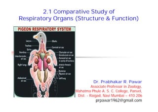
- 21. 2.1.5 Respiration in Pigeon 2.1.5 Respiration in Pigeon Respiratory system is highly developed to supply more oxygen for aerial mode of life. Respiratory system is extensively modified. Respiratory system has 2 unique features: Presence of non-elastic, compact & smaller size lungs. Possession of several air sacs to increase functional efficiency. Respiratory system consists of respiratory tract, the lungs & the air sacs. Respiratory tract consists of nares, nasal sacs, glottis, larynx, trachea & syrinx. Respiratory system is highly developed to supply more oxygen for aerial mode of life. Respiratory system is extensively modified. Respiratory system has 2 unique features: Presence of non-elastic, compact & smaller size lungs. Possession of several air sacs to increase functional efficiency. Respiratory system consists of respiratory tract, the lungs & the air sacs. Respiratory tract consists of nares, nasal sacs, glottis, larynx, trachea & syrinx.
- 22. 2.1.5 Respiratory system of Pigeon 2.1.5 Respiratory system of Pigeon Nares & nasal sacs: Pair of slit like openings present at the base of upper beak are called, external nares. Nares are covered by a sensitive pad of skin called cere. External nares open into the nasal sacs which open into the pharynx by internal nares. Glottis: Present near the base of the tongue. Opens into a larynx, present at the anterior part of trachea. Larynx opens into trachea. Nares & nasal sacs: Pair of slit like openings present at the base of upper beak are called, external nares. Nares are covered by a sensitive pad of skin called cere. External nares open into the nasal sacs which open into the pharynx by internal nares. Glottis: Present near the base of the tongue. Opens into a larynx, present at the anterior part of trachea. Larynx opens into trachea.
- 23. 2.1.5 Respiratory system of Pigeon 2.1.5 Respiratory system of Pigeon Trachea: Long, cylindrical & flexible tube. Runs through the neck & present ventral to the oesophagus. Supported by complete rings of cartilage. Divides into two bronchi. Each bronchi enters into the lung of its corresponding side. Syrinx: Sound producing organ of birds present at the posterior end of trachea where it divides into two. Presence of syrinx is a unique characteristics of birds. Syrinx can dilate & acts as a resonating chamber. Trachea: Long, cylindrical & flexible tube. Runs through the neck & present ventral to the oesophagus. Supported by complete rings of cartilage. Divides into two bronchi. Each bronchi enters into the lung of its corresponding side. Syrinx: Sound producing organ of birds present at the posterior end of trachea where it divides into two. Presence of syrinx is a unique characteristics of birds. Syrinx can dilate & acts as a resonating chamber.
- 24. 2.1.5 Respiratory system of Pigeon 2.1.5 Respiratory system of Pigeon Lungs: One pair, bright red colour & spongy. Present in plural cavities. Bronchus enters into the lungs & is known as pulmonary bronchus. Bronchus continues as a main trunk to the distal end of the lungs & is known as mesobronchus. Mesobronchus branches into secondary bronchi. Secondary bronchi divides into tubes with uniform diameter & are called parabronchi. Parabronchi are branched fine tubules or air capillaries. Parabronchi & air capillaries works as a respiratory surface for gases exchange. Lungs: One pair, bright red colour & spongy. Present in plural cavities. Bronchus enters into the lungs & is known as pulmonary bronchus. Bronchus continues as a main trunk to the distal end of the lungs & is known as mesobronchus. Mesobronchus branches into secondary bronchi. Secondary bronchi divides into tubes with uniform diameter & are called parabronchi. Parabronchi are branched fine tubules or air capillaries. Parabronchi & air capillaries works as a respiratory surface for gases exchange.
- 25. 2.1.5 Respiratory system of Pigeon 2.1.5 Respiratory system of Pigeon Air sacs: Presence of air sacs is an important feature of respiratory system of birds. Secondary bronchi pass through the walls of lungs to form air sacs. Air sacs: Bladder-like, thin walled, membranous, non-muscular & non- vascular structures. Air sacs are present in communication with pneumatic cavities of bones. 9 air sacs of 5 different types are present in Pigeon. Air sacs are accessory respiratory organs used as reservoir of gases. Acts as balloons giving buoyancy during flight. Air sacs: Presence of air sacs is an important feature of respiratory system of birds. Secondary bronchi pass through the walls of lungs to form air sacs. Air sacs: Bladder-like, thin walled, membranous, non-muscular & non- vascular structures. Air sacs are present in communication with pneumatic cavities of bones. 9 air sacs of 5 different types are present in Pigeon. Air sacs are accessory respiratory organs used as reservoir of gases. Acts as balloons giving buoyancy during flight.
- 26. 2.1.5 Types of Air Sacs in Pigeon 2.1.5 Types of Air Sacs in Pigeon Interclavicular: Unpaired & median air sac of large size. Has two ducts, one opening into each lung. Each side of this sac gives off 2 extensions: clavicular air sac & humeral air sac. Cervical: Paired air sacs present near the base of the neck & in front of the lungs. Each sac sends diverticula into the cervical vertebra & the skull. Interclavicular: Unpaired & median air sac of large size. Has two ducts, one opening into each lung. Each side of this sac gives off 2 extensions: clavicular air sac & humeral air sac. Cervical: Paired air sacs present near the base of the neck & in front of the lungs. Each sac sends diverticula into the cervical vertebra & the skull.
- 27. 2.1.5 Types of Air Sacs in Pigeon 2.1.5 Types of Air Sacs in Pigeon Anterior thoracic: Paired & present at the ventral side of lungs. Present in close contact with ribs. Posterior thoracic: Paired & overlap the posterior end of the corresponding lung. Abdominal: Paired & arise from distal end of the lung. Present along dorsal wall of the abdomen. Present ventral to kidneys & in close contact with small intestine. Anterior thoracic: Paired & present at the ventral side of lungs. Present in close contact with ribs. Posterior thoracic: Paired & overlap the posterior end of the corresponding lung. Abdominal: Paired & arise from distal end of the lung. Present along dorsal wall of the abdomen. Present ventral to kidneys & in close contact with small intestine.
- 28. Mechanism of Respiration in Pigeon Mechanism of Respiration in Pigeon At rest, inspiration is brought about by intercostal muscles. By contraction of these muscles, pressure on lungs is reduced & air is drawn in into the lungs & air sacs. Exchange of gases takes place in the capillaries. Expiration is brought about by movements of thoracic & abdominal muscles. By compression of air sacs, fresh air goes into the capillaries. During flight, movement of sternum & vertebral column, helps in respiration. Rate of respiration is rapid when the bird moves faster. At rest, inspiration is brought about by intercostal muscles. By contraction of these muscles, pressure on lungs is reduced & air is drawn in into the lungs & air sacs. Exchange of gases takes place in the capillaries. Expiration is brought about by movements of thoracic & abdominal muscles. By compression of air sacs, fresh air goes into the capillaries. During flight, movement of sternum & vertebral column, helps in respiration. Rate of respiration is rapid when the bird moves faster.
- 29. Thank you