Protein – DNA interactions, an overview
•Als PPTX, PDF herunterladen•
59 gefällt mir•34,385 views

A presentation on the various interactions occurring during gene expression between proteins and DNA

Empfohlen
Weitere ähnliche Inhalte
Was ist angesagt?
Was ist angesagt? (20)
Andere mochten auch
Andere mochten auch (20)
Ähnlich wie Protein – DNA interactions, an overview
Ähnlich wie Protein – DNA interactions, an overview (20)
Mehr von Dariyus Kabraji
Mehr von Dariyus Kabraji (9)
Kürzlich hochgeladen
Kürzlich hochgeladen (20)
Protein – DNA interactions, an overview
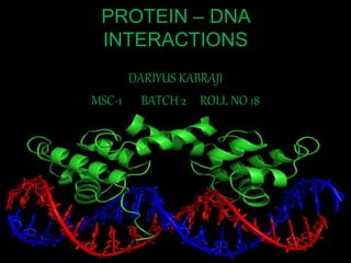
- 1. PROTEIN – DNA INTERACTIONS DARIYUS KABRAJI MSC-1 BATCH 2 ROLL NO 18
- 2. INTRODUCTION • In the late 19th century, scientists microscopically observed the association of proteins with DNA strands. • Since then, researchers have used a variety of procedures to demonstrate that proteins interact with DNA and RNA to influence the structure and function of the corresponding nucleic acid. • Protein–nucleic acid interactions therefore play a crucial role in central biological processes, ranging from the mechanism of replication, transcription and recombination to enzymatic events utilizing nucleic acids as substrates.
- 3. DNA BINDING PROTEIN • DNA-binding proteins (DBPs), such as transcription factors, constitute about 10% of the protein-coding genes in eukaryotic genomes and play pivotal roles in the regulation of chromatin structure and gene expression by binding to short stretches of DNA. • Sequencing of eukaryote genomes disclosed that about 10% of all genes encode potential DBPs. Hence, every higher plant or vertebrate genome harbours over 2000 of these DBP genes. • Despite their importance in many fundamental processes, e.g. during stress or disease, throughout development and in controlling yield or growth, our knowledge on this tremendous number of putative DBPs and their interaction with DNA is limited
- 5. INTERACTION TYPES SPECIFIC • The sequence of nucleotides directly affects the interaction outcome • Control transcription in prokaryotes and eukaryotes, mediated by hydrogen bonding, ionic interactions and Van der Waal’s forces NON SPECIFIC • The sequence of nucleotides does not matter, as far as the binding interactions are concerned • Histone (protein) - DNA interactions are an example of such interactions, and they occur between functional groups on the protein and the sugar-phosphate backbone of DNA
- 6. SPECIFIC INTERACTION • In humans, Replication protein A is the best-understood member of this family and is used in processes where the double helix is separated • The DNA binding proteins in these processes include transcription factors • These binding proteins seem to stabilize single-stranded DNA and protect it from forming stem-loops or being degraded by nucleases • This process is used in DNA replication, Transcription and repair
- 8. LEVELS OF SPECIFICITY The factors considered to assess the levels of specificity in a DNA- protein interaction are: • Site specification • Recognition • Affinity • Equilibrium selection
- 9. NON SPECIFIC INTERACTIONS • Within chromosomes, DNA is held in complexes with structural proteins. These proteins organize it into chromatin. In eukaryotes this structure involves DNA binding to a complex of small basic proteins (histones), while in prokaryotes multiple types of proteins are involved. • The histones form a disk-shaped complex nucleosome, which contains two turns of ds DNA wrapped around its surface. These non-specific interactions are formed through basic residues in the histones making ionic bonds to the acidic sugar-phosphate backbone of the DNA, and are independent of the base sequence. • Chemical modifications of these basic amino acid residues include methylation, phosphorylation and acetylation. These chemical changes alter the interaction strength & make the DNA more or less accessible to transcription factors and changing the rate of transcription
- 11. CHROMATIN REMODELLING • It s the modification of chromatin architecture to allow access of condensed genomic DNA to the regulatory transcription machinery proteins, and thus control gene expression • Two types: Covalent Histone Modifying Complexes & ATP dependant remodelling complexes • Histone Acetyl Transferases (HAT) bring about covalent histone modifications by binding themselves to the DNA • This is an important interaction since dynamic remodelling of chromatin imparts an epigenetic regulatory role in several key biological processes, and irregularities can result in inconveniences
- 12. PROCEDURE • Specific histone-modifying complexes catalyse the addition or deletion of various chemical elements on histones. • These enzymatic modifications include acetylation, methylation, phosphorylation, and ubiquitination and occur at the nucleosome • Such modifications affect the binding affinity between histones and DNA, and thus loosening and tightening the condensed DNA wrapped around histones • Observed by mass spectrometry (measures the mass-to- charge ratio and abundance of gas-phase ions)
- 13. PROCEDURE
- 14. DETECTION METHODS 1. Chromatin Immunoprecipitation (ChIP)Assays: • The ChIP method can be used to monitor transcriptional regulation through histone modification. • The ChIP assay method allows analysis of DNA-Protein interactions in living cells by treating the cells with formaldehyde to stabilize the interactions for detection. • It requires knowledge of the target protein and DNA sequence which will be analysed, to provide an antibody against the protein of interest to selectively precipitate the protein-DNA complex from the other genomic DNA fragments and protein-DNA complexes, which can be amplified by PCR.
- 15. DETECTION METHODS 2. DNA Electrophoretic Mobility Shift Assay (EMSA): • The EMSA studies proteins binding to known DNA oligonucleotide probes and assesses the specificity of the interaction. • The technique is based on the principle that protein-DNA complexes migrate more slowly than free DNA molecules when subjected to polyacrylamide or agarose gel electrophoresis. Because the rate of DNA migration is retarded upon protein binding, the assay is also called a gel retardation assay. • Adding a protein-specific antibody to the binding components creates an even larger complex (antibody-protein-DNA) which migrates even slower during electrophoresis, this is known as a supershift and can be used to confirm protein identities.
- 16. DETECTION METHODS 3. DNA Pull-down Assay: • Pull-down assays use a DNA probe labelled with a high affinity tag, such as biotin, which allows the probe to be recovered or immobilized. A DNA probe can be complexed with a protein from a cell lysate in a reaction similar to that used in the EMSA and then used to purify the complex using agarose or magnetic beads. • The proteins are then eluted from the DNA and detected by Western blot or identified by mass spectrometry. • Alternatively, the protein may be labelled with an affinity tag or the DNA-protein complex may be isolated using an antibody against the protein of interest (similar to a supershift assay). In this case, the unknown DNA sequence bound by the protein is detected by Southern blotting or through PCR analysis.
- 17. DETECTION METHODS 4. Reporter Assay: • Reporter assays provide a real-time in vivo read-out of translational activity for a promoter of interest. • Reporter genes are fusions of a target promoter DNA sequence and a reporter gene DNA sequence which is customized by the researcher and the DNA sequence codes for a protein with detectable properties like firefly /Renilla luciferase or alkaline phosphatase. These genes produce enzymes only when the promoter of interest is activated. • The enzyme, in turn, catalyses a substrate to produce either light or a colour change that can be detected by spectroscopic instrumentation. The signal from the reporter gene is used as an indirect determinant for the translation of endogenous proteins driven from the same promoter.
- 18. DETECTION METHODS 5. Microplate Capture and Detection Assays: • A mix of the pull down assay and ELISA, microplate capture assays use immobilized DNA probes to capture specific protein-DNA interactions and confirm protein identities and relative amounts with target specific antibodies. • Typically, a DNA probe is immobilized on the surface of 96- or 384-well microplates coated with streptavidin. A cellular extract is prepared and added to allow the binding protein to bind to the oligonucleotide. The extract is then removed and each well is washed several times to remove non-specifically bound proteins. • Finally, the protein is detected using a specific antibody labelled for detection. This method can be extremely sensitive , detecting less than 0.2pg of the target protein per well. This method may also be utilized for oligonucleotides labelled with other tags, such as primary amines that can be immobilized on microplates coated with an amine-reactive surface chemistry.
- 19. DETECTION METHODS 6. DNA Footprinting: • Footprinting is one of the most widely used methods for obtaining detailed information on the individual nucleotides in protein–DNA complexes, even inside living cells. In such an experiment, chemicals or enzymes are used to modify or digest the DNA molecules. • When sequence specific proteins bind to DNA they can protect the binding sites from modification or digestion. This can subsequently be visualized by denaturing gel electrophoresis, where unprotected DNA is cleaved more or less at random. • Therefore it appears as a ‘ladder’ of bands and the sites protected by proteins have no corresponding bands and look like foot prints in the pattern of bands. The foot prints there by identify specific nucleosides at the protein–DNA binding sites
- 20. CONCLUSION • The entire hierarchy of specificity criteria must be considered during examination of DNA - protein interactions. • There are several methods used to specifically detect the DNA binding proteins (DPB) and isolate them from the sample. Identifying these proteins and their manner of interaction with the nucleic acid sheds light on the genes expressed and the functions that arise from the expression • However, each method has their own limitations and new extraction methods are being tested for more sensitive and accurate results
- 21. FUTURE PROSPECTS Confirming the Functional Importance of a Protein–DNA Interaction: • Correlation between nucleotides required for protein binding and those required for activity of the control element • Trans-activation of a reporter gene or endogenous gene by over-expression of a DNA-binding protein • Cooperative binding and synergistic function of proteins bound to adjacent control elements • Comparison of genomic and in vitro Footprinting patterns