SlideShare ist ein Scribd-Unternehmen logo
1 von 42
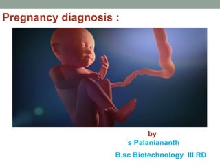
Pregnancy diagnosis :
by
s Palaniananth
B.sc Biotechnology III RD
Pregnancy:
 It can define as the condition of having a
developing a embryo or fetus in the body after
successful conception
Duration of pregnancy :
 Calculated in term of 10 lunar months /9
calender months and 7days  280 days 40 weeks.
 Calculated from 1st day of last menstrual
period
 Also called menstrual or gestational age
 First trimester : first 12 weeks
 Second trimester : 13 -28 weeks
 Last trimester : 29 -40 weeks
LIFE IN WOMB 9 MONTHS
PRESUMPTIVE SIGN :
 AMENORRHEA : ABSENCES OF MENSTRUATION MORE
THAN 3 MONTHS .
 MORNING SICKNESS : NAUSEA AND VOMITING .THESE
SYMPTOMES DISAPPER SPANTANEOUSLY FROM 6 TO 12
WEEKS (FIRST TRIMESTER)
 ENLARGEMENT OF BREASTS : INCREASED SIZE &SENSITIVITY
OF BREASTS AND NIPPLS INCREASED VASCULARITY AND
APPERANCE OF SUBCUTANEOUS VENIS.
 DARK PIGMENT OF THE NIPPLS AND AREOLA.
 SECRETION OF CLOSTRUM
 FREQUENCY OF MICTURITION :
 INCREASED DURING 8TH TO 12TH WEEK OF PREGNANCY.
RESTING OF BULKY UTERUS ON FUNDUS OF BLADDER
CONGESTION OF BLADDER MUCOSA
CHANGES IN MATERNAL OSMOREGULATION CAUSES
INCREASED THIST & POLYURIA .
Hyperpigmentation : MELASMA ,LINCANIGRA
Quickening : 12 to14 weeks women feel fetus moment movement
Probable evidence :
GOODELL’S SIGN :Tips of cervix become soft and swollen
CHADWICK’S SIGN: cervix and vagina a violet bluish colour
BALLOTTEMENT :its an bimanually evident from 16th week .
Uterine ‘s sign : The uterus become large in size ,globular in
shape and soft in consistence. By the end of third month the funds
of the uterus can be felt just above symphysis pubis.
Hegar ‘s sign: It is the sofening of isthmus of the uterus the area between the
cervix and body of uterus ,which occur at 6to 8 weeks of pregnancy. This area
may become so soft that on bimanual examination the anterior fornix finger and
abdominally fingers meet each other
HEGAR SIGN
Positive sign:
Fetal heart sounds : These are usually heard by the 24th
week. The presence is sue sign of pregnancy, their rate
of fetal heart sounds is between 120 to 140 under
normal condition.
Visualization of fetus : By ultra sound scanning.
HCG (HUMAN CHRONIC GONADATROPHIN )
Hormone found in blood and urine of pregnant women
*(HCG is an embryonic hormone that maintains secretion of progesterone and estrogen by
the uterus lining through the first trimester. The level of HCG in maternal blood is so high
that it is present in the urine, which allows it to be detected for pregnancy tests.)*
Prenatal diagnosis employs a variety of techniques to
determine the health and condition of an unborn fetus.
Without knowledge gained by prenatal diagnosis, there could
be an untoward outcome for the fetus or the mother or both.
Congenital anomalies account for 20 to 25% of perinatal
deaths. Specifically, prenatal diagnosis is helpful for:
 Managing the remaining weeks of the pregnancy
 Determining the outcome of the pregnancy
 Planning for possible complications with the birth
process
 Planning for problems that may occur in the newborn
infant
 Deciding whether to continue the pregnancy
 Finding conditions that may affect future pregnancies
There are a variety of non-invasive and invasive techniques
available for prenatal diagnosis. Each of them can be applied
only during specific time periods during the pregnancy for
greatest utility. The techniques employed for prenatal
diagnosis include:
 Ultrasonography
 Amniocentesis
 Chorionic villus sampling
 Fetal blood cells in maternal blood
 Maternal serum alpha-fetoprotein
 Maternal serum beta-HCG
 Maternal serum estriol
 Karyotyping
 Radiography
 Ultrasonography :
This is a non-invasive procedure that is harmless
to both the fetus and the mother. High frequency
sound waves are utilized to produce visible
images from the pattern of the echos made by
different tissues and organs, including the baby in
the amniotic cavity. The developing embryo can
first be visualized at about 6 weeks gestation.
Recognition of the major internal organs and
extremities to determine if any are abnormal can
best be accomplished between 16 to 20 weeks
gestation
o Although an ultrasound examination can be quite
useful to determine the size and position of the
fetus, the size and position of the placenta, the
amount of amniotic fluid, and the appearance of
fetal anatomy, there are limitations to this
procedure. Subtle abnormalities may not be
detected until later in pregnancy, or may not be
detected at all.
o A good example of this is Down syndrome
(trisomy 21) where the morphologic abnormalities
are often not marked, but only subtle, such as
nuchal thickening.
Ultrasonography
AMNIOCENTESIS :
This is an invasive procedure in which a needle is passed
through the mother's lower abdomen into the amniotic cavity
inside the uterus.
 Enough amniotic fluid is present for this to be
accomplished starting about 14 weeks gestation.
 For prenatal diagnosis, most amniocenteses are
performed between 14 and 20 weeks gestation. However,
an ultrasound examination always proceeds
amniocentesis in order to determine gestational age, the
position of the fetus and placenta, and determine if enough
amniotic fluid is present.
o Within the amniotic fluid are fetal cells (mostly derived
from fetal skin) which can be grown in culture for
chromosome analysis, biochemical analysis, and
molecular biologic analysis.
In the third trimester of pregnancy, the amniotic
fluid can be analyzed for determination of fetal lung
maturity.
 This is important when the fetus is below 35 to 36
weeks gestation, because the lungs may not be
mature enough to sustain life.
 This is because the lungs are not producing
enough surfactant. After birth, the infant will
develop respiratory distress syndrome from
hyaline membrane disease.
 The amniotic fluid can be analyzed by
fluorescence polarization (fpol), for
lecithin:sphingomyelin (LS) ration, or for
phosphatidyl glycerol (PG).
Risks with amniocentesis are uncommon, but include fetal
loss and maternal Rh sensitization.
 The increased risk for fetal mortality following
amniocentesis is about 0.5% above what would normally
be expected.
 Rh negative mothers can be treated with RhoGam.
Contamination of fluid from amniocentesis by maternal
cells is highly unlikely.
 If oligohydramnios is present, then amniotic fluid cannot
be obtained.
 It is sometimes possible to instill saline into the amniotic
cavity and then remove fluid for analysis
CHORIONIC VILLUS SAMPLING (CVS)
In this procedure, a catheter is passed via the vagina through
the cervix and into the uterus to the developing placenta
under ultrasound guidance.
 Alternative approaches are transvaginal and
transabdominal.
 The introduction of the catheter allows sampling of cells
from the placental chorionic villi.
 These cells can then be analyzed by a variety of
techniques.
 The most common test employed on cells obtained by CVS
is chromosome analysis to determine the karyotype of the
fetus.
 The cells can also be grown in culture for biochemical or
molecular biologic analysis. CVS can be safely performed
between 9.5 and 12.5 weeks gestation.
CVS has the disadvantage of being an invasive
procedure, and it has a small but significant rate of
morbidity for the fetus; this loss rate is about 0.5 to
1% higher than for women undergoing
amniocentesis.
 Rarely, CVS can be associated with limb defects
in the fetus. The possibility of maternal Rh
sensitization is present.
 There is also the possibility that maternal blood
cells in the developing placenta will be sampled
instead of fetal cells and confound chromosome
analysis
KARYOTYPING
 Tissues must be obtained as fresh as possible
for culture and without contamination.
 A useful procedure is to wash the tissue samples
in sterile saline prior to placing them into cell
culture media.
 Tissues with the best chance for growth are
those with the least maceration: placenta, lung,
diaphragm.
 Obtaining tissue from more than one site can
increase the yield by avoiding contamination or
by detection of mosaicism
DNA PROBES : (is single standard seqences that is complementary to know the region od dna)
 Fetal cells obtained via amniocentesis or CVS can be analyzed by
probes specific for DNA sequences.
 One method employs restriction fragment length polymorphism
(RFLP) analysis.
 This method is useful for detection of mutations involving genes that
are closely linked to the DNA restriction fragments generated by the
action of an endonuclease. The DNA of family members is analyzed to
determine differences by RFLP analysis.
 In some cases, if the DNA sequence of a gene is known, a probe to a
DNA sequence specific for a genetic marker is available, and the
polymerase chain reaction (PCR) technique can be applied for
diagnosis.
 There are many genetic diseases, but only in a minority have
particular genes been identified, and tests to detect them have been
developed in some of these. Thus, it is not possible to detect all
genetic diseases. Moreover, testing is confounded by the presence of
different mutations in the same gene, making testing more complex.
and then detect the matches by using x-ray flim
MATERNAL SERUM ESTRIOL
 The amount of estriol in maternal serum is dependent
upon a viable fetus, a properly functioning placenta, and
maternal well-being.
 The substrate for estriol begins as
dehydroepiandrosterone (DHEA) made by the fetal adrenal
glands. This is further metabolized in the placenta to
estriol.
 The estriol crosses to the maternal circulation and is
excreted by the maternal kidney in urine or by the
maternal liver in the bile. The measurement of serial estriol
levels in the third trimester will give an indication of
general wellbeing of the fetus.
 If the estriol level drops, then the fetus is threatened and
delivery may be necessary emergently.
 Estriol tends to be lower when Down syndrome is present
and when there is adrenal hypoplasia with anencephaly.22
FISH (PERFORMED ON FRESH TISSUE OR PARAFFIN
BLOCKS)
 In addition to karyotyping, fluorescence in situ
hybridization (FISH) can be useful. A wide variety of probes
are available. It is useful for detecting aneuploid conditions
(trisomies, monosomies).
 Fresh cells are desirable, but the method can be applied
even to fixed tissues stored in paraffin blocks, though
working with paraffin blocks is much more time consuming
and interpretation can be difficult. The ability to use FISH
on paraffin blocks means that archival tissues can be
examined in cases where karyotyping was not performed,
or cells didn't grow in culture.
RADIOGRAPHY
 Standard anterior-posterior and lateral radiographic views are essential
for analysis of the fetal skeleton. Radiographs are useful for
comparison with prenatal ultrasound, and help define anomalies when
autopsy consent is limited, or can help to determine sites to be
examined microscopically. Conditions diagnosed by postmortem
radiography may include:
 Skeletal anomalies (dwarfism, dysplasia, sirenomelia, etc.)
 Neural tube defects (anencephaly, iniencephaly, spina bifida, etc.)
 Osteogenesis imperfecta (osteopenia, fractures)
 Soft tissue changes (hydrops, hygroma, etc.)
 Teratomas or other neoplasms
 Growth retardation
 Orientation and audit of fetal parts (with D&E specimens)
 Assessment of catheter or therapeutic device placement
dysplasia
Radiograph
MICROBIOLOGIC CULTURE
 Culture can aid in diagnosis or confirmation of congenital
infections. Examples of congenital infection include:
 T - toxoplasmosis
 O - other, such as Listeria monocytogenes, group B
streptococcus, syphilis
 R - rubella
 C - cytomegalovirus
 H - herpes simplex or human immunodeficiency virus
(HIV)
 Cultures have to be appropriately obtained with the proper
media and sent with the proper requisitions ("routine"
includes aerobic and anaerobic bacteria; fungal and viral
cultures must be separately ordered).
 Viral cultures are difficult and expensive. Separate media
and collection procedures may be necessary depending
upon what virus is being sought.
 Bacterial contamination can be a problem
THANK YOU

Weitere ähnliche Inhalte

Was ist angesagt?

Chorionic villus sampling
Chorionic villus    samplingChorionic villus    sampling
Chorionic villus samplingHafiz M Waseem
 
Prenatal diagnosis of congenital anomalies 3
Prenatal diagnosis of congenital anomalies 3Prenatal diagnosis of congenital anomalies 3
Prenatal diagnosis of congenital anomalies 3DrAhmed Badr
 
Chorion biopsy ( Chorion Villus Sampling)
Chorion biopsy ( Chorion Villus Sampling)Chorion biopsy ( Chorion Villus Sampling)
Chorion biopsy ( Chorion Villus Sampling)Shreshtha Aggarwal
 
methods of Chromosomal Evaluation in Amniocentesis- Define, Time for test, C...
methods of Chromosomal Evaluation in Amniocentesis-  Define, Time for test, C...methods of Chromosomal Evaluation in Amniocentesis-  Define, Time for test, C...
methods of Chromosomal Evaluation in Amniocentesis- Define, Time for test, C...sonal patel
 
First trimester screening Down's
First trimester screening Down's First trimester screening Down's
First trimester screening Down's ajay dhawle
 
Prenatal diagnosis test
Prenatal diagnosis testPrenatal diagnosis test
Prenatal diagnosis testqussai abbas
 
Assessment of fetal wellbeing
Assessment of fetal wellbeingAssessment of fetal wellbeing
Assessment of fetal wellbeingShrooti Shah
 
• Rate of Miscarriage Following Chorionic Villus Sampling on 315 for Prenatal...
•	Rate of Miscarriage Following Chorionic Villus Sampling on 315 for Prenatal...•	Rate of Miscarriage Following Chorionic Villus Sampling on 315 for Prenatal...
• Rate of Miscarriage Following Chorionic Villus Sampling on 315 for Prenatal...Sujoy Dasgupta
 
Prenatal and preimplantation diagnosis 22.04.2021
Prenatal and preimplantation diagnosis 22.04.2021Prenatal and preimplantation diagnosis 22.04.2021
Prenatal and preimplantation diagnosis 22.04.2021Shazia Iqbal
 

Was ist angesagt? (20)

Genetic testing
Genetic testingGenetic testing
Genetic testing
 
Screening
ScreeningScreening
Screening
 
Second trimestric soft markers of aneuploidy
Second trimestric soft markers of aneuploidySecond trimestric soft markers of aneuploidy
Second trimestric soft markers of aneuploidy
 
Chorionic villus sampling
Chorionic villus    samplingChorionic villus    sampling
Chorionic villus sampling
 
Early diagnosis of pregnancy
Early diagnosis of pregnancyEarly diagnosis of pregnancy
Early diagnosis of pregnancy
 
Prenatal diagnosis of congenital anomalies 3
Prenatal diagnosis of congenital anomalies 3Prenatal diagnosis of congenital anomalies 3
Prenatal diagnosis of congenital anomalies 3
 
Chorion biopsy ( Chorion Villus Sampling)
Chorion biopsy ( Chorion Villus Sampling)Chorion biopsy ( Chorion Villus Sampling)
Chorion biopsy ( Chorion Villus Sampling)
 
methods of Chromosomal Evaluation in Amniocentesis- Define, Time for test, C...
methods of Chromosomal Evaluation in Amniocentesis-  Define, Time for test, C...methods of Chromosomal Evaluation in Amniocentesis-  Define, Time for test, C...
methods of Chromosomal Evaluation in Amniocentesis- Define, Time for test, C...
 
Fetal DNA in maternal serum
Fetal DNA in maternal serumFetal DNA in maternal serum
Fetal DNA in maternal serum
 
First trimester screening Down's
First trimester screening Down's First trimester screening Down's
First trimester screening Down's
 
Prenatal diagnosis
Prenatal diagnosisPrenatal diagnosis
Prenatal diagnosis
 
Prenatal diagnosis
Prenatal diagnosisPrenatal diagnosis
Prenatal diagnosis
 
Prenatal diagnosis test
Prenatal diagnosis testPrenatal diagnosis test
Prenatal diagnosis test
 
Ncm [Recovered]
Ncm [Recovered]Ncm [Recovered]
Ncm [Recovered]
 
Chorionic villus sampling
Chorionic villus samplingChorionic villus sampling
Chorionic villus sampling
 
Maternal screening in Pregnancy (Double & quadruple marker)
Maternal screening in Pregnancy (Double & quadruple marker)Maternal screening in Pregnancy (Double & quadruple marker)
Maternal screening in Pregnancy (Double & quadruple marker)
 
Assessment of fetal wellbeing
Assessment of fetal wellbeingAssessment of fetal wellbeing
Assessment of fetal wellbeing
 
• Rate of Miscarriage Following Chorionic Villus Sampling on 315 for Prenatal...
•	Rate of Miscarriage Following Chorionic Villus Sampling on 315 for Prenatal...•	Rate of Miscarriage Following Chorionic Villus Sampling on 315 for Prenatal...
• Rate of Miscarriage Following Chorionic Villus Sampling on 315 for Prenatal...
 
Genetic screening Dr.Padmesh
Genetic screening  Dr.PadmeshGenetic screening  Dr.Padmesh
Genetic screening Dr.Padmesh
 
Prenatal and preimplantation diagnosis 22.04.2021
Prenatal and preimplantation diagnosis 22.04.2021Prenatal and preimplantation diagnosis 22.04.2021
Prenatal and preimplantation diagnosis 22.04.2021
 

Ähnlich wie pregnancy &prenatal diagnosisi

Genetictesting 140305094745-phpapp01
Genetictesting 140305094745-phpapp01Genetictesting 140305094745-phpapp01
Genetictesting 140305094745-phpapp01t7260678
 
PRENATAL_DIAGNOSIS[1].pptx
PRENATAL_DIAGNOSIS[1].pptxPRENATAL_DIAGNOSIS[1].pptx
PRENATAL_DIAGNOSIS[1].pptxAnandSGiri
 
screening for down syndrome
screening for down syndromescreening for down syndrome
screening for down syndromeSadaf Khan
 
Seminar prenatal genetic screening
Seminar prenatal genetic screeningSeminar prenatal genetic screening
Seminar prenatal genetic screeningSreelasya Kakarla
 
Genetic screening..dr.padmesh
Genetic screening..dr.padmeshGenetic screening..dr.padmesh
Genetic screening..dr.padmeshv_padmesh
 
Matenal and fetal weebeing
Matenal and fetal weebeingMatenal and fetal weebeing
Matenal and fetal weebeingHarikrishnanR76
 
BIOCHEMICAL FOETAL MEASURES PPT.pptx
BIOCHEMICAL FOETAL MEASURES PPT.pptxBIOCHEMICAL FOETAL MEASURES PPT.pptx
BIOCHEMICAL FOETAL MEASURES PPT.pptxsunnyalvakharshandi
 
Sridhar prenatal diagnosis
Sridhar prenatal diagnosisSridhar prenatal diagnosis
Sridhar prenatal diagnosisSridhar Mulaka
 
genetic tests.pptx
genetic tests.pptxgenetic tests.pptx
genetic tests.pptxmkniranda
 
Practical 5 07
Practical 5 07Practical 5 07
Practical 5 07medik.cz
 
Ectopic Pregnancy.pptx
Ectopic Pregnancy.pptxEctopic Pregnancy.pptx
Ectopic Pregnancy.pptxAbdela8
 
Recurrent pregnancy loss.pptx
Recurrent pregnancy loss.pptxRecurrent pregnancy loss.pptx
Recurrent pregnancy loss.pptxPuiteaChhangte
 
PRENATAL DIAGNOSIS TECHNIQUES.pptx
PRENATAL DIAGNOSIS TECHNIQUES.pptxPRENATAL DIAGNOSIS TECHNIQUES.pptx
PRENATAL DIAGNOSIS TECHNIQUES.pptxssusera7617d
 
radiology.Obst lec 3 & 4.(dr.nasreen)
radiology.Obst lec 3 & 4.(dr.nasreen)radiology.Obst lec 3 & 4.(dr.nasreen)
radiology.Obst lec 3 & 4.(dr.nasreen)student
 
Radiology 5th year, 8th & 9th lectures (Dr. Nasrin Alatrushi)
Radiology 5th year, 8th & 9th lectures (Dr. Nasrin Alatrushi)Radiology 5th year, 8th & 9th lectures (Dr. Nasrin Alatrushi)
Radiology 5th year, 8th & 9th lectures (Dr. Nasrin Alatrushi)College of Medicine, Sulaymaniyah
 

Ähnlich wie pregnancy &prenatal diagnosisi (20)

Genetictesting 140305094745-phpapp01
Genetictesting 140305094745-phpapp01Genetictesting 140305094745-phpapp01
Genetictesting 140305094745-phpapp01
 
PRENATAL_DIAGNOSIS[1].pptx
PRENATAL_DIAGNOSIS[1].pptxPRENATAL_DIAGNOSIS[1].pptx
PRENATAL_DIAGNOSIS[1].pptx
 
screening for down syndrome
screening for down syndromescreening for down syndrome
screening for down syndrome
 
Seminar prenatal genetic screening
Seminar prenatal genetic screeningSeminar prenatal genetic screening
Seminar prenatal genetic screening
 
Fetal therapy
Fetal therapyFetal therapy
Fetal therapy
 
Genetic screening..dr.padmesh
Genetic screening..dr.padmeshGenetic screening..dr.padmesh
Genetic screening..dr.padmesh
 
Matenal and fetal weebeing
Matenal and fetal weebeingMatenal and fetal weebeing
Matenal and fetal weebeing
 
UNIT 3 GENETICS.pptx
UNIT 3 GENETICS.pptxUNIT 3 GENETICS.pptx
UNIT 3 GENETICS.pptx
 
Prenatal cytogenetic
Prenatal cytogenetic Prenatal cytogenetic
Prenatal cytogenetic
 
prenatal diagnosis.ppt..pptx
prenatal diagnosis.ppt..pptxprenatal diagnosis.ppt..pptx
prenatal diagnosis.ppt..pptx
 
BIOCHEMICAL FOETAL MEASURES PPT.pptx
BIOCHEMICAL FOETAL MEASURES PPT.pptxBIOCHEMICAL FOETAL MEASURES PPT.pptx
BIOCHEMICAL FOETAL MEASURES PPT.pptx
 
Sridhar prenatal diagnosis
Sridhar prenatal diagnosisSridhar prenatal diagnosis
Sridhar prenatal diagnosis
 
genetic tests.pptx
genetic tests.pptxgenetic tests.pptx
genetic tests.pptx
 
Biochemical markers of prenatal diagnosis
Biochemical markers of prenatal diagnosisBiochemical markers of prenatal diagnosis
Biochemical markers of prenatal diagnosis
 
Practical 5 07
Practical 5 07Practical 5 07
Practical 5 07
 
Ectopic Pregnancy.pptx
Ectopic Pregnancy.pptxEctopic Pregnancy.pptx
Ectopic Pregnancy.pptx
 
Recurrent pregnancy loss.pptx
Recurrent pregnancy loss.pptxRecurrent pregnancy loss.pptx
Recurrent pregnancy loss.pptx
 
PRENATAL DIAGNOSIS TECHNIQUES.pptx
PRENATAL DIAGNOSIS TECHNIQUES.pptxPRENATAL DIAGNOSIS TECHNIQUES.pptx
PRENATAL DIAGNOSIS TECHNIQUES.pptx
 
radiology.Obst lec 3 & 4.(dr.nasreen)
radiology.Obst lec 3 & 4.(dr.nasreen)radiology.Obst lec 3 & 4.(dr.nasreen)
radiology.Obst lec 3 & 4.(dr.nasreen)
 
Radiology 5th year, 8th & 9th lectures (Dr. Nasrin Alatrushi)
Radiology 5th year, 8th & 9th lectures (Dr. Nasrin Alatrushi)Radiology 5th year, 8th & 9th lectures (Dr. Nasrin Alatrushi)
Radiology 5th year, 8th & 9th lectures (Dr. Nasrin Alatrushi)
 

Mehr von PALANIANANTH.S

Host pathogen interactions
 Host pathogen interactions Host pathogen interactions
Host pathogen interactionsPALANIANANTH.S
 
Next generation sequencing
Next generation sequencingNext generation sequencing
Next generation sequencingPALANIANANTH.S
 
Plant disease and its importance
Plant disease and its importancePlant disease and its importance
Plant disease and its importancePALANIANANTH.S
 
Uv visible spectroscopy
Uv visible spectroscopyUv visible spectroscopy
Uv visible spectroscopyPALANIANANTH.S
 
color blindness and haemophilia
color blindness and haemophiliacolor blindness and haemophilia
color blindness and haemophiliaPALANIANANTH.S
 
Application recombinant dna technology
Application recombinant dna technologyApplication recombinant dna technology
Application recombinant dna technologyPALANIANANTH.S
 
Sterilization techniques .TYPES .MERTIES. AND DIMERTIES AND APPLICATION......
Sterilization techniques  .TYPES .MERTIES. AND DIMERTIES AND APPLICATION......Sterilization techniques  .TYPES .MERTIES. AND DIMERTIES AND APPLICATION......
Sterilization techniques .TYPES .MERTIES. AND DIMERTIES AND APPLICATION......PALANIANANTH.S
 

Mehr von PALANIANANTH.S (14)

Host pathogen interactions
 Host pathogen interactions Host pathogen interactions
Host pathogen interactions
 
Oriental foods
Oriental foodsOriental foods
Oriental foods
 
Gm crops
Gm cropsGm crops
Gm crops
 
Nitrogen fixation
Nitrogen fixationNitrogen fixation
Nitrogen fixation
 
Next generation sequencing
Next generation sequencingNext generation sequencing
Next generation sequencing
 
Autoimmunity
AutoimmunityAutoimmunity
Autoimmunity
 
Plant disease and its importance
Plant disease and its importancePlant disease and its importance
Plant disease and its importance
 
Uv visible spectroscopy
Uv visible spectroscopyUv visible spectroscopy
Uv visible spectroscopy
 
color blindness and haemophilia
color blindness and haemophiliacolor blindness and haemophilia
color blindness and haemophilia
 
Haptens
HaptensHaptens
Haptens
 
Archaebacteria
ArchaebacteriaArchaebacteria
Archaebacteria
 
Trp operon
Trp operonTrp operon
Trp operon
 
Application recombinant dna technology
Application recombinant dna technologyApplication recombinant dna technology
Application recombinant dna technology
 
Sterilization techniques .TYPES .MERTIES. AND DIMERTIES AND APPLICATION......
Sterilization techniques  .TYPES .MERTIES. AND DIMERTIES AND APPLICATION......Sterilization techniques  .TYPES .MERTIES. AND DIMERTIES AND APPLICATION......
Sterilization techniques .TYPES .MERTIES. AND DIMERTIES AND APPLICATION......
 

Kürzlich hochgeladen

❤Jammu Kashmir Call Girls 8617697112 Personal Whatsapp Number 💦✅.
❤Jammu Kashmir Call Girls 8617697112 Personal Whatsapp Number 💦✅.❤Jammu Kashmir Call Girls 8617697112 Personal Whatsapp Number 💦✅.
❤Jammu Kashmir Call Girls 8617697112 Personal Whatsapp Number 💦✅.Nitya salvi
 
Pests of mustard_Identification_Management_Dr.UPR.pdf
Pests of mustard_Identification_Management_Dr.UPR.pdfPests of mustard_Identification_Management_Dr.UPR.pdf
Pests of mustard_Identification_Management_Dr.UPR.pdfPirithiRaju
 
9654467111 Call Girls In Raj Nagar Delhi Short 1500 Night 6000
9654467111 Call Girls In Raj Nagar Delhi Short 1500 Night 60009654467111 Call Girls In Raj Nagar Delhi Short 1500 Night 6000
9654467111 Call Girls In Raj Nagar Delhi Short 1500 Night 6000Sapana Sha
 
Labelling Requirements and Label Claims for Dietary Supplements and Recommend...
Labelling Requirements and Label Claims for Dietary Supplements and Recommend...Labelling Requirements and Label Claims for Dietary Supplements and Recommend...
Labelling Requirements and Label Claims for Dietary Supplements and Recommend...Lokesh Kothari
 
VIRUSES structure and classification ppt by Dr.Prince C P
VIRUSES structure and classification ppt by Dr.Prince C PVIRUSES structure and classification ppt by Dr.Prince C P
VIRUSES structure and classification ppt by Dr.Prince C PPRINCE C P
 
Biological Classification BioHack (3).pdf
Biological Classification BioHack (3).pdfBiological Classification BioHack (3).pdf
Biological Classification BioHack (3).pdfmuntazimhurra
 
Physiochemical properties of nanomaterials and its nanotoxicity.pptx
Physiochemical properties of nanomaterials and its nanotoxicity.pptxPhysiochemical properties of nanomaterials and its nanotoxicity.pptx
Physiochemical properties of nanomaterials and its nanotoxicity.pptxAArockiyaNisha
 
Zoology 4th semester series (krishna).pdf
Zoology 4th semester series (krishna).pdfZoology 4th semester series (krishna).pdf
Zoology 4th semester series (krishna).pdfSumit Kumar yadav
 
Animal Communication- Auditory and Visual.pptx
Animal Communication- Auditory and Visual.pptxAnimal Communication- Auditory and Visual.pptx
Animal Communication- Auditory and Visual.pptxUmerFayaz5
 
Botany krishna series 2nd semester Only Mcq type questions
Botany krishna series 2nd semester Only Mcq type questionsBotany krishna series 2nd semester Only Mcq type questions
Botany krishna series 2nd semester Only Mcq type questionsSumit Kumar yadav
 
Spermiogenesis or Spermateleosis or metamorphosis of spermatid
Spermiogenesis or Spermateleosis or metamorphosis of spermatidSpermiogenesis or Spermateleosis or metamorphosis of spermatid
Spermiogenesis or Spermateleosis or metamorphosis of spermatidSarthak Sekhar Mondal
 
TEST BANK For Radiologic Science for Technologists, 12th Edition by Stewart C...
TEST BANK For Radiologic Science for Technologists, 12th Edition by Stewart C...TEST BANK For Radiologic Science for Technologists, 12th Edition by Stewart C...
TEST BANK For Radiologic Science for Technologists, 12th Edition by Stewart C...ssifa0344
 
Pests of cotton_Borer_Pests_Binomics_Dr.UPR.pdf
Pests of cotton_Borer_Pests_Binomics_Dr.UPR.pdfPests of cotton_Borer_Pests_Binomics_Dr.UPR.pdf
Pests of cotton_Borer_Pests_Binomics_Dr.UPR.pdfPirithiRaju
 
Presentation Vikram Lander by Vedansh Gupta.pptx
Presentation Vikram Lander by Vedansh Gupta.pptxPresentation Vikram Lander by Vedansh Gupta.pptx
Presentation Vikram Lander by Vedansh Gupta.pptxgindu3009
 
Lucknow 💋 Russian Call Girls Lucknow Finest Escorts Service 8923113531 Availa...
Lucknow 💋 Russian Call Girls Lucknow Finest Escorts Service 8923113531 Availa...Lucknow 💋 Russian Call Girls Lucknow Finest Escorts Service 8923113531 Availa...
Lucknow 💋 Russian Call Girls Lucknow Finest Escorts Service 8923113531 Availa...anilsa9823
 
Forensic Biology & Its biological significance.pdf
Forensic Biology & Its biological significance.pdfForensic Biology & Its biological significance.pdf
Forensic Biology & Its biological significance.pdfrohankumarsinghrore1
 
Unlocking the Potential: Deep dive into ocean of Ceramic Magnets.pptx
Unlocking  the Potential: Deep dive into ocean of Ceramic Magnets.pptxUnlocking  the Potential: Deep dive into ocean of Ceramic Magnets.pptx
Unlocking the Potential: Deep dive into ocean of Ceramic Magnets.pptxanandsmhk
 
Asymmetry in the atmosphere of the ultra-hot Jupiter WASP-76 b
Asymmetry in the atmosphere of the ultra-hot Jupiter WASP-76 bAsymmetry in the atmosphere of the ultra-hot Jupiter WASP-76 b
Asymmetry in the atmosphere of the ultra-hot Jupiter WASP-76 bSérgio Sacani
 
Nightside clouds and disequilibrium chemistry on the hot Jupiter WASP-43b
Nightside clouds and disequilibrium chemistry on the hot Jupiter WASP-43bNightside clouds and disequilibrium chemistry on the hot Jupiter WASP-43b
Nightside clouds and disequilibrium chemistry on the hot Jupiter WASP-43bSérgio Sacani
 
GBSN - Microbiology (Unit 1)
GBSN - Microbiology (Unit 1)GBSN - Microbiology (Unit 1)
GBSN - Microbiology (Unit 1)Areesha Ahmad
 

Kürzlich hochgeladen (20)

❤Jammu Kashmir Call Girls 8617697112 Personal Whatsapp Number 💦✅.
❤Jammu Kashmir Call Girls 8617697112 Personal Whatsapp Number 💦✅.❤Jammu Kashmir Call Girls 8617697112 Personal Whatsapp Number 💦✅.
❤Jammu Kashmir Call Girls 8617697112 Personal Whatsapp Number 💦✅.
 
Pests of mustard_Identification_Management_Dr.UPR.pdf
Pests of mustard_Identification_Management_Dr.UPR.pdfPests of mustard_Identification_Management_Dr.UPR.pdf
Pests of mustard_Identification_Management_Dr.UPR.pdf
 
9654467111 Call Girls In Raj Nagar Delhi Short 1500 Night 6000
9654467111 Call Girls In Raj Nagar Delhi Short 1500 Night 60009654467111 Call Girls In Raj Nagar Delhi Short 1500 Night 6000
9654467111 Call Girls In Raj Nagar Delhi Short 1500 Night 6000
 
Labelling Requirements and Label Claims for Dietary Supplements and Recommend...
Labelling Requirements and Label Claims for Dietary Supplements and Recommend...Labelling Requirements and Label Claims for Dietary Supplements and Recommend...
Labelling Requirements and Label Claims for Dietary Supplements and Recommend...
 
VIRUSES structure and classification ppt by Dr.Prince C P
VIRUSES structure and classification ppt by Dr.Prince C PVIRUSES structure and classification ppt by Dr.Prince C P
VIRUSES structure and classification ppt by Dr.Prince C P
 
Biological Classification BioHack (3).pdf
Biological Classification BioHack (3).pdfBiological Classification BioHack (3).pdf
Biological Classification BioHack (3).pdf
 
Physiochemical properties of nanomaterials and its nanotoxicity.pptx
Physiochemical properties of nanomaterials and its nanotoxicity.pptxPhysiochemical properties of nanomaterials and its nanotoxicity.pptx
Physiochemical properties of nanomaterials and its nanotoxicity.pptx
 
Zoology 4th semester series (krishna).pdf
Zoology 4th semester series (krishna).pdfZoology 4th semester series (krishna).pdf
Zoology 4th semester series (krishna).pdf
 
Animal Communication- Auditory and Visual.pptx
Animal Communication- Auditory and Visual.pptxAnimal Communication- Auditory and Visual.pptx
Animal Communication- Auditory and Visual.pptx
 
Botany krishna series 2nd semester Only Mcq type questions
Botany krishna series 2nd semester Only Mcq type questionsBotany krishna series 2nd semester Only Mcq type questions
Botany krishna series 2nd semester Only Mcq type questions
 
Spermiogenesis or Spermateleosis or metamorphosis of spermatid
Spermiogenesis or Spermateleosis or metamorphosis of spermatidSpermiogenesis or Spermateleosis or metamorphosis of spermatid
Spermiogenesis or Spermateleosis or metamorphosis of spermatid
 
TEST BANK For Radiologic Science for Technologists, 12th Edition by Stewart C...
TEST BANK For Radiologic Science for Technologists, 12th Edition by Stewart C...TEST BANK For Radiologic Science for Technologists, 12th Edition by Stewart C...
TEST BANK For Radiologic Science for Technologists, 12th Edition by Stewart C...
 
Pests of cotton_Borer_Pests_Binomics_Dr.UPR.pdf
Pests of cotton_Borer_Pests_Binomics_Dr.UPR.pdfPests of cotton_Borer_Pests_Binomics_Dr.UPR.pdf
Pests of cotton_Borer_Pests_Binomics_Dr.UPR.pdf
 
Presentation Vikram Lander by Vedansh Gupta.pptx
Presentation Vikram Lander by Vedansh Gupta.pptxPresentation Vikram Lander by Vedansh Gupta.pptx
Presentation Vikram Lander by Vedansh Gupta.pptx
 
Lucknow 💋 Russian Call Girls Lucknow Finest Escorts Service 8923113531 Availa...
Lucknow 💋 Russian Call Girls Lucknow Finest Escorts Service 8923113531 Availa...Lucknow 💋 Russian Call Girls Lucknow Finest Escorts Service 8923113531 Availa...
Lucknow 💋 Russian Call Girls Lucknow Finest Escorts Service 8923113531 Availa...
 
Forensic Biology & Its biological significance.pdf
Forensic Biology & Its biological significance.pdfForensic Biology & Its biological significance.pdf
Forensic Biology & Its biological significance.pdf
 
Unlocking the Potential: Deep dive into ocean of Ceramic Magnets.pptx
Unlocking  the Potential: Deep dive into ocean of Ceramic Magnets.pptxUnlocking  the Potential: Deep dive into ocean of Ceramic Magnets.pptx
Unlocking the Potential: Deep dive into ocean of Ceramic Magnets.pptx
 
Asymmetry in the atmosphere of the ultra-hot Jupiter WASP-76 b
Asymmetry in the atmosphere of the ultra-hot Jupiter WASP-76 bAsymmetry in the atmosphere of the ultra-hot Jupiter WASP-76 b
Asymmetry in the atmosphere of the ultra-hot Jupiter WASP-76 b
 
Nightside clouds and disequilibrium chemistry on the hot Jupiter WASP-43b
Nightside clouds and disequilibrium chemistry on the hot Jupiter WASP-43bNightside clouds and disequilibrium chemistry on the hot Jupiter WASP-43b
Nightside clouds and disequilibrium chemistry on the hot Jupiter WASP-43b
 
GBSN - Microbiology (Unit 1)
GBSN - Microbiology (Unit 1)GBSN - Microbiology (Unit 1)
GBSN - Microbiology (Unit 1)
 

pregnancy &prenatal diagnosisi

  • 1. Pregnancy diagnosis : by s Palaniananth B.sc Biotechnology III RD
  • 2. Pregnancy:  It can define as the condition of having a developing a embryo or fetus in the body after successful conception Duration of pregnancy :  Calculated in term of 10 lunar months /9 calender months and 7days 280 days 40 weeks.  Calculated from 1st day of last menstrual period  Also called menstrual or gestational age
  • 3.  First trimester : first 12 weeks  Second trimester : 13 -28 weeks  Last trimester : 29 -40 weeks
  • 4.
  • 5. LIFE IN WOMB 9 MONTHS
  • 6. PRESUMPTIVE SIGN :  AMENORRHEA : ABSENCES OF MENSTRUATION MORE THAN 3 MONTHS .  MORNING SICKNESS : NAUSEA AND VOMITING .THESE SYMPTOMES DISAPPER SPANTANEOUSLY FROM 6 TO 12 WEEKS (FIRST TRIMESTER)  ENLARGEMENT OF BREASTS : INCREASED SIZE &SENSITIVITY OF BREASTS AND NIPPLS INCREASED VASCULARITY AND APPERANCE OF SUBCUTANEOUS VENIS.  DARK PIGMENT OF THE NIPPLS AND AREOLA.  SECRETION OF CLOSTRUM  FREQUENCY OF MICTURITION :  INCREASED DURING 8TH TO 12TH WEEK OF PREGNANCY. RESTING OF BULKY UTERUS ON FUNDUS OF BLADDER CONGESTION OF BLADDER MUCOSA CHANGES IN MATERNAL OSMOREGULATION CAUSES INCREASED THIST & POLYURIA . Hyperpigmentation : MELASMA ,LINCANIGRA Quickening : 12 to14 weeks women feel fetus moment movement
  • 7. Probable evidence : GOODELL’S SIGN :Tips of cervix become soft and swollen CHADWICK’S SIGN: cervix and vagina a violet bluish colour BALLOTTEMENT :its an bimanually evident from 16th week . Uterine ‘s sign : The uterus become large in size ,globular in shape and soft in consistence. By the end of third month the funds of the uterus can be felt just above symphysis pubis. Hegar ‘s sign: It is the sofening of isthmus of the uterus the area between the cervix and body of uterus ,which occur at 6to 8 weeks of pregnancy. This area may become so soft that on bimanual examination the anterior fornix finger and abdominally fingers meet each other
  • 9. Positive sign: Fetal heart sounds : These are usually heard by the 24th week. The presence is sue sign of pregnancy, their rate of fetal heart sounds is between 120 to 140 under normal condition. Visualization of fetus : By ultra sound scanning. HCG (HUMAN CHRONIC GONADATROPHIN ) Hormone found in blood and urine of pregnant women *(HCG is an embryonic hormone that maintains secretion of progesterone and estrogen by the uterus lining through the first trimester. The level of HCG in maternal blood is so high that it is present in the urine, which allows it to be detected for pregnancy tests.)*
  • 10.
  • 11.
  • 12.
  • 13. Prenatal diagnosis employs a variety of techniques to determine the health and condition of an unborn fetus. Without knowledge gained by prenatal diagnosis, there could be an untoward outcome for the fetus or the mother or both. Congenital anomalies account for 20 to 25% of perinatal deaths. Specifically, prenatal diagnosis is helpful for:  Managing the remaining weeks of the pregnancy  Determining the outcome of the pregnancy  Planning for possible complications with the birth process  Planning for problems that may occur in the newborn infant  Deciding whether to continue the pregnancy  Finding conditions that may affect future pregnancies
  • 14. There are a variety of non-invasive and invasive techniques available for prenatal diagnosis. Each of them can be applied only during specific time periods during the pregnancy for greatest utility. The techniques employed for prenatal diagnosis include:  Ultrasonography  Amniocentesis  Chorionic villus sampling  Fetal blood cells in maternal blood  Maternal serum alpha-fetoprotein  Maternal serum beta-HCG  Maternal serum estriol  Karyotyping  Radiography
  • 15.  Ultrasonography : This is a non-invasive procedure that is harmless to both the fetus and the mother. High frequency sound waves are utilized to produce visible images from the pattern of the echos made by different tissues and organs, including the baby in the amniotic cavity. The developing embryo can first be visualized at about 6 weeks gestation. Recognition of the major internal organs and extremities to determine if any are abnormal can best be accomplished between 16 to 20 weeks gestation
  • 16. o Although an ultrasound examination can be quite useful to determine the size and position of the fetus, the size and position of the placenta, the amount of amniotic fluid, and the appearance of fetal anatomy, there are limitations to this procedure. Subtle abnormalities may not be detected until later in pregnancy, or may not be detected at all. o A good example of this is Down syndrome (trisomy 21) where the morphologic abnormalities are often not marked, but only subtle, such as nuchal thickening.
  • 18.
  • 19.
  • 20. AMNIOCENTESIS : This is an invasive procedure in which a needle is passed through the mother's lower abdomen into the amniotic cavity inside the uterus.  Enough amniotic fluid is present for this to be accomplished starting about 14 weeks gestation.  For prenatal diagnosis, most amniocenteses are performed between 14 and 20 weeks gestation. However, an ultrasound examination always proceeds amniocentesis in order to determine gestational age, the position of the fetus and placenta, and determine if enough amniotic fluid is present. o Within the amniotic fluid are fetal cells (mostly derived from fetal skin) which can be grown in culture for chromosome analysis, biochemical analysis, and molecular biologic analysis.
  • 21. In the third trimester of pregnancy, the amniotic fluid can be analyzed for determination of fetal lung maturity.  This is important when the fetus is below 35 to 36 weeks gestation, because the lungs may not be mature enough to sustain life.  This is because the lungs are not producing enough surfactant. After birth, the infant will develop respiratory distress syndrome from hyaline membrane disease.  The amniotic fluid can be analyzed by fluorescence polarization (fpol), for lecithin:sphingomyelin (LS) ration, or for phosphatidyl glycerol (PG).
  • 22. Risks with amniocentesis are uncommon, but include fetal loss and maternal Rh sensitization.  The increased risk for fetal mortality following amniocentesis is about 0.5% above what would normally be expected.  Rh negative mothers can be treated with RhoGam. Contamination of fluid from amniocentesis by maternal cells is highly unlikely.  If oligohydramnios is present, then amniotic fluid cannot be obtained.  It is sometimes possible to instill saline into the amniotic cavity and then remove fluid for analysis
  • 23.
  • 24. CHORIONIC VILLUS SAMPLING (CVS) In this procedure, a catheter is passed via the vagina through the cervix and into the uterus to the developing placenta under ultrasound guidance.  Alternative approaches are transvaginal and transabdominal.  The introduction of the catheter allows sampling of cells from the placental chorionic villi.  These cells can then be analyzed by a variety of techniques.  The most common test employed on cells obtained by CVS is chromosome analysis to determine the karyotype of the fetus.  The cells can also be grown in culture for biochemical or molecular biologic analysis. CVS can be safely performed between 9.5 and 12.5 weeks gestation.
  • 25. CVS has the disadvantage of being an invasive procedure, and it has a small but significant rate of morbidity for the fetus; this loss rate is about 0.5 to 1% higher than for women undergoing amniocentesis.  Rarely, CVS can be associated with limb defects in the fetus. The possibility of maternal Rh sensitization is present.  There is also the possibility that maternal blood cells in the developing placenta will be sampled instead of fetal cells and confound chromosome analysis
  • 26.
  • 27.
  • 28. KARYOTYPING  Tissues must be obtained as fresh as possible for culture and without contamination.  A useful procedure is to wash the tissue samples in sterile saline prior to placing them into cell culture media.  Tissues with the best chance for growth are those with the least maceration: placenta, lung, diaphragm.  Obtaining tissue from more than one site can increase the yield by avoiding contamination or by detection of mosaicism
  • 29.
  • 30.
  • 31. DNA PROBES : (is single standard seqences that is complementary to know the region od dna)  Fetal cells obtained via amniocentesis or CVS can be analyzed by probes specific for DNA sequences.  One method employs restriction fragment length polymorphism (RFLP) analysis.  This method is useful for detection of mutations involving genes that are closely linked to the DNA restriction fragments generated by the action of an endonuclease. The DNA of family members is analyzed to determine differences by RFLP analysis.  In some cases, if the DNA sequence of a gene is known, a probe to a DNA sequence specific for a genetic marker is available, and the polymerase chain reaction (PCR) technique can be applied for diagnosis.  There are many genetic diseases, but only in a minority have particular genes been identified, and tests to detect them have been developed in some of these. Thus, it is not possible to detect all genetic diseases. Moreover, testing is confounded by the presence of different mutations in the same gene, making testing more complex.
  • 32.
  • 33. and then detect the matches by using x-ray flim
  • 34. MATERNAL SERUM ESTRIOL  The amount of estriol in maternal serum is dependent upon a viable fetus, a properly functioning placenta, and maternal well-being.  The substrate for estriol begins as dehydroepiandrosterone (DHEA) made by the fetal adrenal glands. This is further metabolized in the placenta to estriol.  The estriol crosses to the maternal circulation and is excreted by the maternal kidney in urine or by the maternal liver in the bile. The measurement of serial estriol levels in the third trimester will give an indication of general wellbeing of the fetus.  If the estriol level drops, then the fetus is threatened and delivery may be necessary emergently.  Estriol tends to be lower when Down syndrome is present and when there is adrenal hypoplasia with anencephaly.22
  • 35. FISH (PERFORMED ON FRESH TISSUE OR PARAFFIN BLOCKS)  In addition to karyotyping, fluorescence in situ hybridization (FISH) can be useful. A wide variety of probes are available. It is useful for detecting aneuploid conditions (trisomies, monosomies).  Fresh cells are desirable, but the method can be applied even to fixed tissues stored in paraffin blocks, though working with paraffin blocks is much more time consuming and interpretation can be difficult. The ability to use FISH on paraffin blocks means that archival tissues can be examined in cases where karyotyping was not performed, or cells didn't grow in culture.
  • 36.
  • 37.
  • 38. RADIOGRAPHY  Standard anterior-posterior and lateral radiographic views are essential for analysis of the fetal skeleton. Radiographs are useful for comparison with prenatal ultrasound, and help define anomalies when autopsy consent is limited, or can help to determine sites to be examined microscopically. Conditions diagnosed by postmortem radiography may include:  Skeletal anomalies (dwarfism, dysplasia, sirenomelia, etc.)  Neural tube defects (anencephaly, iniencephaly, spina bifida, etc.)  Osteogenesis imperfecta (osteopenia, fractures)  Soft tissue changes (hydrops, hygroma, etc.)  Teratomas or other neoplasms  Growth retardation  Orientation and audit of fetal parts (with D&E specimens)  Assessment of catheter or therapeutic device placement
  • 41. MICROBIOLOGIC CULTURE  Culture can aid in diagnosis or confirmation of congenital infections. Examples of congenital infection include:  T - toxoplasmosis  O - other, such as Listeria monocytogenes, group B streptococcus, syphilis  R - rubella  C - cytomegalovirus  H - herpes simplex or human immunodeficiency virus (HIV)  Cultures have to be appropriately obtained with the proper media and sent with the proper requisitions ("routine" includes aerobic and anaerobic bacteria; fungal and viral cultures must be separately ordered).  Viral cultures are difficult and expensive. Separate media and collection procedures may be necessary depending upon what virus is being sought.  Bacterial contamination can be a problem