Comparative anatomy integumentary system
•Als PPTX, PDF herunterladen•
23 gefällt mir•11,225 views

This document provides information on the integumentary system of several chordates including humans, fish, birds, cattle/horses, and ruminants. It describes the basic structure of skin which consists of an epidermis, dermis, and hypodermis layers across these species. Key differences are highlighted such as fish having scales instead of hair and birds having feathers. The structure and function of hair, nails, hooves and horns are also reviewed for different animals.

Empfohlen
Weitere ähnliche Inhalte
Was ist angesagt?
Was ist angesagt? (20)
Ähnlich wie Comparative anatomy integumentary system
Ähnlich wie Comparative anatomy integumentary system (20)
Kürzlich hochgeladen
Kürzlich hochgeladen (20)
Comparative anatomy integumentary system
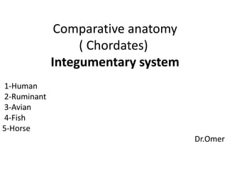
- 1. Comparative anatomy ( Chordates) Integumentary system 1-Human 2-Ruminant 3-Avian 4-Fish 5-Horse Dr.Omer
- 3. Human integumentary system The Integument Is the largest system of the body The integument is made up of two parts: 1. Cutaneous membrane a. Epidermis– Superficial epithelium b. Dermis – underlying CT with blood supply 2. Accessory structures a. Hair b. Nails c. Exocrine Glands
- 4. 5-4 Functions of Skin 1-Protection 2-Prevention of water loss 3-Temperature regulation 4-Metabolic regulation 5-Immune defense 6-Sensory reception 7-Excretion
- 5. STRUCTURE OF SKIN The layers of the skin:- The Epidermis Epithelial tissue Dermis Dense connective tissue proper – irregular Hypodermis Subcutaneous tissue- loose connective tissue proper and adipose tissue
- 7. Skin Structure: Epidermis The Epidermis Is a vascular stratified squamous epithelium Nutrients and oxygen diffuse from capillaries in the dermis There are four or five layers of the epidermis, depending upon the degree of friction and mechanical pressure applied to the skin
- 8. Four Principle Cells of the Epidermis • Keratinocytes Produce the protein keratin, which helps protect the skin and underlying tissue from heat, microbes, and chemicals, and lamellar granules, which release a waterproof sealant • Melanocytes Produce the pigment melanin which contributes to skin color and absorbs damaging ultraviolet (UV) light • Langerhans cells – Derived from bone marrow – Participate in immune response • Merkel cells – contact a sensory structure called a tactile (Merkel) disc and function in the sensation of touch 8
- 10. Skin Structure: Epidermis Thin Skin Covers most of the body Has four layers of keratinocytes Thick Skin Covers the palms of the hands and soles of the feet Has five layers of keratinocytes
- 11. Skin Structure: Epidermis Structures of the Epidermis The five strata of keratinocytes in thick skin From basal lamina to free surface 1. Stratum basale 2. Stratum spinosum 3. Stratum granulosum 4. Stratum lucidum 5. Stratum corneum
- 12. Epidermal Layers Stratum corneum - nuclei and organelles are destroyed by lysosomes and the cells fill with keratin Stratum lucidum - only found in the palms and soles of feet 3-5 layers of clear, flat, dead keratinocytes -Dense packed intermediate filaments thick plasma membranes Stratum granulosum - cells start to become keritanized --Secretes lipid-rich secretion that acts as a water sealant Stratum spinosum - 8-10 layers of keratinocytes skin both strength and flexibility Stratum basale - Also referred to as stratum germinatum -where new cells are formed -
- 13. Thick skin LM 210 Surface Stratum corneum Stratum lucidum Stratum granulosum Stratum spinosum Stratum basale Basement membrane DermisPapillary layer of dermis E P I D E R M I S
- 14. Sources of Skin Color • Most significant factor is melanin • Black or brown color due to eumelanin produced by melanocytes and transferred to keratinocytes in follicle. • Red color produced by pigment called pheomelanin • Blonde color produced by intermediate levels of pheomelanin and low levels of eumelanin • Other factors include hemoglobin (red to pink) and carotene (yellow) • Different races have the same of melanocytes, but amount of melanin produced differs • Melanin accumulates in keratinocytes Copyright © 2007 Pearson Education, Inc., publishing as Benjamin Cummings
- 15. Skin Structure: Dermis The Dermis Located between epidermis and subcutaneous layer Anchors epidermal accessory structures (hair follicles, sweat glands) Two components 1. Outer papillary layer 2. Deep reticular layer Dermis
- 16. Skin Structure: Dermis The Papillary Layer Consists of areolar tissue Contains smaller capillaries, lymphatics, and sensory neurons Has dermal papillae projecting between epidermal ridges The Reticular Layer Consists of dense irregular connective tissue Contains larger blood vessels, lymphatic vessels, and nerve fibers Contains collagen and elastic fibers Contains connective tissue proper
- 18. Skin Structure: Hypodermis The Hypodermis (Subcutaneous Layer) Lies below the integument Stabilizes the skin Allows separate movement Made of elastic areolar and adipose tissues Connected to the reticular layer of integument by connective tissue fibers
- 22. Functions – Hair & Nails • Functions of Hair – Hair on the head protects scalp from injury and sunlight – Eyelashes and eyebrows protect eyes – Nostril and ear hairs protect from foreign particles – Help in sensing light touch due to the touch receptors associated with the hair root plexuses. • Functions of the Nails – Grasping objects – Manipulating objects – Protects ends of digits from trauma – Scratching
- 23. Hair Anatomy Shaft: portion of hair that projects from skin surface Root: portion of hair deep to the shaft penetrating the dermis •Has 3 layers: medulla : Core, dead cells contain soft keratin and air to provide flexible Cortex: Middle layer, dead cells contain hard keratin to provide stiffness Cuticle : Outermost, overlapping dead keratinized cells form shiny surface Base of the hair follicle •Bulb: houses the papilla which contains the blood vessels that nourishes the growing hair follicle. •Matrix: responsible for hair growth and produces new hair Arrector pili: smooth muscle •Extends from the dermis to the side of hair follicle. Hair root plexus - dendrites of neurons which are sensitive to touch
- 24. Anatomy of the hair
- 25. Nails • Made of tightly packed, hard, keratinized epidermal cells • Consist of: Nail body: portion of the nail that is visible- Free edge: part that exten Nail root: the distal end of the digit portion buried in a fold of skin Lunula: means little moon - Crescent shaped area of the nail Hyponychium: secures the nail to the fingertip -Thickened stratum corneum Eponychium or cuticle: narrow band of epidermis-Growth of nails is in the nail matrix.
- 27. Aging Skin •In our 20s, the effects of aging begin to be visible in the skin. •Stem cell activity declines: skin thin, repair difficult •Epidermal dendritic cells decrease: reduced immune response •Vitamin D3 production declines: calcium absorption declines and brittle bones •Glandular activity declines: skin dries, body can overheat •Blood supply to dermis declines: tend to feel cold •Hair follicles die or produce thinner hair •Dermis thins and becomes less elastic – wrinkles
- 28. Fish integumentary system Fish like other vertebrates have Three layers of skin 1-Outer epidermis 2- Inner dermis. 3-Hypodermis • Multicellular glands also occur in some fishes, e.g. electric organs of eels and electric rays, • luminescent glands in deep sea fishes. •Mucous glands are all unicellular. • Important modifications of skin are dermal scales that cover the body for protection.
- 32. Epidermis • Generally the epiderm of vertebrates consists of five layers • stratum corneum In fish keratin is replaced with mucous ( glycoprotein = mucin)
- 33. Dermis • Thicker than epidermis • Contains blood vessels, nerves, sense organs, connective tissue and pigment cells Dermis consists of two layers 1- Upper looser layer - Stratum spongioso in this layer Scales are imbeded with their bases 2- Lower tighter layer ‐ Stratum compactum contains lipid cells and connective tissue • Fish dermis connects directly onto myosepta of the muscles and to the caudal fin.
- 34. Colors of the skin • Melanophores – Black • Erythrophores – Red • Xanthaphores – Yellow • Cyanophores – blue • Leucophores – light scattering • Iridiophores – light reflecting ‐ silver
- 35. Avian intgumentary system • Birds possess thin skin that is loosely attached to body to allow free movement of wings during flight. • Feathers are characteristic modifications of bird skin, which not only cover the entire body but also help in flight. The skin is composed of three layers : 1-The Epidermis 2-The dermis or corium 3-The hypodermis .
- 36. The fowl’s skin is divided into a number of separate areas These areas are: • The feathered skin. • The scale covered skin on the lower legs and feet. • The hard, horny areas of the beak and toenails. • The pad of the foot (or plantar). • The skin of the comb and wattles • The areas where they do grow are called pterylae • The areas where they do not grow are called apteria.
- 38. Glands • Birds do not possess sweat glands. • The uropygial gland secretes an oil (preen oil) bird will typically transfer preen oil to its body during preening by rubbing its beak • Feathers are an extremely efficient insulator. • Heat is lost from the respiratory tract and by radiation from featherless surfaces. • The feet and legs of starlings loose a substantial amount of the body’s heat. • Heat loss at night is also a probable reason for sleeping with their head (really their beak) under their wing, and one leg raised.
- 39. cattle and Horse • The skin of animals consists of three layers, • The epidermis • The dermis. • Hypodermis • Epidermis of thick keratinized stratified squamous epithelium • Special keratinized features include hair, nails, claws, horns, hooves. • Numerous multicellular glands • Chromophores present
- 40. Hoofs and Horns • True horns are made of keratin and are found in sheep, goats and cattle. • They consist of a core of bone arising in the dermis of the skin and are fused with the skull. • Hoofs are found in sheep, cows, horses . • These are animals that have lost toes in the process of evolution and walk on the “nails” of the remaining toes. • The hoof is a cylinder of horny ,material that surrounds and protects the tip of the toe
- 41. 1. Heel bulb 2. Periople at the heel 3. Heel 4. Quarter 5. Toe 6. Periople 7. Coronary band