Hyoid bone
•Als PPTX, PDF herunterladen•
20 gefällt mir•10,988 views

forensic medicine

Empfohlen
Weitere ähnliche Inhalte
Was ist angesagt?
Was ist angesagt? (20)
Ähnlich wie Hyoid bone
Ähnlich wie Hyoid bone (20)
Mehr von Dr Nikita Prabhakaran
Mehr von Dr Nikita Prabhakaran (20)
Kürzlich hochgeladen
Kürzlich hochgeladen (20)
Hyoid bone
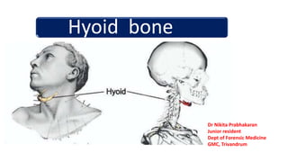
- 1. Hyoid bone Dr Nikita Prabhakaran Junior resident Dept of Forensic Medicine GMC, Trivandrum
- 2. Name • Shape: “U” shaped • Letter ‘upsilon’ in Greek ~hyoideus
- 4. Anatomy • Horizontally • Superior to the larynx • At the level of C 3
- 7. •Independent •Interface floor of the oral cavity larynx pharynx
- 8. Parts • Horizontal body • Lesser horns • Greater horns
- 10. Surface marking 3 cm below angle of mandible 1.5 cm outer to midline Connecting a line 3 cm 1.5 cm
- 11. Joints • Fibrous joint • Sometimes with synovium • Gets calcified only > 40 yrs • Often confused with fractures
- 12. Development • 2nd pharyngeal arch~ 7th • 3rd pharyngeal arch ~9th
- 13. Ossification centres • 2 centres for the body • 4 centres for each cornua • Greater cornua complete ossfn(20-30 yrs) • Fuses completely with the body at 40-60 yrs 10 m IU 20-30 yrs 16 yrs Just after birth
- 14. Calcification • Young age – cartilagenous • joints mobile • >30 yrs - ends of the horns calcify • becomes more brittle • hence <30yrs - less chance for # • ↓ common in females – calcifies only at old age
- 15. Muscle attachments • Body upper border • anterior surface • posterior border • Greater horn medial surface • lateral surface • • Lesser horn
- 16. Upper border of the body
- 17. • Anterior surface-body • Geniohyoid • Mylohyoid • hyoglossus
- 18. Lower border of the body
- 19. Greater Cornua: Medial border Digastric pulley Middle constrictor Lateral border Thyrohyoid Lesser Cornua: stylohyoid
- 20. Ligaments attached • Stylohyoid ligament Lesser horn • Thyrohoid memberane • Hyoepiglottic ligament Medial surface of greater horn
- 21. Hyoid fractures According to Displacement of fractured ends • Inward / lateral compression # • Outward (AP) compression # • One side inward & other side outward Mechanism of fracture • Direct pressure: outward • inward • Avulsion
- 22. Inward /lateral compression fractures • Force- inward • Eg: throttling • Fingers of the grasping hands squeeze the greater horns posterior fragment – displaced inwards • U/l or B/L • Periosteum~ torn outer side • Fragment can be seen lying medially
- 25. Outward /Anteroposterior compression # • Force – inward • Eg : hanging /ligature strangulation • Hyoid forced backwards • Fixed on to the vertebrae • ↑ divergence of the greater horns • Periosteum – torn on inner side • DD: RTA runover ( multiple #) • blows on the front ~ other signs
- 26. One side inward and other side outward • During violent neck violence • Backwards and sideways • One end gets caught up between the paravertebral structures • So that end- inward # • Other –outward #
- 27. According to the mechanism of fracture • Direct pressure ~ outward • inward • Avulsion ~ muscular stretch or overactivity • usually in hanging • hyoid is drawn up and held rigid • sudden suspn~ downward displacement of TC • traction through TH lig • usually outward
- 28. Demonstration of the fracture • Palpatory method ~ • body held stable in one hand • distal fragment between index and thumb • assess its mobility • antemortem ~ infiltration Advanced putrefaction hard to determine the type periosteum is completely destroyed
- 29. • Tests • 1% tolidene blue • Leave for 15 sec • Clean with water • Under stereomicroscope: • # end stains blue
- 30. Semi-microradiography • Method to visualise • Soft tissue structures of neck • To diagnose microtrauma Focal spot 1*1 mm 125 cm special photoelectronic printers
- 32. X ray and CT ~ best &most certain not feasible
- 33. Medicolegal importance • Hanging • Strangulation • Throttling • Blow to the neck • RTA ~ runover injuries • Identification~ skeletal remians
- 34. Case study •35 year old man •h/o strangulation * 12 days back •Persistent neck pain •Tenderness+++ •USG and X-ray WNL
- 35. •Flexible nasal endoscopy: right vocal cord hematoma •CT was advised •???
- 37. Thank you
- 38. References: 1. Krogman, W. M. and Iscan, M. Y. Human Skeleton in Forensic Medicine. 2nd Edition, Charles C. Thomas, Springfield, 1986. 2. Langmanns textbook of osteology 3. Poddar’s handbook of osteology, 13 th edition 4. Grays anatomy for students 3rd edition 5. Gradwohl’s legal medicine 6. Guharaj’s forensic medicine 7. Anil Aggarwal’s textbook of forensic medicine 8. B Umadathen’s textbook of forensic medicine 9. Essentials of forensic medicine by KS Narayana Reddy