2. PRACTICE
434 BRITISH DENTAL JOURNAL VOLUME 195 NO. 8 OCTOBER 25 2003
3. To eliminate occlusion that could damage the
long-term health of the teeth and periodontium
DENTO FACIAL APPEARANCE
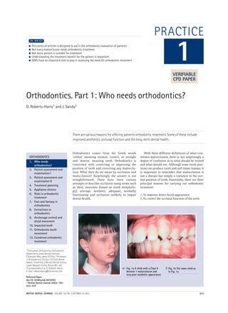
Improving the appearance of the teeth is without
question the main reason why most orthodontic
treatment is undertaken. Although it might be
tempting to dismiss this as a trivial need, there is
little doubt that a poor dental appearance can
have a profound psychosocial effect on children.
Figure 1 illustrates such a case with a child who
has a substantial aesthetic need for treatment.
The case is shown before (Fig. 1a, b) and after
(Fig 2a, b) orthodontic treatment. Few would
question that there has been an improvement in
both the dental and facial appearance of this
child. Indeed, orthodontic treatment can have a
beneficial psychosocial effect. For example
Shaw et al.1 found that children were teased
more about their teeth than anything else, such
as the clothes they wear or their weight and
height (Table 1).
OCCLUSAL FUNCTION
Teeth, which do not occlude properly, can make
eating difficult and may predispose to temporo-
mandibular joint (TMJ) dysfunction. However,
the association with TMJ dysfunction and mal-
occlusion is a controversial subject and will be
discussed in more detail in a later section. Indi-
viduals who have poor occlusion, such as shown
in Figure 3, may find it difficult and embarrass-
ing to eat because they cannot bite through food
using their incisors. They can only chew food
using their posterior teeth.
DENTAL HEALTH
Surprisingly there is no strong association
between dental irregularity and dental caries or
periodontal disease. It seems that dietary factors
are much more important than the alignment of
the teeth in the aetiology of caries. Although
straight teeth may be easier to clean than
crooked ones, patient motivation and dental
Table 1 Features children most dislike or are teased
about (Shaw et al.1)
Feature Disliked appearance
or teased (%)
Teeth 60.7
Clothes 53.8
Ears 51.7
Weight 41.5
Brace 33.3
Nose 29.3
Height 25.3
Fig. 2a Same child as in
Fig. 1 after orthodontic
treatment
Fig. 2b Occlusion of the same
patient as in Fig. 2a, there has
been a significant
improvement in the buccal
segment relation and overjet
compared with the initial
presentation in Fig. 1b
➠
➠
Fig. 3 This patient
has a severe
anterior open bite
with contact only
on the molars
Fig. 4 Class II
Division 1 with an
increased overjet.
The anterior teeth
are at risk of
potential trauma
with an overjet of
10 mm or greater
3. PRACTICE
BRITISH DENTAL JOURNAL VOLUME 195 NO. 8 OCTOBER 25 2003 435
hygiene seems to be the overriding factor in pre-
venting gingivitis and periodontitis. That said,
few of the studies that have investigated the link
between crowding and periodontal disease have
been longitudinal, over a long term and included
older adults. It would appear that aligned teeth
confer no benefit to those who clean their teeth
well because they can keep their teeth clean
regardless of any irregularity. Similarly, align-
ment will not help bad brushers. If there is poor
tooth brushing, periodontal diseases will devel-
op no matter how straight the teeth are. Howev-
er, having straight teeth may help moderate
brushers, although there is no firm evidence to
support or refute this statement. This is an area
that requires further study.
Some malocclusions may damage both the
teeth and soft tissues if they are left untreated. It is
well known that the more prominent the upper
incisors are the more prone they are to trauma2,3
(Table 2).
When the overjet is 9 mm or more the risk of
damaging the upper incisors increases to over
40%. Reducing a large overjet is not only bene-
ficial from an aesthetic point of view but min-
imises the risk of trauma and long-term com-
plications to the dentition. Fig. 4 shows a child
with a large overjet and it is not difficult to
imagine the likely dental trauma that would
result if he or she fell over.
Certain other occlusal relationships are also
liable to cause long-term problems. Figure 5a
and b show a case where there is an anterior
cross-bite with an associated mandibular dis-
placement in a 60-year-old man. The constant
attrition of the lower incisors against the upper
when the patient bites together, have produced
some substantial wear. If allowed to continue
then the long-term prognosis for these teeth is
extremely poor. In order to preserve the teeth,
the patient accepted fixed appliance treatment
that eliminated the cross bite and helped prevent
further wear Figure 5c and d.
Another example of problems caused by an
anterior cross bite is shown in Figure 6. A trau-
matic anterior occlusion produced a displacing
force on the lower incisors with apical migration
of the gingival attachment as a consequence. Pro-
vided this situation is remedied early (Fig. 7) the
soft tissue damage stops and as the rest of the
gingivae matures the situation often resolves
Table 2 Relation between size of overjet and
prevalence of traumatised anterior teeth
Overjet (mm) Incidence %
5 22
9 24
> 9 44
Fig. 5a Anterior crossbite in a 60-year-old man occluding
in the intercuspal position
Fig. 5b Shows the retruded contact position of the patient.
To reach full intercuspation the mandible displaces forward
and this movement is probably associated with the wear on
the incisors
Fig. 5c The patient in fixed appliances in order to
correct the displacement and the position of his upper
anterior teeth
Fig. 5d After correction and space reorganisation the patient
is wearing a prosthesis to replace the missing lateral incisors
4. PRACTICE
436 BRITISH DENTAL JOURNAL VOLUME 195 NO. 8 OCTOBER 25 2003
spontaneously and no long-term problems usu-
ally develop.
Deep overbites can occasionally cause strip-
ping of the soft tissues as shown in Figure 8a
and b. This is a case where there is little aes-
thetic need for treatment but because of the
deep overbite there is substantial damage to
the soft tissues. Clearly if this is allowed to
continue there is a risk of early loss of the
lower incisors that would produce a difficult
restorative problem.
WHO SHOULD BE TREATED?
Dental irregularity alone is not an indication for
treatment. Most orthodontic treatment is carried
out for aesthetic reasons and the benefit an indi-
vidual will receive from this will depend on the
severity of the presenting malocclusion as well as
the patients own perception of the problem.
Some individuals can have a marked degree of
dento-facial deformity and be unconcerned with
their appearance. Although a practitioner may
suggest treatment for such an individual,
patients should not be talked into treatment and
must be left to make the final decision them-
selves. Mild malocclusions should be treated with
caution. Not only will the net improvement in the
appearance of the teeth be small, but also as
nearly all teeth move to some degree after ortho-
dontic treatment the risk of relapse in these cases
is high. Whilst minor movements after the cor-
rection of severe malocclusions will still produce
a substantial net overall improvement for the
patients, the same is not true of minor problems.
Many practitioners will have encountered the
parent who can spot a 5-degree rotation of an
upper lateral incisor from fifty metres and is con-
vinced this will be the social death of their child.
Regardless of how insistent the parent or child is,
the practitioner should approach such problems
Fig. 6 A traumatic
anterior occlusion
is displacing the
lower right central
incisor labially and
there is an
associated
dehiscence
Fig. 7 The same
patient as in Fig. 6,
but the cross bite
has been corrected
with a removable
appliance and
there has been
an improvement
in the gingival
condition
Fig. 8a This malocclusion has an extremely deep bite which
can be associated with potential periodontal problems
Fig. 8b The same patient as in Fig. 8a, but not in
occlusion. The deep bite has resulted in labial stripping
of the periodontium on the lower right central incisor
Table 3 Index of Treatment Need
Dental health component Treatment need
1 No need
2 Little need
3 Moderate need
4 Great need
5 Very great need
Aesthetic component Treatment need
1
2 Little need
3
4
5
6 Moderate need
7
8
9 Great need
10
5. PRACTICE
BRITISH DENTAL JOURNAL VOLUME 195 NO. 8 OCTOBER 25 2003 437
with care and only carry out the treatment if it is in
the best interests of the patient. It is essential that
the patient and parent are fully aware of the limi-
tations of treatment and that long term, ie perma-
nent retention is currently the only way to ensure
long-term alignment of the teeth.
In order to assess the need for orthodontic
treatment, various indices have been developed.
The one used most commonly in the United King-
dom is the Index of Orthodontic Treatment Need
(IOTN).4 This index attempts to rank malocclu-
sion, in order, from worst to best. It comprises two
parts, an aesthetic component and a dental health
component (Table 3). The aesthetic component
consists of a series of ten photographs ranging
from most to least attractive. The idea is to match
the patient’s malocclusion as closely as possible
with one of the photographs. It is unlikely that a
perfect match will be found but the practitioner
should use his or her best guess to match to the
nearest equivalent photograph. The dental health
component consists of a series of occlusal traits
that could affect the long-term dental health of
the teeth. Various features are graded from 1–5
(least severe — worst). The worst feature of the
presenting malocclusion is matched to the list and
given the appropriate score.
Many hospital orthodontic services will not
accept patients in categories 1–3 of the dental
health component or grade 6 or less of the aes-
thetic component of the IOTN unless they are suit-
able for undergraduate teaching purposes.
Whilst the IOTN is a useful guide in prioritising
treatment and determining treatment need it
takes no account of the degree of treatment diffi-
culty. For example, class II division 2 malocclu-
sions are notoriously difficult to treat yet they
might have a low IOTN. Figure 9 illustrates such a
case. The IOTN of this patient is only 2 but it is a
difficult case to manage and treatment requires
a high level of expertise.
1. Shaw W C, Meek S C, Jones D S. Nicknames, teasing,
harassment and the salience of dental features among school
children.BrJOrthod 1980; 7: 75-80.
2. Office of Population Censuses and Surveys (1994). Children’s
dentalhealthintheUnitedKingdom1993.London: HMSO
0116916079.
3. Office of Population Censuses and Surveys (1985).Children’s
dentalhealthintheUnitedKingdom1983. London: HMSO
0116911360.
4. Brook P, Shaw W C. The development of an index of
orthodontic treatment priority. EurJOrthod 1989; 11:
309-320.
Fig. 9 The Index of
Treatment Need
for this patient
is 2. Although this
is low, the level of
expertise required
to treat it is high
BDA Information Centre Services
Did you know?
• As a BDA member you can gain
access to one of the best dental infor-
mation services in the world
• You don’t have to be based in London
to use the service
• You can borrow books, videos and
information packages
• You can borrow up to eight items via
the postal system
The only cost to you is the cost of the
return postage. If you’re not sure
what to request then telephone us and
we can advise you.
• You are entitled to free MEDLINE searches
Telephone us with a subject and we
will send you a list of relevant refer-
ences with abstracts.
• You can request photocopies of journal
articles
There is a small charge for this service
and you need to fill in a Photocopy
Request Form first. Telephone us if
you would like one of these forms.
• BDA Members can view the latest
Current Dental Titles on our web site
free of charge.
These are Medline-based lists of refer-
ences on eight areas of dentistry which
are available to BDA members only on
the web site and which are updated
twice yearly. Just use your password
with which you have been issued.
For further details of any of these services
dial 020 7563 4545.
Contact us via e-mail at:
Infocentre@bda.org
Visit the Information Centre web
pages at:
http://www.bda.org
7. PRACTICE
320 BRITISH DENTAL JOURNAL VOLUME 196 NO. 6 MARCH 27 2004
is a retained deciduous canine and the perma-
nent canine lies buccal which moves the root
of the lateral incisor palatally and the crown
labially.
Any general dental examination of a patient
from the age of 10 years should include palpa-
tion for the permanent canine on the buccal
aspect. It is possible to locate the canines with
palpation, but this will lead to some false observa-
tions. For instance, the buccal root of a decidous
canine, if it is not resorbing, can feel like the
crown of the permanent tooth. It is therefore
important to back up clinical examination with
radiographs. Failure to make these observations
will eventually result in patients complaining of
loose incisors; inevitably some permanent
canines will resorb adjacent teeth with devastat-
ing efficiency as shown in Figures 3 and 4.
WHAT ARE THE BEST RADIOGRAPHIC VIEWS
TO LOCATE CANINES?
Most patients undergoing routine orthodontic
screening will have a dental pantomogram.
Location of tooth position requires two radi-
ographs in different positions. In the interests of
radiation hygiene it is sensible to use this as
a base x-ray and to take further location radi-
ographs in relation to the dental pantomogram.
An anterior occlusal radiograph allows this and
the principle of vertical parallax can be used to
locate the position of the canine. The tube has to
shift in order to take an anterior occlusal and it
moves in a vertical direction. If the tooth crown
appears to move in the direction of the tube shift
(ie vertically) then the tooth will be positioned
on the palatal aspect. If the crown appears to
move in the opposite direction it is buccal and if
it shows no movement at all it is in the line of
the arch (Figures 5 to 7). This also provides a rea-
sonably detailed intra-oral view in cases of root
resorption.
Although there are other radiographic tech-
niques which can be used to locate canines, this
method works well. If there are difficulties in
being sure of the exact location then two peri-
apicals taken of the region with a horizontal
shift of the tube may give slightly better preci-
sion. The periapical radiographs will also give
good detail of the roots of adjacent teeth, partic-
ularly the lateral incisors. The proximity of the
canine crown to incisor roots does make them
vulnerable to root resorption. The percentage of
teeth adjacent to the crown of the canine which
undergo some form of resorption is probably
quite high at the microscopic level. No extrac-
tions should be contemplated until the canines
have been located. Figure 8 shows a dental pan-
tomogram of a patient who had both upper and
lower first premolars removed as part of a treat-
ment plan, but where the canines had not been
located beforehand. The dental pantomogram
clearly shows the poor position of the canines
and subsequent treatment involved the removal
of the permanent canines since their position
was deemed hopeless and movement of the
canines may well have resulted in root resorp-
Fig. 2 There are clues as to the whereabouts of the upper right
permanent canine in this patient. The upper right lateral crown
is proclined because the upper right permanent canine is
buccally positioned and therefore places pressure on the root
moving the crown of the lateral incisor labially and the root of
the lateral incisor palatally
Fig. 3 Consequences of failure to diagnose impacted canines. This
radiograph shows the roots of the upper lateral incisor to be resorbing
Fig. 4 The patient shown in Figure 3 had both lateral incisors
removed because of the severe root resorption caused by the
unerupted permanent canines
6p319-327.qxd 23/02/2004 10:06 Page 320
8. PRACTICE
BRITISH DENTAL JOURNAL VOLUME 196 NO. 6 MARCH 27 2004 321
tion of the incisors. The canines had to be
replaced prosthetically and it would be difficult
to see how a legal defence of this situation could
be raised.
INTERCEPTIVE MEASURES FOR CANINES
One of the most significant publications in the
orthodontic literature came from Ericson &
Kurol (1988)1 who demonstrated that extrac-
tion of upper deciduous canines where the
upper permanent canines were developing on
the palatal aspect, resulted in nearly an 80%
chance of correcting the impaction. The paper
was very specific about what types of maloc-
clusions this could be applied to. Nearly all the
cases were Class I with no incisor crowding.
This is important to emphasise, a subsequent
follow-up paper2 confirmed their original
observation and also indicated that the tech-
nique could not be applied easily to crowded
cases and in some cases this would result in a
worsening of the situation rather than an
improvement. The dental pantomogram of a
patient is shown in Figure 9. The canines are
palatally positioned. Since this was an
uncrowded case, the deciduous canines were
extracted and Figure 10 shows that both the
Fig. 5 Orthopantomograph of patient where the two canines are clearly impacted Fig. 6 Anterior occlusal radiograph of the same
patient shown in Figure 5. Both canines are now
more apically positioned and therefore these
teeth have moved with the tube shift and are
palatally positioned
Fig. 7 Same patient as in Figures 5 and 6 with
both permanent canines exposed surgically and
on the palatal aspect
Fig. 8 Dental pantomogram of a patient who had had all four first premolars
removed without sufficient diagnostic information. The upper permanent canines are
in a very poor position and with the distinct possibility of some root resorption it was
felt that the upper permanent canines should be removed and replaced prosthetically
Fig. 9 Dental pantomogram of a patient with palatally impacted canines
6p319-327.qxd 23/02/2004 10:08 Page 321
9. PRACTICE
322 BRITISH DENTAL JOURNAL VOLUME 196 NO. 6 MARCH 27 2004
permanent canines erupted with no orthodon-
tic assistance. Clearly this saved the patient a
considerable amount of treatment and early
appropriate referral would be wise if a general
dental practitioner is unsure. The interceptive
measure of extracting deciduous canines
works well if carried out between the ages of
11–13 years. The closer the crowns are to the
mid-line the worse the prognosis. It is worth
re-emphasising that this works best in Class I
uncrowded cases.
TREATMENT OF CANINES
The treatment of buccally or palatally impact-
ed canines involves exposure and then a form
of traction to pull the tooth into the correct
position in the arch. Palatally impacted teeth
can be exposed and allowed to erupt. This
tends to form a better gingival attachment
since the tooth is erupting into attached
mucosa. This cuff may be lost on the palatal
aspect as the tooth is brought into line. Some
operators prefer to raise a flap, attach a bracket
pad with a gold chain to the tooth in theatre
and then replace the flap. Traction is subse-
quently applied to the chain and the tooth
pulled through the mucosa (Fig. 11). There is
some evidence that this procedure is less suc-
cessful than a straight forward exposure. It
also has a disadvantage that if the bonded
attachment fails then a further operation,
either to expose or reattach, is needed. The
advantage with this technique is that the root
usually needs less buccal torque once the
crown is in position.
If the canine is moderately high and buccal,
it will not be possible to expose the tooth since it
will then erupt through unattached mucosa and
an apically repositioned flap should be consid-
ered. If it is very high, it is not possible to apical-
ly reposition the flap and therefore it is better in
this situation to raise a flap and bond an attach-
ment with a gold chain. It is critical that the
chain passes underneath the attached mucosa
and exits in the space where the permanent
canine will eventually be placed as in Figure 11.
If it is not placed in this situation and exits out of
non-keratinised mucosa the final gingival
attachment will be poor (Fig. 12).
Other options include:
Accept and observe
Leaving the deciduous canine in place and either
observing the impacted canine or removing it.
Long term, the deciduous canine will need pros-
thetic replacement.
Extract the impacted canine
If there is a good contact between the lateral
incisor and the first premolar then it has to be
carefully considered whether this should be
accepted. The purists of occlusion will argue that
the premolar is not capable of providing good
canine guidance. Aesthetically there are prob-
lems since the palatal cusp hangs down. This can
be disguised by grinding or rotating the tooth
Fig. 10 The same patient as in Figure 9. The dental pantomogram clearly shows that
both canines have disimpacted and are now erupting in the line of the arch. The only
active treatment was extraction of both upper deciduous canines
Fig. 12 A bucally positioned canine has had a gold chain attached
but in an incorrect position. The chain should exit mid alveolus and
from keratinised mucosa
Fig. 11 Palatally impacted canines which have had a flap raised and
gold chain bonded to the crowns of the permanent canines
6p319-327.qxd 23/02/2004 10:09 Page 322
10. PRACTICE
BRITISH DENTAL JOURNAL VOLUME 196 NO. 6 MARCH 27 2004 323
orthodontically so that the palatal cusp is posi-
tioned more distally. The placement of a veneer
on the premolar is another way of improving the
appearance.
Transplantation
Canine transplantation has received poor press
in the past. Many of the problems arose because
the canines were transplanted with a closed
apex. These teeth were seldom followed up with
root fillings on the basis that they would revas-
cularise. This is unlikely through a closed apex
and it is preferable to treat them as if they were
non-vital. Transplantation is an option which
should only be reserved for teeth that are in
almost an impossible position and where there is
extensive hypodontia or other tooth loss.
Implants
Implants are also an option and as single tooth
implants improve, this may become more
favoured in future. It is important to remember
that implants in a growing child will ankylose
and appear to submerge as the alveolus contin-
ues to develop. These are not therefore an option
until the patient is at least 20 years of age.
Correction of canine position
Favourable indications for correction of
impacted canines.
Canines are moved most easily into their correct
position if the root apex is in a favourable posi-
tion. If the tooth lies horizontally it is extremely
difficult to correct this and generally the closer
the tooth to the midline the more difficult the
correction will be. Treatment is nearly always
lengthy and can damage adjacent teeth. Figure
13 shows a lateral incisor adjacent to a palatally
impacted canine where the opposite reaction to
pulling the palatal canine out is the labial posi-
tioning of the lateral incisor root. Obviously this
is not favourable and the gingival recession will
worsen. The force to move the canines can be
obtained from elastomeric chain or thread.
Figure 14 shows elastomeric chain being used to
pull the canine labially. An attachment has been
bonded to the tooth, but as the tooth moves to its
correct position it will be necessary to rebond it.
Moving the tooth over the bite sometimes
requires the occlusion to be disengaged with a
bite plane or glass ionomer cement build ups on
posterior teeth, for a few weeks.
An alternative is to use a smaller diameter
nickel titanium ‘piggy back’ wire with a stiff
base wire to align the tooth (Fig. 15). The thicker
base wire maintains the archform by resisting
local distortion caused by the traction on the
canine. The nickel titanium piggy back wire pro-
duces flexibility and a constant low force, unlike
elastomeric chain or thread which have a high
initial force and then a rapid decay of this force.
It is better not to tie the piggy back in fully as the
wire needs to be able to slide distally as the
canine moves labially. If tied in fully the friction
does not allow this function. It also helps if the
Fig. 13 A lateral incisor adjacent to a palatally
impacted canine. When the canine is pulled
labially the reaction will be for the lateral root
to move labially. It is essential therefore to use
thick rectangular wires during the movement of
palatally positioned canines. Further labial
movement of the lateral root would be
potentially damaging to the periodontal
attachment
Fig. 15 A ‘piggy back’ flexible wire has been
deflected in order to apply traction to the gold
chain which has been attached to a palatally
positioned canine. There is a stiff base wire
which prevents unwanted reactions to this
traction
Fig. 14 Palatally positioned canine being moved
with power chain into the correct position
6p319-327.qxd 23/02/2004 10:11 Page 323
11. PRACTICE
324 BRITISH DENTAL JOURNAL VOLUME 196 NO. 6 MARCH 27 2004
piggy back wires run through auxiliary tubes on
the first molar bands. One further method which
has gained some popularity is the use of a sec-
tional archwire made of beta-titanium alloy.
This wire is formable and flexible, it can be
deflected as a sectional arm and pulls the canine
labially. It is important to use a palatal arch
which cross braces the molars to prevent them
moving into crossbite as an opposite force reac-
tion to the buccal movement of the canines
(Fig. 16).
The use of heat activated nickel titanium
alloys has also done much to improve the effi-
ciency of moving impacted canines into the
correct position. Figures 17 and 18 show a
sequence of tooth movements where a nickel
titanium alloy has been deflected, after cooling
with a refrigerant, into the bracket. The length
of time between the two slides was 8 weeks and
it can be seen how much tooth movement has
occurred. The transpalatal arch is also useful
anchorage for vertical and antero-posterior
tooth movements.
WHAT CAN GO WRONG?
There are a number of problems with moving
permanent canines from either a buccal or a
palatal position. By and large, the older the
patient the less chance there is of succeeding,
and certainly moving canines in adults
requires caution. If the canines have to be
moved a considerable distance then ankylosis
is a distinct possibility as well as loss of vascu-
lar supply and therefore pulp death. Treatment
often takes in excess of 2 years and it is impor-
tant to maintain a motivated and co-operative
patient. It is necessary to create sufficient
space for the canine to be aligned and this is
usually around 9 mm.
The periodontal condition of canines that
have been moved into the correct position in the
arch can deteriorate, this is particularly true if
care has not been taken to ensure that the canine
either erupts or is positioned into keratinised
mucosa. There may also be damage to adjacent
teeth during surgery, or indeed the surgeons can
damage the canine itself with burs or other
instruments. Figure 19 shows the crown of a
canine which has clearly been grooved by a bur
which was used for bone removal when the
canine was exposed. It is quite easy to induce
root resorption of adjacent teeth (either the lat-
eral incisor or the first premolar), particularly if
care is not taken in the direction of traction
applied to the impacted canine. Loss of blood
supply of adjacent teeth can also occur. It is
quite common at the end of treatment to see a
slightly darker crown of the permanent canine,
this probably results from either a change in
vascularity and vitality of the canines, or poten-
tially haemoglobin products can be produced
and seep into the dentine thus changing the
colour of the overlying enamel. The worst sce-
nario of all is that the canine ankyloses and will
not move. The protracted length of treatment
also results in patients abandoning treatment.
Despite all of our improvements in treatment
mechanics and diagnosis for impacted canines,
the eruption path is often unpredictable.
Canines which have a seemingly hopeless prog-
Fig. 16 Beta titanium sectional arch which is both
formable and flexible can be deflected to apply
traction to move the impacted canine buccally
Fig. 17 The impacted canine has had a heat
activated nickel titanium wire deflected into
the bracket
Fig. 18 Same patient as shown in Figure 17
nearly 8 weeks later where significant tooth
movement has taken place
Fig. 19 The crown of this canine was grooved by
a bur used during bone removal when the canine
was exposed
6p319-327.qxd 23/02/2004 10:12 Page 324
12. PRACTICE
BRITISH DENTAL JOURNAL VOLUME 196 NO. 6 MARCH 27 2004 325
nosis can sometimes correct their position and
erupt. Nevertheless, to sit and observe a patient
where the canines are clearly in difficulty with-
out referral to a specialist would be difficult to
defend legally. Hopefully the days of patients
arriving in orthodontic departments with
retained deciduous teeth at the age of 16 will
diminish as the profession takes on the chal-
lenge of life long learning.
OTHER IMPACTED TEETH
The most common cause of unerupted maxillary
incisors is the presence of a supernumerary
tooth. These are often typed as follows:
• Conical
• Tuberculate
• Odontomes (complex or compound)
• Supplemental teeth
There are some conditions which have a
genetic basis where impacted teeth are seen
more frequently and this includes cleidocranial
dysplasia, cleft lip and palate, gingival fibro-
matosis and Down's Syndrome.
It is worth remembering that most central
incisors should have erupted by the age of 7 and
lateral incisors by the age of 8. Surprisingly, most
referrals for impacted maxillary incisors are
when the patient is 9 years of age. This delay in
diagnosis could potentially influence the out-
come and it is important that when the contralat-
eral incisor has erupted 6 months previously
there is likely to be a problem. Similarly, if the
lateral incisors erupt well before the central inci-
sor then consideration should be given to inves-
tigating further (Fig. 20).
It is perfectly possible that a supernumerary
tooth may be present and not affect the erup-
tion of the incisors (Fig. 21). Indeed one of the
clinical signs that a supernumerary may be
present is the evidence of spacing where
a supernumerary is in the midline and causing
a diastema between the upper incisors. The dif-
ferent types of supernumerary teeth seem to
have different implications for treatment.
Conical supernumerary teeth are small and
peg-shaped, they usually have a root and they
do not often affect incisor eruption (Fig. 21), but
if they are in the midline they can cause a medi-
an diastema. They should only be removed if
they are adjacent to incisors which need to
undergo root movement. Potentially the move-
ment of the root against the supernumerary
tooth could cause resorption of an incisor root.
Where the supernumerary teeth are tubercu-
late these usually have no roots and develop
palatally. They often prevent the eruption of
central incisors and if they do, they need to be
removed (Fig. 22). Complex and compound
odontomes are rare, but can similarly prevent
eruption of the permanent incisors and also
need to be removed. Obviously, radiographs are
needed to confirm any clinical observations
about impacted teeth and parallax used in the
same way as for canines in order to locate the
position of the supernumerary teeth. Eighty per
Fig. 20 This patient has both her lateral incisors fairly well erupted
but retained deciduous teeth. This could easily have been diagnosed
sooner and may influence the outcome of final tooth position
Fig. 21 Conical supernumerary which has
not inhibited the eruption of the
permanent incisors. The supernumerary
is in the midline
Fig. 22 Anterior occlusal radiograph which shows both upper
lateral incisors to have erupted, both upper deciduous central
incisors are retained and the upper permanent central incisors are
unerupted. There are two tuberculate supernumeraries present
which are associated with the non-eruption of these upper
permanent central incisors
6p319-327.qxd 23/02/2004 10:13 Page 325
13. PRACTICE
326 BRITISH DENTAL JOURNAL VOLUME 196 NO. 6 MARCH 27 2004
cent of supernumerary teeth occur in the anteri-
or part of the maxilla and there is a male to
female ratio of 2:1. The incidence in the popula-
tion as a whole varies, but is somewhere in the
region of 1–2%.
Treatment of impacted supernumeraries
Although guidelines have been published from
the Royal College of Surgeons,3 these review
what the options are rather than defining what
the best treatment is. In part this is due to a
lack of good research to show what the best
methods are.
Obviously if there is an obstruction the sooner
it is removed the better. Some suggest exposing
the incisor at the same time or attachment of a
gold chain in order to prevent re-operation if the
tooth fails to erupt. However, this is potentially
damaging, particularly if bone has to be
removed in order to expose or bond an attach-
ment to the tooth with a gold chain brought out
through the mucosa in order to place traction
and move the impacted incisor.
If diagnosed early and the supernumerary is
removed when the apex of the incisor is open
then eruption of the tooth can be anticipated.
Even if there is a need to re-operate at a later
date, if the tooth has come further down it is
much easier to either expose the tooth or raise a
flap and place an attachment on an incisal edge
that is now much closer to its correct position.
Often incisors will erupt quite a long way and
then become impacted in fibrous tissue (Fig. 23).
In this situation it only requires a small exposure
usually on the palatal aspect to allow the tooth
to come down. Apically repositioned flaps are
often disastrous and produce poor mucosal
attachment. In the main they should be avoided.
Fixed appliances are usually needed in order
to regain lost space where adjacent teeth have
drifted and these appliances are also useful if
traction does need to be applied to the impacted
teeth. Removable appliances in this situation are
often cumbersome, although they have been
used with a magnet bonded to the unerupted
tooth and a further magnet embedded into the
removable appliance in order to bring the tooth
down. The use of fixed appliances in this situa-
tion allows alignment, space management and
overbite correction. Ultimately the early diagno-
sis of unerupted teeth is the biggest contribution
a practitioner can make to the management of
impacted teeth.
IMPACTED PREMOLARS
Where crowding exists or where there has been
early loss of deciduous molars, premolars are
sometimes unable to erupt. Often relief of
crowding (usually extraction of first premolars)
allows impacted second premolars to erupt. Sec-
ond premolars do seem to have enormous poten-
tial to erupt and given time these teeth often find
their way into the arch (Figs 24 and 25). Often in
the upper arch they displace palatally. There may
also be clues from the deciduous teeth. If the
deciduous teeth become ankylosed they often
Fig. 24 Dental pantomogram of a patient who appears to have
severe impaction of her lower left second premolar
Fig. 25 The same patient as in Figure 24, 9 months later where
there has been good eruption of the lower left second premolar.
Eventually this tooth made its way fully into the line of the arch
and it was possible to upright the lower left first molar
Fig. 23 Typical fibrous tissue impaction of the permanent incisor. In this patient a
supernumerary had been removed 9 months earlier. The tooth will only require a
small exposure on the palatal aspect to enable it to erupt. The bulge in the labial
mucosa is clearly evident and this is where the crown will sit
6p319-327.qxd 23/02/2004 10:14 Page 326
14. PRACTICE
BRITISH DENTAL JOURNAL VOLUME 196 NO. 6 MARCH 27 2004 327
appear to submerge as alveolar bone growth
continues. This may indicate the absence of a
second premolar, but sometimes this submer-
gence can be seen when permanent successors
are present. Most of these situations will resolve,
but it is thought wise to consider removing the
second deciduous molar if it slips below the con-
tact points and there is then space loss as the
molar tips forward. Where first molar mesial
migration compromises the contact point rela-
tionship, space maintenance might be consid-
ered. Figure 26 shows a radiograph of a patient
who appears to have generalised submergence
since all second deciduous molars are seen to be
submerging in all four quadrants. In this situa-
tion continued observation of the development
of the occlusion with appropriate loss of decidu-
ous molars is essential. With the extensive
restorations and caries, an argument could be
made for loss of all four first molars in this case.
OTHER IMPACTIONS
The only other impactions to be considered in a
general form are first molars. These may impact
in soft tissue and it is sometimes worth consider-
ing occlusal exposure where a first molar has not
erupted. This usually occurs in the upper arch
and can be accepted if the oral hygiene is good
with minimal caries experience. Impacted molars
of this type quite frequently self correct before or
during eruption of the second premolar. There
may also be primary failure of eruption and if the
tooth fails to move with orthodontic traction this
is usually a good indication that the tooth will
not move. First molars may also impact into sec-
ond deciduous molars as they erupt and the
options then are to try and move the molar dis-
tally with a headgear or removable appliance, to
consider using separators (brass wire) to relieve
the impaction or ultimately to remove the second
deciduous molar if any of these methods fail to
relieve the impaction.
It is clear that the biggest single contribution
that can be made to the treatment of impacted
teeth is to improve diagnostic skills and define
care pathways with clinical protocols. Early
referral does not harm, a late referral will
increase the burden of care for patients and
practitioners.
1. Ericson S, Kurol J. Early treatment of palatally erupting
maxillary canines by extraction of the primary canine. EurJ
Orthod1988; 10: 283-295.
2. Power S, Short M B E. An investigation into the response of
palatally displaced canines to the removal of deciduous
canines and an assessment of factors contributing to
favourable eruption. BrJOrthod1993; 20: 215-223.
3. NationalClinicalGuidelines. Faculty of Dental Surgery, Royal
College of Surgeons of England, 1997.
Fig. 26 Dental pantomogram of a patient with all four second
deciduous molars submerging. Those which are below the contact
point (upper right second deciduous molar) should probably be
removed in order to aid eruption. The others should be observed
BDA Information Centre Services
Did you know?
• As a BDA member you can gain access to one of
the best dental information services in the world
• You don’t have to be based in London to use
the service
• You can borrow books, videos and information
packages
• You can borrow up to eight items via the postal system
The only cost to you is the cost of the return postage.
If you’re not sure what to request then telephone us
and we can advise you.
• You are entitled to freeMEDLINE searches
Telephone us with a subject and we will send you a list
of relevant references with abstracts.
• You can request photocopies of journal articles.
There is a small charge for this service and you need to
fill in a Photocopy Request Form first. Telephone us if
you would like one of these forms.
• BDA Members can view the latest Current Dental
Titles on our web site free of charge.
For further details of any of these services dial
020 7563 4545.
Contact us via e-mail at:
Infocentre@bda.org
Visit the Information Centre web pages at:
http://www.bda.org
Please note: The Information Centre is temporarily relocated in the basement Lecture Theatre
during its major refurbishment — please ring first if you plan to visit
6p319-327.qxd 26/02/2004 15:16 Page 327
16. PRACTICE
392 BRITISH DENTAL JOURNAL VOLUME 196 NO. 7 APRIL 10 2004
increase in ionised blood calcium within
1 hour. These finding have done much to
unravel the final links between bone forma-
tion and resorption.
One other role that osteoblasts have in bone
resorption is removal of the non-mineralised
osteoid layer. In response to bone resorbing hor-
mones, the osteoblast secretes MMPs which are
responsible for removal of osteoid. This exposes
the mineral layer to osteoclasts for resorption. It
has been suggested that the mineral is also
chemotactic for osteoclast recruitment and
function.
How mechanical forces stimulate bone
remodelling remains a mystery but some key
facts are known. First, intermittent forces
stimulate more bone remodelling than contin-
uous forces. It is likely that during orthodontic
tooth movement intermittent forces are gener-
ated because of ‘jiggling’ effects as teeth come
into occlusal contact. Second, the key regula-
tory cell in bone metabolism is the osteoblast.
It is therefore relevant to examine what effects
mechanical forces have on these cells. The
application of a force to a cell membrane trig-
gers off a number of responses inside the cell
and this is usually mediated by second mes-
sengers. It is known that cyclic AMP, inositol
phosphates and intracellular calcium are all
elevated by mechanical forces. Indeed the
entry of calcium to the cell may come from
G-protein controlled ion channels or release
of calcium from internal cellular stores. These
messengers will evoke a nuclear response
which will either result in production of fac-
tors responsible for osteoclast recruitment and
activation, or bone forming growth factors.
An indirect pathway of activation also exists
whereby membrane enzymes (phospholipase
A2) make substrate (arachidonic acid) avail-
able for the generation of prostaglandins and
leukotrienes. These compounds have both
been implicated in tooth movement.
The main theories of tooth movement are
now summarised:
BIOMECHANCIAL ORTHODONTIC TOOTH
MOVEMENT
This theory simply states that mechanically dis-
torting a cell membrane activates PLA2 making
arachidonic acid available for the action of cyclo
and lipoxygenase enzymes. This produces
prostaglandins which feed back onto the cell
membrane binding to receptors which then
stimulate second messengers and elicit a cell
response. Ultimately, these responses will
include bone being laid down in tension sites
and bone being resorbed at pressure sites. It is
not clear how tissues discriminate between ten-
sion and pressure. It is worth remembering that
cells which are rounded up show catabolic
changes whereas flattened cells (? under tension)
have anabolic effects.
BONE BENDING, PIEZOELECTRIC AND
MAGNETIC FORCES
There was considerable interest in piezoelectricity
as a stimulus for bone remodelling during the
1960s. This arose because it was noted that distor-
tion of crystalline structures generated small elec-
trical charges, which potentially may have been
responsible for signalling bone changes associat-
ed with mechanical forces. The interest therefore
in ‘electricity’ and bone was considerable.
Magnets have been used to provide the force
needed for orthodontic tooth movement. Classi-
cally an unerupted tooth has a magnet attached
to it and a second magnet is placed on an ortho-
dontic appliance with the poles orientated to
provide an attractive force. It is unlikely that the
magnetic forces alone have any actions on tis-
sues. If magnetic fields are broken (as in pulsed
electromagnetic fields) then there is some evi-
dence that tissues will respond. It is worth mak-
ing the following points about the effects of
magnetic and electric fields on tooth movement:
• The periodontal ligament is unlikely to trans-
fer forces to bone. If the periodontal ligament
is disrupted, orthodontic tooth movement still
occurs
Intermittent forces
appear to move teeth
and stimulate bone
remodelling more
efficiently than
continuous forces
Fig. 1 Pressure side of a tooth being moved. The very vascular
periodontal ligament has cementum on one side and bone on the
other where frontal resorption is occurring. Osteoclasts can be seen
in their lacunae resorbing bone on it's ‘frontal edge’
Fig. 2 This is a tension site where the bone adjacent to the
periodontal ligament has surface lining osteoblasts and no sign of
any osteoclasts. New bone is laid down as the tooth moves
07p391-394.qxd 09/03/2004 16:56 Page 392
17. PRACTICE
BRITISH DENTAL JOURNAL VOLUME 196 NO. 7 APRIL 10 2004 393
• Magnetic fields alone have little, if any, effect
on tissues
• Pulsed magnetic fields (which induce electric
fields) can increase the rate and amount of
tooth movement
• When an orthodontic force is applied, the
tooth is displaced many times more than the
periodontal ligament width. Bone bending
must therefore occur in order to account for
the tooth movement over and above the width
of the periodontal ligament
• Physically distorting dry bone produces
piezoelectric forces which have been implicat-
ed in tooth movement. Piezoelectric forces are
those charges which develop as a consequence
of distorting any crystalline structure. The
magnitude of the charges is very small and
there is some doubt whether they are suffi-
cient to induce cellular change.
• It must also be remembered that in hydrated
tissues, streaming potential and nerve impuls-
es produce larger electrical fields and thus it is
unlikely that piezoelectric forces alone are
responsible for tooth movement.2
A wider application of the phenomenon of
mechanically induced bone remodelling is
seen where sutures are stretched. In young
orthodontic patients the midline palatal
suture can be split using rapid maxillary
expansion techniques. The resulting tension
generates new bone which fills in between the
distracted maxillary shelves. A similar tech-
nique is also used to lengthen limbs. This
method, known as distraction osteogenesis,
can be used in any situation where it is hoped
that new bone will be generated. Originally
this was described in Russia where many sol-
diers returning from war faced the problem of
non-union limb fractures. Initially attempts
were made to induce new bone formation by
compressing bone ends. It was only when a
patient inadvertently turned the screw for
compression of bone ends in the wrong direc-
tion that it was noted excessive new bone for-
mation was seen where bone ends were dis-
tracted rather than compressed.
This may also have application in patients
whose sutures fuse prematurely (craniosynos-
toses such as Crouzon's or Aperts Syndrome).
In this situation continued growth of the brain
results in a characteristic appearance of the
cranium but more importantly the eyes
become protuberant with possible damage to
the optic nerve. Treatment involves surgically
opening the prematurely fused sutures and
burring out to enable normal brain growth. If
distraction forces are applied prior to this early
fusion then bony infill could occur at a con-
trolled rate. The phenomenon of pressure
resulting in bone loss is also seen in pathologi-
cal lesions. Much work was done to examine
pressures within cystic lesions and to equate
this with the rate of bone destruction. It is now
recognised that cytokines and bone resorbing
factors produced by cystic and malignant
lesions are more likely to be responsible for the
associated bone resorption.
Tension results in
bone formation, this
can be used to
generate new bone
for digit lengthening
or suture distraction
Fig. 3 This is an area of excessive pressure
where the periodontal ligament has been
crushed or ‘hylanized’ and the periodontal
ligament has lost its structure. There is a large
cell lying in a lacunae behind the frontal edge
which is probably an area of undermining
resorption
Fig. 4 Area of root resorption associated with
orthodontic tooth movement. The apex of
the tooth has a large excavation of the root
surface and this is typical of excessive
tipping forces that are placed on the apices
of the teeth
07p391-394.qxd 09/03/2004 16:58 Page 393
18. PRACTICE
394 BRITISH DENTAL JOURNAL VOLUME 196 NO. 7 APRIL 10 2004
ROOT RESORPTION
The ability to move teeth through bone is
dependent on bone being resorbed and tooth
roots remaining intact. It is highly probable that
all teeth which have undergone orthodontic
tooth movement exhibit some degree of micro-
scopic root resorption (Fig. 4). Excessive root
resorption is found in 3–5% of orthodontic
patients. Some teeth are more susceptible than
others, upper lateral incisors can, on average,
lose 2 mm of root length during a course of fixed
orthodontic treatment. There are specific fea-
tures of appliances which can increase the risk of
root resorption. The following are considered
risk factors:
• Fixed appliances
• Class II elastics
• Rectangular wires
• Orthognathic surgery
There is also some evidence that the use of
functional appliances appears to cause less
resorption than fixed appliances and may be
used to reduce increased overjet where there are
recognised risks of root resorption which include
pre-existing features such as:
• Short roots
• Blunt root apices
• Thin conical roots
• Root filled teeth
• Teeth which have been previously traumatised
What prevents roots from resorbing is not
known but the following have been suggested:
• Cementum has anti-angiogenic properties.
This means blood vessels are inhibited from
forming adjacent to cementum and osteo-
clasts have less access for resorption.
• Periodontal ligament fibres are inserted more
densely in cementum than alveolar bone and
thus osteoclasts have less access to the cemen-
tal layer.
• Cementum is harder than bone and more
densely mineralised.
• Cemental repair may be by a material which is
intermediate between bone and cementum.
These semi-bone like cells may be more
responsive to systemic factors such as parathy-
roid hormone and thus where roots are already
short (and repaired with a bone/cementum like
material) the teeth are more susceptible to fur-
ther root resorption.
The exact reason why roots generally do not
resorb is not known but without this property it
would not be possible to move teeth orthodonti-
cally. A number of reviews are available which
cover bone remodelling and tooth movement in
greater depth.3,4
1. Waddington R J, Embery G, Samuels R H. Characterization of
proteoglycan metabolites in human gingival fluid during
orthodontic tooth movement. ArchOralBiol1994; 39: 361-
368.
2. McDonald F. Electrical effects at the bone surface. EurJ
Orthod1993; 15: 175-183.
3. Hill P A. Bone remodelling. BrJOrthod 1998; 25: 101-107.
4. Sandy J R, Farndale R W, Meikle M C. Recent advances in
understanding mechanically-induced bone remodelling and
their relevance to orthodontic theory and practice.AmJ
OrthodDento-facOrthop1993; 103: 212-222.
The British Dental Association Research Foundation
invites applications for awards from the Shirley Glasstone
Hughes Memorial Prize Fund.
The Prize may be awarded as a single three year project
grant commencing in 2004, to a maximum of £16,000
including all salary ‘on costs' and running expenses.
Alternatively, smaller grants may be made to more
projects, to the same total. Applications will be
considered from dentists in all fields of practice.
Where applications are made by dentists who are not
in university employment, the Foundation advises that
applications should include appropriate supervisory
arrangements involving an independent experienced
researcher.
The Foundation will favour projects, which will yield
results of direct clinical relevance.
Shirley Glasstone Hughes Memorial Prize for Dental Research
Application forms
and further
information are
available from:
BDA Awards Officer,
Members’ Services
Department
British Dental
Association,
64 Wimpole Street,
London W1G 8YS
Tel: 020 7563 4174
Email: awards@bda.org
The closing date for applications is Friday 30th April 2004.
07p391-394.qxd 09/03/2004 16:59 Page 394
20. PRACTICE
450 BRITISH DENTAL JOURNAL VOLUME 196 NO. 8 APRIL 24 2004
important because if they are pointed they will
look unsightly adjacent to the central incisor.
Although the tips of the teeth can be trimmed
to improve their appearance, this is not always
the best choice. Figure 2 shows a case with
spacing in the upper arch due to developmen-
tally absent upper lateral incisors. The upper
canines have very pointed tips and it would be
difficult to modify the shape of these teeth to
make them resemble lateral incisors. In addi-
tion, the buccal occlusion would make space
closure very difficult. Therefore, space in the
upper arch was recreated to allow prosthetic
replacement. An upper fixed appliance with
coil springs at the upper lateral incisor sites
accomplished this task. At the completion of
treatment, an upper retainer with denture teeth
was used to restore the missing sites. This
retainer was worn for a year prior to definitive
restoration with adhesive bridgework.
If the canine teeth are more amenable to
masking, and the buccal occlusion is not well
inter-cuspated with less spacing in the upper
arch, then consideration can be given to space
closure. Figure 3 shows a case where this was
accomplished, again using a fixed appliance and
the tips of the canines subsequently trimmed. A
good aesthetic appearance was achieved, but it
is worth noting the slightly different colour of
the canines in relation to the central incisors. If
necessary, this can then be masked with veneers.
Before the decision to open or close spaces is
made, consultation with a restorative dentist or
the patient's GDP is a pre-requisite.
Fig.1a,b A patient with a missing upper left central incisor,
which has been replaced with an inadequate denture
Fig. 1c A fixed appliance with a denture tooth to mask the space
Fig. 1e A retainer with a denture tooth. Note the proximal
metal stops. If these are not used there is a risk of the teeth
sliding past the denture tooth.
Fig. 1f A bonded bridge was placed 1 year after the removal of
the fixed appliance
Fig. 1d The space has been redistributed
08p449-455.qxd 24/03/2004 10:29 Page 450
21. PRACTICE
BRITISH DENTAL JOURNAL VOLUME 196 NO. 8 APRIL 24 2004 451
TRAUMATISED TEETH
Traumatised, fractured, intruded or avulsed
teeth may sometimes benefit from an orthodon-
tic input. Teeth, which are fractured or intruded,
may need extrusion, and this can be accom-
plished by using a number of different appli-
ances and techniques. Figure 4 is an example of
an upper appliance being used to extrude two
unerupted upper incisors as an interceptive form
of treatment. The upper permanent lateral inci-
sors had already erupted; a clear sign that some-
thing was wrong. A supernumerary tooth, pre-
venting the eruption of the central incisors, was
first removed and brackets bonded to the central
incisors. A modified palatal arch was then fitted
and attached to the central incisor brackets with
wire ligatures. The ligatures were gently activated
to extrude the teeth. Once the teeth had erupted
the remaining dentition was then allowed to
develop prior to definitive orthodontic treat-
ment. A similar technique can also be used to
extrude fractured roots so that post-crowns can
be placed on the teeth.
If upper incisors are traumatised and have a
poor prognosis it is occasionally possible to
transplant teeth to restore these sites. The main
principles of transplantation have been well
documented by Andreasen1 and provided these
are followed, success rates in excess of 90% can
be expected. Premolars are good teeth to replace
upper central incisors because they often have
the same width at the gingival margin as the
teeth they are replacing. Figure 5 shows an
Fig. 2a A patient with missing upper lateral incisors and
spacing
Fig. 2c Following removal of the fixed appliance Fig. 2d A retainer with denture teeth was fitted and worn for
one year prior to definitive restorative treatment
Fig.2b A fixed appliance with coil springs to re-open the
spaces for the lateral incisors
Fig. 3a Another case with missing upper lateral incisors
Fig. 3b Because the spaces were small these were closed up using a
fixed appliance
08p449-455.qxd 24/03/2004 10:30 Page 451
22. PRACTICE
452 BRITISH DENTAL JOURNAL VOLUME 196 NO. 8 APRIL 24 2004
example of a case where the upper incisors had a
poor prognosis and were extracted. The lower
first premolars were then transplanted into the
extraction sites. Veneers were then placed on the
premolars to produce a satisfactory treatment
outcome. The advantage of transplantation over
implants is that transplantation can be under-
taken at an early age and will grow as the patient
grows. If an implant were placed at this stage it
would, as the child grows, become gradually
submerged. There is also a risk of ridge resorp-
tion by waiting until the patient is old enough to
have an implant placed. In addition the cost of
transplantation is also considerably less than for
implants.
PERIODONTAL PROBLEMS
With advanced periodontal disease, teeth are
prone to drift producing an unsightly appear-
ance. The teeth can be realigned orthodontically,
but prior to this it is essential that all pre-exist-
ing periodontal disease is eliminated and the
patient can maintain a meticulous standard of
oral hygiene. If treatment is undertaken in the
presence of active disease, very rapid bone loss
can result.
Figure 6 shows a patient who had substantial
vertical and horizontal bone loss, and as a con-
sequence, drifting of the upper teeth had
occurred, in particular the upper lateral incisor.
Alignment of the teeth was achieved using fixed
appliances. Near the completion of treatment a
residual black triangle was left between the
upper incisors. This is quite a common problem
in adults and is caused by the inability of the
gingival tissue to regenerate and re-form an
inter-dental papilla. In order to reduce the size of
the black triangle, some inter-proximal reduc-
tion was undertaken to reshape the mesial con-
tact points of the incisors allowing the teeth to
be brought more closely together. Permanent
retention is needed in situations like this because
the tooth will drift as soon as the appliances are
removed.
OCCLUSAL PROBLEMS
Orthodontics can be used to try and produce an
optimal occlusion, and there are many situa-
tions in which this can be used.2 The occlusion
can be adjusted to provide canine guidance,
and eliminate non-working side interferences.
In situations where anterior open bites exist, it
is occasionally possible to close these down
without the need to resort to surgery.3
Sometimes the occlusion can damage the
teeth and supporting tissues. Figure 8 is an
example of a patient with a unilateral cross bite
extending from the upper central incisor to the
terminal molar on the right hand side. This
traumatic occlusion had produced substantial
tooth wear. Treatment was carried out using an
upper fixed appliance in conjunction with a
quad helix to expand the upper arch, correct
the cross-bite and align the teeth. At the com-
pletion of treatment the incisal tips were
restored with composite.
SURGERY
There is a limit to how much tooth movement
can be achieved, and in cases with severe skele-
tal discrepancies, orthodontics alone is not
capable of correcting the incisor relationship,
or improving facial aesthetics. In these circum-
stances close liaison with an oral and maxillo-
facial surgeon will be required. An outline of
the processes involved and the orthodontists
role in orthognathic surgery has recently been
reviewed.4
Figure 7 shows an example of a patient with a
Class III skeletal pattern. There has been some
dento-alveolar compensation with the lower
incisors retroclined and the upper incisors pro-
clined in an attempt to make incisal contact.
There is no scope for correcting the incisor rela-
tionship further with orthodontics alone. A com-
bined orthodontic/surgical protocol was estab-
lished and the patient started treatment with
fixed appliances, in order to decompensate the
incisors. This made the incisor relationship and
the facial profile worse. Clearly, patients need to
Fig. 4a The
presence of a
supernumerary
tooth prevented
the eruption of the
upper central
incisors
Fig. 4b The
supernumerary was
surgically removed
and brackets bonded
to the upper
incisors. A modified
trans-palatal bar
with wire ligatures
was used to extrude
the teeth
Fig. 4c Once the
teeth were
successfully
extruded the
dentition was
allowed to develop
prior to
comprehensive
treatment in the
permanent
dentition
08p449-455.qxd 24/03/2004 10:31 Page 452
23. PRACTICE
BRITISH DENTAL JOURNAL VOLUME 196 NO. 8 APRIL 24 2004 453
Fig. 6a The patient complained that her teeth had moved and
were getting worse. She had extensive periodontal disease
that needed addressing prior to any orthodontic treatment
Fig. 6c A dark triangle between the anterior teeth is a
common complication of treatment in adults. This is because
the inter-dental papilla fails to regenerate
Fig. 6d Inter-proximal reduction (slenderizarition) of the
contact points helped to substantially reduce the gap and
improve the aesthetics
Fig. 6b Fixed appliances were then used to realign the teeth
Fig. 5a Both the upper central incisors had been badly
damaged after a fall
Fig. 5c The teeth were extracted and two lower premolars
transplanted into the extraction sites. The teeth were then
aligned with fixed appliances
Fig. 5d At the completion of fixed appliance treatment
veneers were placed on the transplanted teeth
Fig. 5b A peri-apical
radiograph indicated
that the teeth had a
hopeless prognosis
08p449-455.qxd 24/03/2004 10:33 Page 453
24. PRACTICE
454 BRITISH DENTAL JOURNAL VOLUME 196 NO. 8 APRIL 24 2004
Fig. 7a-d Pre-treatment
photographs of a patient with a
Class III incisor relationship and
skeletal pattern. The problem is
beyond the scope of
orthodontics alone because of
the skeletal discrepancy
Fig. 7f-i The completed case
Fig. 7e Fixed appliances were used to decompensate the
incisors and co-ordinate the arches prior to bi-maxillary
orthognathic surgery
08p449-455.qxd 24/03/2004 10:35 Page 454
25. PRACTICE
BRITISH DENTAL JOURNAL VOLUME 196 NO. 8 APRIL 24 2004 455
be advised of this prior to the commencement of
treatment. Once the incisors are decompensated
and the arches co-ordinated the patient is ready
for surgery. The maxilla was advanced 7 mm
and the mandible set back by 6 mm, producing
an overall change of 13 mm in the skeletal rela-
tionship. In addition, because the patient had a
facial asymmetry, the mandible was rotated in
order to correct this.
As dentistry becomes increasingly sophisti-
cated with more treatment options available
than ever before, no single specialty in dentistry
can work alone to provide the full range of treat-
ment options. Some of the most interesting
aspects of orthodontic treatment come from
working in a combined approach with one’s col-
leagues and it is important to recognize and
respect the skills of other disciplines. Work of
this nature can be amongst the most satisfying
both for the clinician and the patient.
The authors thank Paul Cook for the use of figures 5(a-d)
1. Andreasen J O, Andreasen F. Textbookandcoloratlasof
traumaticinjuriestotheteeth. 3rd ed. pp671-690.
Munksgaard, Copenhagen: Mosby, 1994.
2. Davies S J, Gray R M J, Sandler P J, O'Brien K D O.
Orthodontics and occlusion. BrDentJ 2001; 191: 539-549.
3. Kim Y H. Anterior openbite and its treatment with multiloop
edgewise archwire. AngleOrthod 1987; 57: 290-321.
4. Sandy J R, Irvine G H, Leach A. Update on orthognathic
surgery. DentUpdate2001; 28: 337-345.
Fig. 8a,b A right-sided cross bite has produced substantial
occlusal wear. This would be impossible to correct
restoratively with this occlusion
Fig. 8c An upper fixed appliance with a quad helix was used
to expand the upper arch, correct the incisor relationship and
align the teeth
Fig. 8d At the completion of orthodontic treatment
the teeth were restored with composite
Guest Leaders
Guest leaders in the BDJ are there to provide an opportunity for anyone involved in
dentistry (including patients) to write an appropriate comment for publication. These
are published to accompany the usual Leader from the Editor
Submissions must be between 200 and 500 words, typed and double-spaced.
Name, address and telephone number should be supplied, as well as your position in
the dental world.
For further help and guidance, please contact:
The Editor, British Dental Journal, 64 Wimpole Street, London W1G 8YS or
E-mail: k.maynard@bda.org
08p449-455.qxd 24/03/2004 10:42 Page 455
27. PRACTICE
490 BRITISH DENTAL JOURNAL VOLUME 195 NO. 9 NOVEMBER 8 2003
Conversely, if the head is tipped down the chin
moves back and the patient appears to be more
Class II. Sit the patient upright in the dental
chair and ask them to occlude gently on their
posterior teeth. Ask them to gaze at a distant
point; this will usually bring them into a fairly
neutral horizontal head position. Look at the
patient in profile and identify the most con-
cave points on the soft tissue profile of the
upper and lower lips (Fig. 1).
The point on the upper lip is called soft tis-
sue A point and on the lower lip soft tissue B
point. In a patient with a class I skeletal pattern
B point is situated approximately 1 mm behind
A point. The further back B point is, the more
the pattern is skeletal II and the more anterior,
the more skeletal III it becomes. Figure 2 shows
a patient with a skeletal III pattern where the
outline of the hard tissues has been superim-
posed on the photograph. This demonstrates
that although we are examining the soft tissue
outline this also gives an indication of the
underlying skeletal pattern. Obviously the soft
tissue thickness may vary and mask the A–P
skeletal pattern to some degree but generally
the thickness of the upper and lower lips is sim-
ilar. The underlying skeletal pattern is therefore
often reflected in the soft tissue pattern. The
more severe the skeletal pattern is the more dif-
ficult treatment of the resulting malocclusion
becomes. Figure 3a and b, shows an adult with
an obvious skeletal III pattern and a malocclu-
Fig. 3a Profile of an
adult who has an
obvious skeletal III
pattern
Fig. 2 Shows a patient with a skeletal III pattern
where a tracing of the lateral cephalostat
radiograph has been superimposed on the
photograph. The soft tissue masks to some
extent a significant skeletal III pattern
Fig. 3b Malocclusions of the same
patient in Figure 3a. The patient
has a Class III malocclusion
which is beyond the scope of
orthodontics alone
➠
➠
A
BFig. 1
A tracing of
a lateral cephalostat
radiograph identifying soft
tissue points A and B
28. PRACTICE
BRITISH DENTAL JOURNAL VOLUME 195 NO. 9 NOVEMBER 8 2003 491
sion that is clearly beyond the scope of ortho-
dontic treatment alone.
Vertical dimension
This dimension gives some indication of the
degree of overbite. The vertical dimension is
usually measured in terms of facial height and
the shorter the anterior facial height the more
likely it is that the patient will have a deep over-
bite. Conversely the longer the facial height the
more the patient is likely to have an anterior
open bite. Deep overbites associated with a short
anterior facial height and open bites with long
face heights are difficult to correct with ortho-
dontics alone. The greater the skeletal difference
the more likely it is that the patient will need a
combination of orthodontics and orthognathic
surgery to correct the occlusion and the underly-
ing skeletal discrepancy.
There are various ways of measuring the
vertical dimension, one of the most common is
to measure the Frankfort Mandibular Planes
Angle. This is not a very easy clinical angle to
measure and the problem is compounded by
the fact that not many clinicians can identify
the Frankfort Plane correctly. A more practical
way of assessing this is simply to measure the
vertical dimension as indicated in Figure 4.
The lower anterior facial height is the dis-
tance from the base of the chin to the base of
the nose. The upper anterior facial height is
the distance from the base of the nose to a
point roughly between the eyebrows. These
dimensions can be measured with a ruler
although the index finger and thumb will do
almost as well. The lower and upper facial
heights are usually equal. If the lower anterior
facial height is reduced, as illustrated in Fig-
ure 5, this can result in a deep overbite that
can be difficult to correct (Fig. 6). Conversely,
if the lower anterior facial height is greater
than 50% this can produce an anterior open-
bite (Fig. 7).
Transverse dimension
To assess this dimension, look at the patient
head-on and assess whether there is any asym-
50%
50%
Fig. 4
Assessment of
facial proportions.
The upper and
lower anterior
face heights
should be
approximately
equal
Fig. 5 Profile of a
patient with a
much reduced
lower anterior
facial height
Fig. 6 Occlusion of the patient
shown in Figure 5. The reduced
lower anterior face height is
often associated with a deep
bite as shown
➠
➠
Fig. 7 Anterior
open bites are
often associated
with an increase in
lower anterior face
height
29. PRACTICE
492 BRITISH DENTAL JOURNAL VOLUME 195 NO. 9 NOVEMBER 8 2003
metry in the facial mid-line. If there appears to
be any mandibular asymmetry this may be
reflected in the position of the teeth as shown
in Fig. 8. If there is asymmetry it is important
to distinguish between false and true asymme-
try. A false asymmetry arises when occlusal
interferences force the patient to displace the
mandible laterally producing a cross-bite in
the anterior or buccal region. If the displace-
ment is eliminated then the mandible will
return to a centric position. A true asymmetry
arises as a consequence of unequal facial
growth on the left or right side of the jaws. In
these cases elimination of any occlusal cross-
bites (which can be very difficult) is unlikely to
improve the facial asymmetry.
SOFT TISSUE EXAMINATION
The soft tissues comprise the lips, cheeks and
tongue and these guide the crowns of the teeth
into position as they erupt. Ultimately, the teeth
will lie in a position of soft tissue balance
between the tongue on one side and the lips and
cheeks on the other (Fig. 9).
In patients with a Class I incisor relation-
ship the soft tissues rarely play an important
part unless there is an anterior open-bite. The
anterior open-bite may be caused by a digit
sucking habit, a large lower anterior facial
height, localised failure of eruption of the
teeth, proclination of the incisors or to an
endogenous tongue thrust. The latter cause is
very rare and is usually identified by a large
thrusting tongue that seems to permanently sit
between the upper and lower incisors. This
type of anterior open-bite is extremely diffi-
cult to correct. It is usually possible to reduce
it, but on completion of treatment the tongue
invariably pushes between the teeth and they
move apart once again.
An important aspect of lip position is seen
in patients with an increased overjet. If the
upper incisor prominence is reduced, stability
usually depends on the lower lip covering the
upper incisors in order to prevent the overjet
increasing post-treatment. Therefore, careful
examination of the position of the lower lip in
relation to the upper incisors is important. If
the lower lip does not cover the upper incisors
sufficiently after treatment, relapse of the
overjet may occur. Similarly, if the overjet is
to be reduced, full reduction is very important
in order to give the lip the best possible
chance of stabilising the incisors. Figure 10
illustrates the point; partial reduction of the
overjet does not allow the lip to cover the
upper incisors and they are likely to return to
their pre-treatment position.
Whilst many young children have incompe-
tent lips, this is often just a normal stage of
development. As they pass through puberty, the
lip length increases relative to the size of the face
and the degree of lip competence gradually
improves (Fig. 11).1
Lip incompetence can be caused by either a
lack of lip tissue or an adverse skeletal pattern. If
Fig. 8 A centre line
shift where the
lower centre line is
to the left
Fig. 9 Teeth are
in soft tissue balance
between the tongue and
the lips
Fig. 10 These diagrams show how partial reduction of the overjet does not allow the
lip to cover the upper incisors. The upper incisors are then quite likely to return to
their pre-treatment position
30. PRACTICE
BRITISH DENTAL JOURNAL VOLUME 195 NO. 9 NOVEMBER 8 2003 493
the skeletal pattern is unfavourable in either the
vertical or anterior-posterior position then even
with normal lip length the soft tissues are still
widely separated.
HABITS
Digit sucking is a well-known factor in pro-
ducing anterior open-bite, proclined upper
incisors and buccal cross-bites. If the habit
ceases while the child is still growing then the
incisors are very likely to return to their nor-
mal position. However, once the teenage years
are passed and facial growth slows down,
spontaneous resolution becomes increasingly
unlikely. If the habit persists into adult life it
may be necessary to use appliance treatment to
correct the habit induced anterior open-bite.
Buccal cross-bite possibly produced by digit
sucking habits, rarely resolve spontaneously
on cessation of the habit because of occlusal
interferences. These buccal cross-bites often
need to be corrected with active appliance
treatment.
TEMPORO-MANDIBULAR JOINT PROBLEMS
A comprehensive review of the literature by
Luther2,3 failed to demonstrate any conclusive
association between TMJ dysfunction, maloc-
clusion and orthodontic treatment. However, it
is important that the joints are palpated and
assessed for signs and symptoms of TMJ dys-
function. Patients who present with TMJ pain
seeking an orthodontic solution to correct the
problems should be treated with caution.
1. Vig P S, Cohen A M. Vertical growth of the lips: a serial
cephalometric study. AmJOrthod 1979; 75: 405-415.
2. Luther F. Orthodontics and the tempromandibular joint:
where are we now? Part 1. Orthodontic treatment and
temporomandibular disorders. AngleOrthod 1998; 68:
305-318.
3. Luther F. Orthodontics and the temporomandibular joint:
where are we now? Part 2. Functional occlusion,
malocclusion, and TMD. AngleOrthod1998; 68: 305-318.
5
50
-5
0
10 15 20
Age (years)
Overlap (mm)
Fig. 11 Lip length is thought to increase as children pass through the pubertal growth
spurt. This will aid retention of overjet reduction