Anatomy of Midbrain and pons
•
21 gefällt mir•7,017 views

well describes the external and internal anatomy of upper brain stem. slide also explain some lesion disorders of pons and midbrain.

Empfohlen
Weitere ähnliche Inhalte
Was ist angesagt?
Was ist angesagt? (20)
Andere mochten auch
Andere mochten auch (20)
Ähnlich wie Anatomy of Midbrain and pons
Ähnlich wie Anatomy of Midbrain and pons (20)
Mehr von Dr Laxman Khanal
Mehr von Dr Laxman Khanal (20)
Kürzlich hochgeladen
Kürzlich hochgeladen (20)
Anatomy of Midbrain and pons
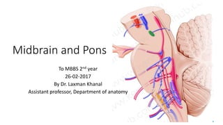
- 1. Midbrain and Pons To MBBS 2nd year 26-02-2017 By Dr. Laxman Khanal Assistant professor, Department of anatomy
- 2. Identify the cranial nerves.
- 3. Identify this swelling in pons. Q. Trigeminal nerve has its nuclei in which part of brain stem? a.Midbrain b.Pons c. Medulla oblongata d.All of the above
- 4. Objectives • To review the anatomy of the upper brainstem • To develop a three-dimensional picture of the interior of the midbrain and pons. • To know the positions of cranial nerve nuclei and the paths of various ascending and descending nerve tracts. • To assess the signs and symptoms presented by a patient and identify the exact location of a structural lesion.
- 5. External feature of midbrain and pons- anterior view
- 6. External feature- posterior view Visible part: Tectum 1. Corpora quadrigemina 2. 4th CN Visible part: Tegmental part 1. Forms upper part of floor of 4th ventricle 2. Median sulcus 3. Medial eminence (facial colliculus) 4. Sulcus limitans
- 7. Pons: • Pontine branches • AICA • Sup cerebellar artery Midbrain: • Posterior cerebral artery • Superior cerebellar artery
- 8. Which sections are important?
- 10. How to study the cross section • Different level of cross sections should be differentiated by- 1. What is the CSF filled cavity ? 2. What are the cranial nerve nuclei? 3. What are the ascending tracts ? 4. What are the descending tracts?
- 11. Descending tract in brainstem Corticospinal fibers Corticonuclear fibers Corticopontocerebellar fibers
- 12. Ascending tract- #MSTL DC/Medial lemniscus Spinal lemniscus Trigeminal lemniscus Lateral lemniscus Spinal lemniscus 1. Anterior spinothalamic tract 2. Lateral spinothalamic tract 3. Spinotectal tract Medial longitudinal fasciculus
- 13. @ Trigeminal nerve @ Facial colliculus
- 14. 66 7 Trapezoid body MLF Tegmentum Basal part Pontine nuclei Transverse pontine fibers (pontocerebellar fibers) Corticospinal and corticonuclear fibers Superior and inferior cerebellar peduncle MSTL Medial L Spinal L Trigeminal L Lateral L 7 FC
- 15. TS – lower pons
- 16. TS – upper pons Cranial nerve nuclei Main sensory nucleus of Vth nerve Motor nucleus of Vth nerve No inferior cerebellar peduncle
- 17. Millard-Gubler syndrome: #P67 Ventral pontine syndrome • Ipsilateral medial squint • Ipsilateral facial palsy • Contralateral hemiplegia Most common tumor of brainstem Astrocytoma of pons
- 19. S1. Tectum 2. Cerebral peduncle Tegmentum Crus cerebri Interpeduncular fossa Parkinson’s disease Substantia nigra
- 21. Crus cerebri (motor zone) • Contains descending fibers 1. Up to pons- FP+TP 2. Up to motor nuclei of CNs- CN 3. Up to spinal cord- CS FP TP CS CN Pontine nucleus Crossing of rubrospinal tract Mesencephalic nucleus MLF MSTL MST Crossing of sup cerebellar peduncle To cerebellum S
- 24. Blockage of cerebral aqueduct Weber syndrome 1. Crus cerebri 2. Oculomotor nerve # COW Benedict syndrome 1. Red nucleus 2. ML 3. Oculomotor nerve