Virus
•Als DOCX, PDF herunterladen•
4 gefällt mir•198 views

Morphology, Structure, types of Viruses

Empfohlen
Weitere ähnliche Inhalte
Was ist angesagt?
Was ist angesagt? (20)
Ähnlich wie Virus
Ähnlich wie Virus (20)
Mehr von Muzna Kashaf
Mehr von Muzna Kashaf (20)
Kürzlich hochgeladen
Kürzlich hochgeladen (20)
Virus
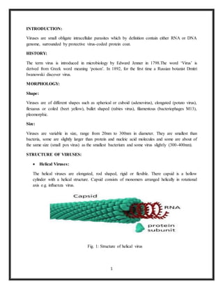
- 1. 1 INTRODUCTION: Viruses are small obligate intracellular parasites which by definition contain either RNA or DNA genome, surrounded by protective virus-coded protein coat. HISTORY: The term virus is introduced in microbiology by Edward Jenner in 1798.The word ‘Virus’ is derived from Greek word meaning ‘poison’. In 1892, for the first time a Russian botanist Dmitri Iwanowski discover virus. MORPHOLOGY: Shape: Viruses are of different shapes such as spherical or cuboid (adenovirus), elongated (potato virus), flexuous or coiled (beet yellow), bullet shaped (rabies virus), filamentous (bacteriophages M13), pleomorphic. Size: Viruses are variable in size, range from 20nm to 300nm in diameter. They are smallest than bacteria, some are slightly larger than protein and nucleic acid molecules and some are about of the same size (small pox virus) as the smallest bacterium and some virus slightly (300-400nm). STRUCTURE OF VIRUSES: Helical Viruses: The helical viruses are elongated, rod shaped, rigid or flexible. There capsid is a hollow cylinder with a helical structure. Capsid consists of monomers arranged helically in rotational axis e.g. influenza virus. Fig. 1: Structure of helical virus
- 2. 2 Polyhedral Viruses: Polyhedral structure has three possible symmetries such as tetrahedral, octahedral and icosahedral. The viruses are more or less spherical, therefore icosahedral symmetry is best one for packaging and bonding of subunits. The capsomers of each face form an equatorial triangles and 12 intersepting point or corners. They consist naked capsid e.g. adenovirus or envelop e.g. herpes simplex virus. Fig. 2. Structure of polyhedral virus Complex Viruses: The viruses which have the unidentifiable capsids or have the capsids with additional structures called complex viruses. Capsids are not clearly identified e.g. vaccinia virus etc. Some other structures are attached to capsids e.g. some bacteriophages. Fig. 3. Structure of complex virus
- 3. 3 Envelop: There are certain plant and animal viruses and bacteriophage both icosahedral and helical, which are surrounded by a thin membranous envelop. This envelop is about 10-15um thick. It is made up of protein, lipids and carbohydrates. They combine to form glycoprotein and lipoprotein. Lipids provide flexibility to the shape, therefore viruses look of variable size and shape. Protein component of the envelop is of viral origin and lipid and carbohydrate may be the feature of host membrane. PLANT VIRUS: Plant viruses are viruses that affect plants like all other viruses plant viruses are obligate intracellular parasites that do not have the molecular machinery to replicate without a pathogenic to higher plants. Example: Tobacco Mosaic Virus (TMV). EFFECT OF VIRUS ON PLANTS: External symptoms: Mosaic: Development of light and dark green area on leaves are due to disturbances of chloroplast and decrease in chlorophyll content. Chlorosis, vein clearing and vein banding: Vein clearing symptoms develop adjacent to veins before chlorosis of tissue. While broader bands of green tissue in chlorosis or necrosis is called vein banding.
- 4. 4 Ringspot: It is characterized by the formation of concentric or broken rings of infected dead cells. The ringspot may be chlorotic rings rather than necrotic rings. Necrosis: Besides localized cell death in necrotic local lesion or ringspots, necrosis in certain areas organs e.g. leaves, fruits, seeds, tubes or entire plant. Affected leaves shows scattered necrotic patches of dead tissue. Leaf abnormalities: Due to virus infection leaves show abnormal growth like leaf curling, leaf rolling, crinkling, puckering etc. the other abnormalities may also develop in leaves such as smaller blistered and thickened leaves. Flower symptoms: The colour breaking means streaks or sector of tissue with such colour that are different from the normal one. This happens due to loss or increase of anthocyanin pigment in petals. Internal Symptoms: Histological abnormalities: In leaves showing mosaic symptoms, mesophyll cells are smaller and less differentiated. Cytological abnormalities: Many cytological abnormalities are seen when virus infected cell are studied cytologically. Moreover some viruses affect specific organelles only. Tymovirus induces formation of marginal verticles in chloroplasts. Tobacco rattle virus modifies mitochondria and aggregates to form inclusion bodies. TMV particles are found in cytoplasm where as TMV of strain U5 are round in chloroplast and nuclei. Chloroplast may be rounded swollen and clumped together in the cells. It may be fragmented and colour may turn to colourless. CLASSIFICATION OF ANIMAL VIRUSES: Baltimore (2008) classified the animal viruses in the following seven groups according to the relationships between virion, nucleic acid and mRNA transcription Table (17.1). The RNA within the virion is known as plus (+) or sense strand because it acts as mRNA, whereas the newly synthesized RNA which is complementary in base-sequence to the original
- 5. 5 infectious strand is called minus (-) or antisense strand. It acts as template to produce additional (+) strand which may act as mRNA. Table. 1. Classification of Animal viruses 1 kb = 1,000 base pairs Class 1. dsDNA viruses: The mRNA is synthesized on a dsDNA genome template (± dsDNA → (+) mRNA) which usually occurs in a cell. Following are the example of some viruses: Papova-viruses: Polyomavirus, SV40 Poxviruses: Vaccinia virus
- 6. 6 Adenoviruses: Human adenovirus Herpes-viruses: Herpes simplex virus type I and type II, Epstein-Barr virus. Class 2. (+) ssDNA viruses: In such viruses an intermediate DNA is synthesized before the synthesis of mRNA transcript (+ ssDNA → + mRNA). The mRNA has the same polarity as the DNA. Parvoviruses: Adeno-associated viruses, mouse minute virus. Class 3. (+) ssRNA viruses: The RNA has similar polarity as the mRNA. Viruses of this class have been grouped into the following two classes: Subclass 3a: Individual mRNA encodes a polyprotein which is broken later on to form viral proteins. Picornaviruses: e.g. polio virus. Subclass 3b: From (+) ss RNA two types of mRNA molecules are transcribed, one is of same length as virion RNA and the other is a fragement of virion RNA. Togaviruses: Alpha viruses (group A), sindbis virus, semliki forest virus, Haviviruses (group B) e.g. dengue virus, yellow fever, St. Louis encephalitis virus are the important examples. Class 4. (-) ssRNA viruses: The virion RNA is complementary to mRNA. Following two types of viruses are found in this class: Subclass 4a: The ssRNA genome encodes a series of monocistronic mRNA. Rhabdoviruses: e.g. Mumps virus, measles virus, sendai virus. Subclass 4b: Each segment molecule of the genome acts as template for the synthesis of mRNA which are monocistronic or encodes polyprotein. Orthomyxo-viruses: e.g. Human influenza virus Bunya viruses: e.g. Bunyawera virus
- 7. 7 Arena-viruses: e.g. Lassa virus Class 5. dsRNA viruses: All the viruses of this class have segmented genome. Each chromosome encodes a single polypeptide. The dsRNA acts as template and asymmetrically synthesize (+) mRNA. Reoviruses: e.g. reovirus of humans. Class 6. (+) ssRNA-RT viruses: In these viruses (+) ssRNA directs the synthesis of (-) DNA which in turn acts as template for the transcription of mRNA (RNA→ (-) DNA→ + RNA). Virion RNA and mRNA are of the same polarity. Retroviruses: e.g. Rous sarcoma virus, mouse leukemia virus. Class 7. dsDNA-RT viruses: This group consists of DNA containing hepatitis B Viruses. CULTIVATION OF ANIMAL VIRUS: Viruses are obligate intracellular parasites so they depend on host for their survival. They cannot be grown in non-living culture media or on agar plates alone, they must require living cells to support their replication. Purpose of virus cultivation: The primary purpose of virus cultivation is: 1. To isolate and identify viruses in clinical samples. 2. To do research on viral structure, replication, genetics and effects on host cell. 3. To prepare viruses for vaccine production. Cultivation : Cultivation of viruses can be discussed under following headings: 1. Animal Inoculation 2. Inoculation into embryonated egg 3. Cell Culture
- 8. 8 ANIMAL INOCULATION Viruses which are not cultivated in embryonated egg and tissue culture are cultivated in laboratory animals such as mice, guinea pig, hamster, rabbits and primates are used. The selected animals should be healthy and free from any communicable diseases. Suckling mice(less than 48 hours old) are most commonly used. Suckling mice are susceptible to togavirus and coxsackie virues, which are inoculated by intracerebral and intranasal route. Viruses can also be inoculated by intraperitoneal and subcutaneous route. After inoculation, virus multiply in host and develops disease. The animals are observed for symptoms of disease and death. Then the virus is isolated and purified from the tissue of these animals. Live inoculation was first used on human volunteers for the study of yellow fever virus. INOCULATION INTO EMBRYONATED EGG Good pasture in 1931 first used the embryonated hen’s egg for the cultivation of virus. The process of cultivation of viruses in embryonated eggs depends on the type of egg which is used. Viruses are inoculated into chick embryo of 7-12 days old. For inoculation, eggs are first prepared for cultivation, the shell surface is first disinfected with iodine and penetrated with a small sterile drill. After inoculation, the opening is sealed with gelatin or paraffin and incubated at 36°c for 2-3 days. After incubation, the egg is broken and virus is isolated from tissue of egg. Viral growth and multiplication in the egg embryo is indicated by the death of the embryo, by embryo cell damage, or by the formation of typical pocks or lesions on the egg membranes
- 9. 9 Viruses can be cultivated in various parts of egg like chorioallantoic membrane, allantoic cavity, amniotic sac and yolk sac. 1. Chorioallantoic Membrane (CAM): Inoculation is mainly for growing poxvirus. After incubation and incubation, visible lesions called pocks are observed, which is grey white area in transparent CAM. Herpes simplex virus is also grown. Single virus gives single pocks This method is suitable for plaque studies. 2. Allantoic cavity: Inoculation is mainly done for production of vaccine of influenza virus, yellow fever, rabies. Most of avian viruses can be isolated using this method. 3. Amniotic sac: Inoculation is mainly done for primary isolation of influenza virus and the mumps virus. Growth and replication of virus in egg embryo can be detected by haemagglutination assay. 4. Yolk sac inoculation: It is also a simplest method for growth and multiplication of virus. It is inoculated for cultivation of some viruses and some bacteria (Chlamydia, Rickettsiae) Immune interference mechanism can be detected in most of avian viruses. CELL CULTURE (TISSUE CULTURE) There are three types of tissue culture; organ culture, explant culture and cell culture. Organ cultures are mainly done for highly specialized parasites of certain organs e.g. tracheal ring culture is done for isolation of coronavirus. Explant culture is rarely done. Cell culture is mostly used for identification and cultivation of viruses. Cell culture is the process by which cells are grown under controlled conditions. Cells are grown in vitro on glass or a treated plastic surface in a suitable growth medium. At first growth medium, usually balanced salt solution containing 13 amino acids, sugar, proteins, salts, calf serum, buffer, antibiotics and phenol red are taken and the host tissue or cell is inoculated. On incubation the cell divide and spread out on the glass surface to form a confluent monolayer.
- 10. 10 CONCLUSION: It is concluded that a virus is a non- cellular particle made up to genetic material and protein that can invade living cell. Virus can replicate its genetic material in the host cell. Virus can naked as well as have envelop. They can infect all type of life- animal, plant, human and cause diseases.
- 11. 11 REFERENCES: R.C DUBEY 2006, MICROBIOLOGY. https://microbiologyinfo.com/techniques-of-virus-cultivation/ VIRUS GENETICS: PLANT AND ANIMAL VIRUSES. Lecturer--J. LEDERBERG. http://www.biologydiscussion.com/viruses/animal-viruses/classification-of-animal- viruses-microbiology/65830