1. Bioengineering fluorescent protein tags from cyanobacteriochromes from T. elongatus: Protein Purification Valerie Metea Summer 2011 CBST Summer Research Project PI: Dr. Susan Spiller
4. Overview: Start to Finish RE & Ligation PCR DNA SDS-PAGE Gel Protein Purification Protein Expression Transformation Site-Directed Mutagenesis Transfection Credit: Monica Bower
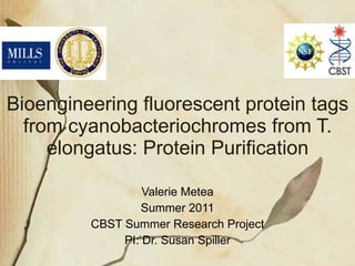
13. SDS-PAGE Glass gel cassettes Electrode assembly Mini Tank Inner chamber Outer Chamber Protein Sample Lid Power Source Blue = Negative Red = Positive http://upload.wikimedia.org/wikipedia/commons/4/46/SDS-PAGE_Electrophoresis.png - Modified Credit to Wikipedia Commons for image
Bioengineer a small, red fluorescent tag from a cyanobacteriochrome found in Thermosynechococcus elongatus A cyanobacteriochrome is a protein possessing only a phytochrome-related GAF domain - Cyanobacteriochromes retain the conserved GAF domain Cys residue to which the bilin chromophore is covalently linked in plant phytochromes Isolated from cyanobacteria and using e. coli to express it in
5 distinct cyanobacteriochrome genes All exhibit multiple protein domains (units of protein structure) We are only interested in the GAF domain cuz these are concerned with the light expression (???) and binding of chromophores My project focused on tlr1999 --> circle Wiki says--> TLRs: type of pattern recognition receptor (PRR) recognize molecules that are broadly shared by pathogens but distinguishable from host molecules are well known to be required for host defence against infection TLRs thus appear to be one of the most ancient, conserved components of the immune system.
Overall process done to visualize a protein in mammalian cells
At the point Monica ended, the result of the Protein Expression is pellets In the unmutated form, the pellets should be yellow indicating photoreversibility
The Pellet containues E.coli with our desired protein inside of it - we wish to purify the protein We use the microfluidizer to (mechanically) lyse the E. coli cells with high pressure to remove the protein from inside (and put them on ice immediately to stop the releazed enzymes from destroying the other cell contents) Centrifuge to keep supernatant and pellet out broken E. coli (extract parts of the cell that we are not interested in) This leads us to one of the final steps in protein purification called column chitin binding.
This is the setup for Chitin Binding Domain Chromatography - our cells were engineered to include a domain that binds to chitin, hence the name
Run buffer over column and beads to prep column for purification Pour supernatant with buffer added onto column over the period of and hour Supernatant still has all the soluble proteins in there, but we only want ours in particular Only our desired protein with the Chitin binding domain will stick (should be yellow for unmutated form, blue for mutated form) Wash other proteins off with column buffer at a faster rate Then add cleavage buffer (w/ DTT: Di-thio-three-i-tol) to cleave our protein from the CBD Let sit for 16 hrs to allow complete cleavage Dialyze samples overnight to remove DTT (toxic and will degrade protein if include in storage) Now have pure protein and can quantify and determine purity via use of the spectrophotomer and SDS-PAGE gels
Because I was unsuccessful in my attempts thus far to isolate pure protein from 1999T, I selected a similar gene with similar properties that the Spiller Lab has had success purifying, 569T - We hypothesize that 569T would display a similar peak in both the blue and green range and would photoreverse Explain that the two peaks show presence of protein - Peaks signify where protein absorbs
SDS-PAGE is a process in which an electric current is used to separate bands of proteins - to determine the purity of our isolated protein Purity of the protein is indicated by a single band of a certain size--> The size is compared to control ladders of known band lengths - The further the band has traveled on the gel, the smaller the size of the protein. - First we will look at SDS, an anionic detergent, that is able to denature a protein to its primary structure without breaking the amino acid chain. - With the presence of negatively charged sulfate ions, the entire linearized protein is covered with a net negative charge. This charge ensures that proteins of similar size are able to migrate at about the same rate. It also allows for the proteins to travel from the negative anode pole to the positive cathode pole when an electric current is applied to the gel.
The gel is SDS-PAGE is composed of two layers: - the 12% acrylamide resolving gel is added first (lower layer) - It is in this bottom layer of the gel where we will see more separation between the bands - The increase amount of acrylamide provides narrow channels in which the proteins can travel (gel matrix) - we will see smaller bands move further down the gel. - the 4% acrylamide stacking gel (added last and forms the upper layer) - This gel is more porous: allowing for the proteins of different size to become “stacked” against each other - when the come to the interface of the resolving gel, stacks proteins so all get into “Starting gate” @ same time - As they are now stacked, the proteins are then able to migrate through the resolving gel (with its thicker matrix) where all proteins move with their size class ~~~~~~~~~~~~~ - Using gel glass plates and a casting frame. The two plates will be place on top of one another. - The resolving gel will be added first to the small space between the plates. Water will soon be added to prevent mixture with the air and create a flat surface at the top of the gel. - After polymerizing for 45 minutes the stacking gel is added. - Immediately after the stacking gel is poured the comb, which forms the gel into wells, is inserted between the plates. This is where the proteins will be loaded. - Once polymerization has occurred, the gel is ready to use.
- flows negative pole to --> positive pole (Trace path of flow!) - We place the gel plates facing toward each other in the electrode assembly, which is then lowered into the mini tank. - Running buffer is added to the inner chamber between the two plates, as well as in the outer chamber. Buffer in these two regions allows for an electric current to flow through the gels. - Now that the buffer is added, the samples can be loaded into the wells. Once this is complete. The mini tank is covered with a lid that is attached to the power source. An electric current travels through the negative anode end, into the inner chamber, down the gels, out through the outer chamber, and back through the positive anode end to the power source.
- Use a vacuum system to dry the gel - Here is a sample of one of our results after drying the gel. You can see the ladders which show the relative sizes of the proteins. - We can determine the purity of the protein by the one band position that is seen across the gel. Each protein is relatively the same size. - We also compare the our proteins to our control chp1. chp1 is a protein that is slightly smaller than the sample proteins used in this gel. - In our lab we want to know if bilin chromophore is covalently bound to our protein, therefore we perform one more test based on the polyacrylamide gel. -- so we Transblot using Zinc Acetate to make sure chromophore is bound (Information on hand….do not state in presentation) 12/27 Lane 1: Ladder used to relate compartative sizes of proteins Lane 2: Cph1 – bacterial phytochrome that we use as a control. We compare protein size. What you see here are the proteins before truncation (shortened) to GAF only domain of the 924 gene. Lane 3: Full length 924 without mutation Lane 4: Full length with mutation (924) of cystine to aspartate Lane 5: Ladder Lane 6: Full length protein with cystine mutated to alanine Lane 7: Blank Lane 8: 924 with cystine to aspartate mutation, but with no chromophore (apoprotein)
SKIP IF DON’T HAVE TIME OR JUST MENTION IF ANA POPOVICH TALKED ABOUT THIS: -binding tag to actin and tubulin of Lymphocyte (White blood cells) - Might allow researchers to obtain insight into the infection process and perhaps progress towards a cure of the viral disease!
- Dr. Susan Spiller for all her direction and advice - Research Associates for their assistance on my experiments - Acknowledge the work of the fellow Spiller lab members - Nathan Rockwell and Clark Lagarias for their direction and support for the Spiller Lab over the many years - Dr.s Corbacho and Molinaro for all their help and support with the CBST Summer Internship Program
![Background ,[object Object],[object Object]](data:image/gif;base64,R0lGODlhAQABAIAAAAAAAP///yH5BAEAAAAALAAAAAABAAEAAAIBRAA7)