Physiology of Chest X-Ray
•Als PPT, PDF herunterladen•
3 gefällt mir•1,847 views

Presentation of students and residents on basics of chest plane film.

Empfohlen
Empfohlen
Weitere ähnliche Inhalte
Andere mochten auch
Andere mochten auch (20)
Ähnlich wie Physiology of Chest X-Ray
Ähnlich wie Physiology of Chest X-Ray (14)
Mehr von Mike Aref
Mehr von Mike Aref (20)
Kürzlich hochgeladen
Kürzlich hochgeladen (20)
Physiology of Chest X-Ray
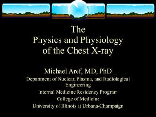
- 1. The Physics and Physiology of the Chest X-ray Michael Aref, MD, PhD Department of Nuclear, Plasma, and Radiological Engineering Internal Medicine Residency Program College of Medicine University of Illinois at Urbana-Champaign
- 2. Röntgen In a dark room Röntgen passed an electrostatic charge through a cathode tube generating a faint shimmering in a nearby barium platinocyanide screen. An invisible ray An x-ray had passed from the tube to the screen. Father of Diagnostic Radiology
- 4. Attenuation I x = Ι0ε −µξ Intensity, I, decreases exponentially with thickness, x. The attenuation coefficient, μ, increases with increasing density, ρ, and vice versa
- 5. Radiological Densities Biological Chemical Density, ρ Composition Composition (kg/L) Air N2, O2, CO2 1.2 CH3(CH2)m(CH=CHCH2)n( Fat CH2)pCOO- 900 Water H2O 1000 (Soft Tissue) Bone Ca10(PO4)6(OH)2 3160 Metal (hydroxyapatite) Blood Minute Fe 7000
- 6. Attenuation Comparison Air Fat Water Intensity Bone Thickness
- 9. Negative Exposure Trickery www.brooksidepress.org
- 10. Projection knitting.designedlykristi.com
- 11. Projection The point-of-view dependent, two-dimensional representation of a three-dimensional object knitting.designedlykristi.com
- 13. What’s this?
- 14. Focus
- 16. PA and AP views www.med.yale.edu
- 17. Lateral View Localizes STENTOR Teaching File
- 18. High IQ ABC’s Identification Quality Airway, aorta, and adenopathy Bones and breast shadow Cardiac silhouette Diaphragm Everything else Fields, fluid, and foreign objects Gastric air bubble History
- 20. Where’s the mass? Middle Mediastinum
- 21. Where’s the mass? Posterior Mediastinum
- 24. 61-year-old woman with dyspnea The inferior margin of the opacity in the right upper thorax is due to the B. major fissure in RUL collapse without a hilar mass. C. minor fissure in RUL collapse with a hilar mass. D. minor fissure in RUL collapse without a hilar mass. E. major fissure in RUL collapse with a hilar mass.
- 25. 61-year-old woman with dyspnea The inferior margin of the opacity in the right upper thorax is due to the B. major fissure in RUL collapse without a hilar mass. C. minor fissure in RUL collapse with a hilar mass. D. minor fissure in RUL collapse without a hilar mass. E. major fissure in RUL collapse with a hilar mass.
- 26. 61-year-old woman with dyspnea The inferior margin of the opacity in the right upper thorax is due to the B. major fissure in RUL collapse without a hilar mass. C. minor fissure in RUL collapse with a hilar mass. D. minor fissure in RUL collapse without a hilar mass. E. major fissure in RUL collapse with a hilar mass.
- 27. 45-year-old woman with chronic cough All of the following are true with regard to right middle lobe collapse except B. a triangular opacity is superimposed on the heart on the lateral radiograph. C. the right heart border is obscured. D. the minor fissure is inferiorly displaced. E. the right heart border is shifted to the left.
- 28. 45-year-old woman with chronic cough All of the following are true with regard to right middle lobe collapse except B. a triangular opacity is superimposed on the heart on the lateral radiograph. C. the right heart border is obscured. D. the minor fissure is inferiorly displaced. E. the right heart border is shifted to the left.
- 29. 45-year-old woman with chronic cough All of the following are true with regard to right middle lobe collapse except B. a triangular opacity is superimposed on the heart on the lateral radiograph. C. the right heart border is obscured. D. the minor fissure is inferiorly displaced. E. the right heart border is shifted to the left.
- 30. 62-year-old man with a cough productive of blood-tinged sputum Signs of left lower lobe collapse include all of the following except B. obscuration of the lateral wall of the descending thoracic aorta. C. inferior displacement of the left hilum. D. obliteration of the posterior aspect of the left hemidiaphragm on the lateral view. E. triangular opacity in the left retrocardiac area on the frontal view. F. shift of the major fissure toward the anterior chest wall on the lateral view.
- 31. 62-year-old man with a cough productive of blood-tinged sputum Signs of left lower lobe collapse include all of the following except B. obscuration of the lateral wall of the descending thoracic aorta. C. inferior displacement of the left hilum. D. obliteration of the posterior aspect of the left hemidiaphragm on the lateral view. E. triangular opacity in the left retrocardiac area on the frontal view. F. shift of the major fissure toward the anterior chest wall on the lateral view.
- 32. 62-year-old man with a cough productive of blood-tinged sputum Signs of left lower lobe collapse include all of the following except B. obscuration of the lateral wall of the descending thoracic aorta. C. inferior displacement of the left hilum. D. obliteration of the posterior aspect of the left hemidiaphragm on the lateral view. E. triangular opacity in the left retrocardiac area on the frontal view. F. shift of the major fissure toward the anterior chest wall on the lateral view.
- 33. 49-year-old woman with cough Signs of left upper lobe collapse include all of the following except B. crescent of air around the transverse section of the aortic arch resulting from hyperexpansion of the superior segment of the left lower lobe. C. anterior displacement of the left major fissure on the lateral view. D. obscuration of the left heart border. E. tracheal deviation to the left. F. inferior displacement of the left hilum.
- 34. 49-year-old woman with cough Signs of left upper lobe collapse include all of the following except B. crescent of air around the transverse section of the aortic arch resulting from hyperexpansion of the superior segment of the left lower lobe. C. anterior displacement of the left major fissure on the lateral view. D. obscuration of the left heart border. E. tracheal deviation to the left. F. inferior displacement of the left hilum.
- 35. 49-year-old woman with cough Signs of left upper lobe collapse include all of the following except B. crescent of air around the transverse section of the aortic arch resulting from hyperexpansion of the superior segment of the left lower lobe. C. anterior displacement of the left major fissure on the lateral view. D. obscuration of the left heart border. E. tracheal deviation to the left. F. inferior displacement of the left hilum.
- 36. Airror sprojects.mmi.mcgill.ca www.surgical-tutor.org.uk
- 37. 40-year-old man with fever and dyspnea The most likely diagnosis is B. massive left pleural effusion. C. total atelectasis of the left lung. D. right pneumothorax. E. aplasia of the left lung. F. mediastinal hematoma.
- 38. 40-year-old man with fever and dyspnea The most likely diagnosis is B. massive left pleural effusion. C. total atelectasis of the left lung. D. right pneumothorax. E. aplasia of the left lung. F. mediastinal hematoma.
- 39. Tall, 21-year-old man who noted the sudden onset of dyspnea, and right-sided pleuritic chest pain The most likely diagnosis is B. pulmonary embolism. C. overinflation associated with asthma. D. pneumothorax. E. normal chest, with a skin fold projected over the right hemithorax.E. left lower lobe atelectasis.
- 40. Tall, 21-year-old man who noted the sudden onset of dyspnea, and right-sided pleuritic chest pain The most likely diagnosis is B. pulmonary embolism. C. overinflation associated with asthma. D. pneumothorax. E. normal chest, with a skin fold projected over the right hemithorax.E. left lower lobe atelectasis.
- 41. 62-year-old man with dyspnea that increased over 2 days The most likely diagnosis is B. left pleural effusion. C. collapse of the left lung. D. right pneumothorax. E. collapse of the right lung. F. mediastinal hematoma.
- 42. 62-year-old man with dyspnea that increased over 2 days The most likely diagnosis is B. left pleural effusion. C. collapse of the left lung. D. right pneumothorax. E. collapse of the right lung. F. mediastinal hematoma.
- 43. COPD STENTOR Teaching File
- 44. Silicosis STENTOR Teaching File
- 45. Cystic Fibrosis
- 46. 64-year-old man, Navy veteran, with a cough productive of blood-tinged sputum The most likely diagnosis is B. progressive massive fibrosis, due to silicosis. C. pneumonia in a patient with chronic interstitial lung disease. D. lung cancer in a patient with asbestosis. E. rounded atelectasis in a patient with asbestosis. F. berylliosis.
- 47. 64-year-old man, Navy veteran, with a cough productive of blood-tinged sputum The most likely diagnosis is B. progressive massive fibrosis, due to silicosis. C. pneumonia in a patient with chronic interstitial lung disease. D. lung cancer in a patient with asbestosis. E. rounded atelectasis in a patient with asbestosis. F. berylliosis.
- 48. 55-year-old man who worked as a coal miner for 30 years The most likely diagnosis is B. progressive massive fibrosis, due to silicosis. C. pneumonia in a patient with chronic interstitial lung disease. D. lung cancer in a patient with asbestosis. E. rounded atelectasis in a patient with asbestosis. F. berylliosis.
- 49. 55-year-old man who worked as a coal miner for 30 years The most likely diagnosis is B. progressive massive fibrosis, due to silicosis. C. pneumonia in a patient with chronic interstitial lung disease. D. lung cancer in a patient with asbestosis. E. rounded atelectasis in a patient with asbestosis. F. berylliosis.
- 50. Pulmonary malignancies Bronchoalveolar Carcinoma Chronic Histoplasmosis Granulomas Metastasized Esophageal SCC 1° SCC
- 51. 53-year-old man scheduled for coronary artery bypass grafting Characteristics suggesting that a nodule is benign are B. size of the nodule does not change over 2 years. C. it contains central calcification. D. CT attenuation values within the nodule are greater than 200 Hounsfield units (Hu). E. all of the above.
- 52. 53-year-old man scheduled for coronary artery bypass grafting Characteristics suggesting that a nodule is benign are B. size of the nodule does not change over 2 years. C. it contains central calcification. D. CT attenuation values within the nodule are greater than 200 Hounsfield units (Hu). E. all of the above.
- 53. 64-year-old man with cough and weight loss and a 50-pack-per-year history of tobacco use The best description of the chest radiograph is B. mass in the left upper lobe. C. left upper lobe collapse. D. mediastinal mass. E. consolidation of the left upper lobe. F. enlargement of the left pulmonary artery.
- 54. 64-year-old man with cough and weight loss and a 50-pack-per-year history of tobacco use The best description of the chest radiograph is B. mass in the left upper lobe. C. left upper lobe collapse. D. mediastinal mass. E. consolidation of the left upper lobe. F. enlargement of the left pulmonary artery.
- 55. 64-year-old man with cough and weight loss and a 50-pack-per-year history of tobacco use The best description of the chest radiograph is B. mass in the left upper lobe. C. left upper lobe collapse. D. mediastinal mass. E. consolidation of the left upper lobe. F. enlargement of the left pulmonary artery.
- 56. 70-year-old woman with uterine carcinoma treated with surgical resection 3 years ago The most likely cause of the multiple pulmonary nodules is B. metastasis. C. herpes simplex pneumonia. D. histoplasmosis. E. Wegener's granulomatosis. F. arteriovenous malformations
- 57. 70-year-old woman with uterine carcinoma treated with surgical resection 3 years ago The most likely cause of the multiple pulmonary nodules is B. metastasis. C. herpes simplex pneumonia. D. histoplasmosis. E. Wegener's granulomatosis. F. arteriovenous malformations
- 58. Airways www.smbs.buffalo.edu www.mevis.de
- 59. Pneumonia STENTOR Teaching File
- 60. Pneumonia STENTOR Teaching File
- 61. 32-year-old man with fever, cough, and hemoptysis Which of the following is not an accurate descriptor of the opacity in the left upper lobe B. Lobar distribution C. Ill-defined margins D. Reticular pattern E. Air bronchograms F. Airspace disease
- 62. 32-year-old man with fever, cough, and hemoptysis Which of the following is not an accurate descriptor of the opacity in the left upper lobe B. Lobar distribution C. Ill-defined margins D. Reticular pattern E. Air bronchograms F. Airspace disease
- 63. 57-year-old man with fever and a cough productive of purulent sputum Which one of the following best explains the opacity in the left hemithorax? B. Collapse of the left upper lobe due to bronchial obstruction C. Airspace consolidation of the lingula D. Empyema loculated within the left major fissure E. Carcinoma in the left upper lobe
- 64. 57-year-old man with fever and a cough productive of purulent sputum Which one of the following best explains the opacity in the left hemithorax? B. Collapse of the left upper lobe due to bronchial obstruction C. Airspace consolidation of the lingula D. Empyema loculated within the left major fissure E. Carcinoma in the left upper lobe
- 65. 69-year-old man with progressive dyspnea, orthopnea, and pedal edema and a history of hypertension Which of the following best describes the chest radiograph? B. Normal heart size, alveolar pulmonary edema C. Cardiomegaly, interstitial pulmonary edema, and small bilateral pleural effusions D. Unilateral interstitial disease E. Cardiomegaly, oligemia in the right lung
- 66. 69-year-old man with progressive dyspnea, orthopnea, and pedal edema and a history of hypertension Which of the following best describes the chest radiograph? B. Normal heart size, alveolar pulmonary edema C. Cardiomegaly, interstitial pulmonary edema, and small bilateral pleural effusions D. Unilateral interstitial disease E. Cardiomegaly, oligemia in the right lung
- 68. Pleural Effusion
- 70. 45-year-old man with increasing dyspnea and abdominal swelling of 1-week duration Which of the following radiographic signs suggest the presence of pleural effusion? B. Meniscus-shaped opacity in a posterior costophrenic angle on the lateral projection C. Biconvex lens-shaped opacity projecting in the midthorax on the lateral projection D. Fluid levels that have different lengths on the PA and lateral views in a hemithorax E. Homogeneous increased density in a hemithorax with preservation of the vascular shadows in the lungs F. Separation of the gastric air bubble from the inferior lung margin by more than 2 cm
- 71. 45-year-old man with increasing dyspnea and abdominal swelling of 1-week duration Which of the following radiographic signs suggest the presence of pleural effusion? B. Meniscus-shaped opacity in a posterior costophrenic angle on the lateral projection C. Biconvex lens-shaped opacity projecting in the midthorax on the lateral projection D. Fluid levels that have different lengths on the PA and lateral views in a hemithorax E. Homogeneous increased density in a hemithorax with preservation of the vascular shadows in the lungs F. Separation of the gastric air bubble from the inferior lung margin by more than 2 cm
- 72. 62-year-old woman with worsening shortness of breath and mild hemoptysis 1 day after receiving IV chemotherapy The most likely cause for her dyspnea and hemoptysis is B. pulmonary metastases. C. malignant pleural effusion. D. pulmonary embolism. E. septic emboli. F. drug-related pneumonitis.
- 73. 62-year-old woman with worsening shortness of breath and mild hemoptysis 1 day after receiving IV chemotherapy The most likely cause for her dyspnea and hemoptysis is B. pulmonary metastases. C. malignant pleural effusion. D. pulmonary embolism. E. septic emboli. F. drug-related pneumonitis.
- 74. 62-year-old woman with worsening shortness of breath and mild hemoptysis 1 day after receiving IV chemotherapy The most likely cause for her dyspnea and hemoptysis is B. pulmonary metastases. C. malignant pleural effusion. D. pulmonary embolism. E. septic emboli. F. drug-related pneumonitis.
- 75. The Final Can you spot the pathology?
- 76. References Paul and Juhl’s Essentials of Radiologic Imaging, 11th edition Felson’s Principles of Chest Roentgenology, 2nd edition Chen MYM, Pope TL, and Ott DJ, Basic Radiology, The McGraw-Hill Companies, 2008