2. VOL. 71, 2005 MUTANTS RESISTANT TO THE LACTOPEROXIDASE SYSTEM 3513
TABLE 1. Primers used for this study the presence of (i) lactoperoxidase (5 g/ml), (ii) KSCN (0.25 mM), (iii) glucose
(0.4%), and (iv) glucose oxidase (0.1 units/ml). The function of the glucose
Primer 5Ј–3Јa
oxidase enzyme and of glucose was to generate H2O2 in situ, which subsequently
Pcat1.................................................AAGCACCGCCGGACATC served as a substrate for the lactoperoxidase enzyme. Cells were inoculated at an
Pcat2.................................................CTCCCAGAGCCTGATAA initial density of approximately 104 CFU/ml by diluting cultures grown at 37°C in
PompmutC1 .....................................AAGGCATATAACAGAGGGTT LB medium in microplates for 21 h. Incubation was done at 25°C for 44 to 64 h,
AATAACATGAAAGTTAAAG and the optical density was measured every 15 min by use of a wide-band filter
TACTGTgtgtaggctggagctgcttc (405 to 600 nm). Under these conditions, the LP system caused complete growth
PompmutC2 .....................................CCAGACCCAGAGCTACGATG inhibition of E. coli MG1655 for at least 40 h, while at 37°C, growth inhibition
TTATCAGTGTTGATGCCAG was achieved for no longer than 2 h. Mutants showing growth within Ͻ40 h were
CGTCACcatatgaatatcctcctta retested in five replicates with the same test, and when their phenotype was
PompC1 ...........................................TGATCGCAACCAACAAAGAA confirmed, their mini-Tn10-induced mutation was transduced via P1 into
PompC2 ...........................................GAATGGACTTGCCGACTGAT MG1655 and the LP tolerance of the transductants was examined once more.
PompF1............................................TCAAACATGACGAGGTTCCA Cloning and identification of gene knockouts. Mini-Tn10-containing genomic
PompF2............................................GCAGTGGCAGGTGTCATAAA DNA fragments were isolated by random cloning of ca. 40-kb large genomic
Pkan .................................................CAGTCATAGCCGAATAGCCT DNA fragments from LP-tolerant mutants into the pWEB cosmid cloning vector
(Biozym, Landgraaf, The Netherlands) and the selection of chloramphenicol-
a
Sequences in lowercase were needed to amplify the kan gene from the pKD4 resistant transformants of E. coli EPI305 (Biozym). The genomic DNA flanking
plasmid. Sequences in uppercase are homologous to the ompC gene. the transposon was then sequenced from the cosmid template by the use of
primers complementary to the respective ends of the cat gene (32) (Pcat1 and
Pcat2; Table 1). Sequence analysis was performed by MWG-Biotech (Ebersberg,
Germany).
strated that the LP system induces a specific and unique stress
Isolation and electrophoretic separation of LPS. For the isolation of lipopoly-
response in E. coli (40). Here we report the isolation and saccharide (LPS), a protocol described by Moller et al. (28) was used. LPS was
characterization of random transposon insertion mutants of E. separated by 18% Tricine–sodium dodecyl sulfate–polyacrylamide gel electro-
coli MG1655 with increased tolerance to the LP system. phoresis (SDS-PAGE) (33) and visualized by silver staining (12).
Downloaded from aem.asm.org by on May 4, 2010
Isolation and electrophoretic separation of outer membrane proteins. Outer
membrane proteins were isolated by freeze-thawing of cell suspensions in the
MATERIALS AND METHODS presence of lysozyme and subsequent washing with 0.5% sarcosyl as described by
Growth media and chemicals. The standard growth medium was Luria-Bertani Mecsas et al. (25). Air-dried (3 h at 37°C) pellets were weighed, dissolved in urea
(LB) medium (10 g/l tryptone, 5 g/l yeast extract, 5 g/l NaCl, 10 g/l agar for solid sample buffer, and loaded in a urea-SDS-PAGE gel, ensuring that equivalent
medium). Antibiotics (Sigma-Aldrich, Bornem, Belgium) were added as appro- amounts of outer membrane material (on a weight basis) were loaded in each
priate at the following concentrations: ampicillin, 100 g/ml; kanamycin, 50 lane. Urea was present in the sample buffer and the SDS-PAGE gel at a con-
g/ml; and chloramphenicol, 30 g/ml. Tryptone soy broth (TSB) (Oxoid, Bas- centration of 8% because this improves the separation of OmpC and OmpF (45).
ingstoke, England) was used as the growth medium for evaluations of the bac- Gels were stained with Coomassie blue.
teriostatic activity of the lactoperoxidase system. Stock solutions of lactoperox- SDS, novobiocin, and ampicillin sensitivity tests. SDS and novobiocin sensi-
idase (10 mg/ml) and of glucose oxidase (100 units/ml) (Sigma-Aldrich) were tivities were tested as described by Moller et al. (28). Twofold serial dilutions of
stored at Ϫ18°C in a 50% glycerol solution in phosphate-buffered saline (2.87 SDS (200 to 0.1 mg/ml) and novobiocin (200 to 1.6 g/ml) in LB broth were
mM KH2PO4, 7.12 mM K2HPO4, 0.151 M NaCl, pH 6.0). Potassium thiocyanate made in microplates, inoculated in a final volume of 200 l with approximately
(KSCN) (Acros Organics, Geel, Belgium) was stored at 4°C as a 25 mM stock 105 CFU/ml from an overnight culture, and incubated on a rotary shaker (200
solution. Filter-sterilized glucose was stored at room temperature as a 20% stock rpm) at 37°C, and after 15 h of growth, their optical densities (at 600 nm) were
solution. measured with a microplate reader (Multiskan RC; Thermo Electron Corpora-
Construction of MG1655 ompC and MG1655 ompF. The one-step inactivation tion). Growth was scored as positive for optical densities of Ն0.1.
method of Datsenko and Wanner (5) was used to make a mutant with an For tests of ampicillin sensitivity, overnight cultures were resuspended to
insertional mutation in ompC. First, the kanamycin resistance gene (kan) from approximately 107 CFU/ml in honeycomb microplates containing LB broth with
plasmid pKD4 was amplified with the primers PompmutC1 and PompmutC2 various concentrations of ampicillin, and growth at 37°C was monitored with a
(Table 1). In addition to a priming sequence of 19 or 20 nucleotides at their 3Ј Bioscreen C automatic growth analyzer using a wide-band filter.
ends, these primers have an ϳ50-nucleotide extension homologous to ompC that Evaluation of outer membrane permeability for small hydrophilic solutes.
will allow an exchange of the PCR product with the genomic ompC gene. The Measurements of outer membrane permeability were based on the membrane
PCR product was then electroporated into MG1655 carrying the pKD46 plas- diffusion barrier model described by Martinez et al. (22). The whole-cell alkaline
mid, which expresses the arabinose-inducible Red recombinase (29), to pro- phosphatase assay of Wang et al. (47) was applied to assess the outer membrane
mote recombination. Kanamycin-resistant transformants were selected and permeability. In this assay, the hydrolysis of p-nitrophenyl phosphate (pNPP) by
cured of pKD46 by growth at 37°C, which is a nonpermissive temperature for an E. coli cell suspension under controlled incubation conditions is measured by
replication of this plasmid. The ompC insertional mutant was verified by PCR the increase in absorption at 420 nm and then corrected for cell density (A420/
with the primers PompC1 and PompC2 (Table 1) and was designated LMM- A600). Since the test is conducted at a low substrate concentration and with cells
PDS1. transformed with the plasmid pIV26 (21) to overexpress PhoA in the periplasm,
The ompF::Tn5 allele from E. coli BE strain BZB1107 (2) was transduced with product formation will be controlled by the diffusion of pNPP through the outer
phage P1 into MG1655 (26). Direct transduction between these two strains did membrane, and thus A420/A600 is a valid measure of outer membrane perme-
not succeed, probably because the E. coli BE DNA was degraded by the K12 ability.
restriction-modification system. Therefore, we first transduced the ompF::Tn5
allele into the restriction-negative E. coli K12 strain MT102, and from there to
MG1655, to yield strain LMM-PDS2. Since the location and orientation of the
transposon in ompF are unknown, the correct replacement of ompF by
RESULTS
ompF::Tn5, which contained the kan gene, was verified by PCR with the primer
Screening for mutants with increased tolerance to the LP
couples PompF1-PompF2, Pkan-PompF1, and Pkan-PompF2 in separate reac-
tions (Table 1). antimicrobial system. In several preparatory experiments, we
Screening for transposon insertion mutants with increased tolerance to the determined suitable screening conditions for the detection of
LP system. Transposon knockout mutants of E. coli MG1655 were constructed mutants with an increased tolerance to the LP system. The
by using NK1324, which carries a mini-Tn10 transposon with a chloramphenicol parameters that were varied include the growth medium,
resistance gene, according to the protocol described by Kleckner et al. (15).
Approximately 5,000 mutants were then screened for tolerance to the LP system
growth temperature, initial cell number, and component con-
by recording their growth curves in a Bioscreen C automatic growth analyzer centrations of the LP system. The conditions that were finally
(Thermo Electron Corporation, Vantaa, Finland) in 300 l of TSB medium in chosen (see Materials and Methods) ensured the complete
3. 3514 DE SPIEGELEER ET AL. APPL. ENVIRON. MICROBIOL.
duction. The resulting strains were designated LMM-PDS3,
LMM-PDS4, LMM-PDS5, and LMM-PDS6. Growth curves
for E. coli MG1655 and the four selected mutants in the ab-
sence of additives, in the presence of the H2O2-generating
glucose oxidase/glucose enzyme system and KSCN, and in the
presence of the entire LP system (glucose oxidase, glucose,
KSCN, and lactoperoxidase) are shown, respectively, in Fig.
1A, B, and C. The LP system completely inhibited the growth
of E. coli MG1655 for at least 40 h. The mutants all grew in the
presence of the LP system within Ͻ40 h, but compared to the
growth of the wild-type strain in TSB without additives, they
exhibited an extended lag phase, a lower exponential growth
rate, and a lower stationary-phase cell density. Exposure to
H2O2 stress from the glucose oxidase/glucose enzyme system in
the absence of the LP enzyme (Fig. 1B) also resulted in partial
growth inhibition, but interestingly, this inhibition was the
same for the wild-type strain and the mutants, indicating that
the mutants had acquired tolerance specifically to the LP sys-
tem.
Identification of mini-Tn10 insertion sites and initial char-
acterization of mutants LMM-PDS3 and LMM-PDS4. Mini-
Downloaded from aem.asm.org by on May 4, 2010
Tn10-containing fragments from the four mutants were cloned,
and a short sequence of genomic DNA flanking the trans-
posons was determined. The insertions were located in waaO,
waaQ, ulaA, and lrp for mutants LMM-PDS3, LMM-PDS4,
LMM-PDS5, and LMM-PDS6, respectively, and their exact
locations are presented in Table 2. The finding that two inser-
tions were in the waa (formerly rfa) operon, which is involved
in core oligosaccharide synthesis of lipopolysaccharide (LPS)
molecules, suggested a role for the LPS layer in the tolerance
to the LP system and prompted us to investigate these two
mutants in more detail. Since mutations in the waa operon
result in truncated LPS molecules and, as a result, can cause
hypersensitivity to SDS and hydrophobic antibiotics such as
novobiocin (49), we isolated LPS, subjected it to SDS-PAGE
analysis, and tested the SDS and novobiocin sensitivities of the
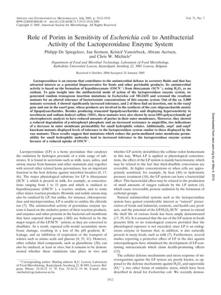
FIG. 1. Growth curves for E. coli strains MG1655 (■), LMM-PDS3
(waaO) (F), LMM-PDS4 (waaQ) (Œ), LMM-PDS5 (ulaA) (E), and mutants. Both waa knockout mutants (LMM-PDS3 and LMM-
LMM-PDS6 (lrp) (ᮀ) incubated at 25°C. (A) Growth in TSB. PDS4) did indeed have LPS molecules that were reduced in
(B) Growth in TSB with the addition of KSCN, glucose, and glucose size (Fig. 2, lanes 2 and 3), while the LPS molecules of both
oxidase. (C) Growth in TSB with the addition of KSCN, glucose, other mutants were indistinguishable from wild-type LPS. Fur-
glucose oxidase, and lactoperoxidase. Only a few measurement points
are labeled with symbols. thermore, both waa mutants also showed increased sensitivi-
ties to SDS and novobiocin. The MICs of SDS and novobiocin,
determined by a 15-h growth test described in Materials and
growth inhibition of E. coli MG1655 for at least 40 h. Out of ca. Methods, were reduced 2-fold for the waaO mutant and 500-
5,000 random mini-Tn10 mutants, 12 showed an increased fold and 12-fold, respectively, for the waaQ mutant compared
tolerance to the LP system after a first screen, and for 4 of to the wild-type strain MG1655 (Table 3). The LMM-PDS5
these, the phenotype could be confirmed with a second test. and LMM-PDS6 mutants had no altered sensitivity to SDS or
These mutants were cotransduced with mini-Tn10 by P1 trans- novobiocin (data not shown).
TABLE 2. Locations of transposon insertions in four lactoperoxidase-tolerant mutants
Orientation of transposon-encoded
Knocked-out Coordinates of gene open Position of transposon
Strain resistance marker relative to gene
gene reading framea on coding strand
coding strand
LMM-PDS3 waaO 3801081–3800062 3801072 Same
LMM-PDS4 waaQ 3806121–3805087 3805205 Same
LMM-PDS5 ulaA 4417946–4419400 4418348 Opposite
LMM-PDS6 lrp 0931818–0932312 0932016 Same
a
From Escherichia coli K-12 MG1655 complete genome (GenBank accession number U00096).
4. VOL. 71, 2005 MUTANTS RESISTANT TO THE LACTOPEROXIDASE SYSTEM 3515
FIG. 3. SDS-PAGE gel of Coomassie blue-stained outer mem-
brane proteins. Lanes: 1, MG1655; 2, LMM-PDS3 (waaO); 3, LMM-
PDS4 (waaQ); 4, LMM-PDS1 (ompC); and 5, LMM-PDS2 (ompF).
FIG. 2. LPS profiles of wild-type MG1655 (lane 1), LMM-PDS3
OmpC and OmpF bands were identified by comparisons to the corre-
(waaO) (lane 2), LMM-PDS4 (waaQ) (lane 3), LMM-PDS5 (ulaA)
sponding knockout reference strains. The OmpA band was presump-
(lane 4), LMM-PDS6 (lrp) (lane 5), LMM-PDS1 (ompC) (lane 6), and
tively identified based on its position relative to OmpC and OmpF and
LMM-PDS2 (ompF) (lane 7).
by comparison with the results of Brissette et al. (3) and Liu and
Ferenci (19).
Outer membrane protein content and permeability to hy-
drophilic molecules. Since truncated LPS molecules can result Tolerance of ompC and ompF knockout mutants to the LP
in reduced amounts of proteins, particularly porins, being in- system. The tolerance of the ompC and ompF mutants to the
corporated into the outer membrane (1, 16), we conducted an LP system was analyzed and compared to that of the wild-type
electrophoretic analysis of the outer membrane proteins of the strain MG1655 and one of the isolated LP-tolerant waa mu-
waa mutants and of ompC and ompF knockout mutants for tants, using the same approach as that for the initial screening
comparison. The latter allowed us to identify the OmpC and of the mutant collection. Both porin mutants showed an in-
creased tolerance to the LP system, to a similar extent as that
Downloaded from aem.asm.org by on May 4, 2010
OmpF proteins on the gel. For the same amount of outer
membrane material, both mutants showed reduced quantities of the waaO mutant shown in Fig. 1C (data not shown).
of the OmpC and OmpF proteins compared to those in strain
MG1655 (Fig. 3, lanes 2 and 3). The amount of a third protein DISCUSSION
of approximately 35 kDa that is present in outer membrane
extracts of MG1655 and that has been proposed to be OmpA For this work, we isolated four transposon insertion mutants
in other studies (3, 19) was slightly increased in both waa of the Escherichia coli K12 strain MG1655 that showed in-
mutants. creased tolerance to the antimicrobial effect of the lactoper-
Since OmpC and OmpF form channels allowing the passive oxidase system. Interestingly, this tolerance was specific to the
diffusion of small hydrophilic molecules through the outer lactoperoxidase system since no increased resistance to H2O2
membrane, we speculated that the increased tolerance of the was observed (Fig. 1). This suggests that hypothiocyanate, the
waa mutants to the LP system might stem from a reduced presumed active antimicrobial component of the lactoperoxi-
uptake of OSCNϪ, the active component generated by the dase system when thiocyanate is used as a substrate, has a
system. Therefore, we compared the outer membrane perme- mode of action which is different from that of H2O2. This idea
abilities of the different strains by using a pNPP assay and an is further supported by the recent finding that the lactoperox-
ampicillin sensitivity test (see Materials and Methods). Figure idase system elicits a distinct stress response in E. coli which is
4 shows that compared to wild-type MG1655, both waa mu- different from the responses induced by H2O2 and the super-
tants exhibited a reduced uptake of pNPP, as was the case for oxide generator plumbagin (40).
porin knockout mutants of ompC and ompF which were in- Two of the four mutants had an insertion in the central waa
cluded in the experiment as controls. Resistance to the hydro- operon, which in E. coli K12 consists of 10 genes whose prod-
philic antibiotic ampicillin was used as an alternative method ucts are involved in the assembly and decoration of the core
to estimate the porin-dependent outer membrane permeabil-
ity. Figure 5 shows that compared to wild-type E. coli MG1655,
the waa mutants and the ompF mutant had an increased re-
sistance to 5 g/ml of ampicillin. In contrast, the ompC mutant
showed a reduced resistance, probably because in this strain
OmpF is still present and other proteins, including (the puta-
tive) OmpA, are overexpressed (Fig. 3).
TABLE 3. MICs of SDS and novobiocin for E. coli MG1655,
LMM-PDS3 (waaO), and LMM-PDS4 (waaQ)
MIC
Strain FIG. 4. Outer membrane permeability of mutant strains relative to
SDS (mg/ml) Novobiocin (g/ml) that of MG1655. The height of each bar is correlated with the absorp-
MG1655 100 100 tion at 420 nm and thus with the degradation of p-nitrophenyl phos-
LMM-PDS3 50 50 phate by alkaline phosphatase. Each bar represents the average of
LMM-PDS4 0.2 12.5 three independent experiments, with the error bars corresponding to
the standard deviation.
5. 3516 DE SPIEGELEER ET AL. APPL. ENVIRON. MICROBIOL.
via porins (21, 24, 53) and that is converted by the periplasmic
alkaline phosphatase into p-nitrophenol, a yellow compound
that can be quantified by its absorption at 420 nm. At low
substrate concentrations and in the presence of excess alkaline
phosphatase (by phoA overexpression from the plasmid pIV26
[21]), the uptake of pNPP rather than the availability of alka-
line phosphatase is the rate-limiting step (23), and the increase
in absorption at 420 nm is a measure of the outer membrane
permeability. Resistance to the hydrophilic antibiotic ampicil-
lin was used as an alternative method to estimate the porin-
dependent outer membrane permeability. The validity of this
approach is based on the findings that ampicillin passes the
outer membrane barrier through the porins, especially OmpF
FIG. 5. Growth curves for E. coli strains MG1655 (■), LMM-PDS3
(waaO) (F), LMM-PDS4 (waaQ) (Œ), LMM-PDS1 (ompC) (ϩ), and (30, 31), and that a reduction in porin content can increase the
LMM-PDS2 (ompF) (ϫ) incubated at 37°C in LB supplemented with resistance to some antibiotics, including ampicillin (36, 37, 38).
5 g/ml ampicillin. Each curve represents the averages of three inde- These methods demonstrated that the waaO and waaQ mu-
pendent experiments. tants had a lower uptake of the hydrophilic molecules pNPP
and ampicillin (Fig. 4 and 5). We believe that this was caused
by the lower porin content of these strains, since a similar
oligosaccharide of LPS (34). The LMM-PDS4 mutant has an reduction in pNPP permeability was apparent for OmpC and
insertion in waaQ, the first gene of the operon, whose gene OmpF knockout strains and a similar ampicillin sensitivity was
Downloaded from aem.asm.org by on May 4, 2010
product adds a heptose side chain to the last heptose of the seen for an OmpF knockout strain. A knockout of OmpC did
inner core oligosaccharide. A nonpolar knockout mutation of not affect the ampicillin sensitivity of E. coli, but this can be
this gene does not produce a particular phenotype (28, 49). explained by the facts that ampicillin preferentially passes the
Therefore, the LPS truncation (Fig. 2) and SDS and novobio- outer membrane via OmpF (30, 31) and that the OmpC knock-
cin sensitivity (Table 3) observed in our work suggest that the out strain still produces the OmpF porin (Fig. 3). Finally, the
mini-Tn10 insertion in waaQ has a polar effect, knocking out finding that both OmpF and OmpC mutants proved to be
the expression of the downstream genes in the operon and similarly resistant as the waa mutants to the lactoperoxidase
producing a phenotype reminiscent of deep rough mutants of system further strengthens the hypothesis that reduced outer
E. coli (28, 49). Specifically, we can anticipate that LPS in the membrane permeability for the hydrophilic OSCNϪ ion is the
waaQ mutant will completely lack an outer core oligosaccha- basis of the tolerance of the waa mutants to the LP system.
ride chain due to the knockout of waaG and, additionally, will A reduced net uptake through the outer membrane is the
lack phosphoryl residues on both inner core heptoses due to basis of several emerging resistances to antimicrobial com-
the knockout of waaP and waaY. The waaO knockout mutant, pounds in gram-negative bacteria (31, 44, 51). Pathogenic E.
on the other hand, still has functional waaG and waaP genes, coli O157:H7 strains, for instance, exhibit an enhanced resis-
and its LPS is thus predicted to retain an outer core oligosac- tance to antimicrobials compared to other E. coli strains, which
charide consisting of one glucose residue and to lack only one is believed to stem from differences in the permeative proper-
phosphoryl group on its inner core heptoses. The lack of an ties of their porins (24). Also, exposure to ampicillin results in
extra phosphoryl residue in the waaQ LPS may explain the the rapid emergence of E. coli mutants lacking OmpF (31).
extreme hypersensitivity of this mutant to the hydrophobic Although this mutation provides only a low level of resistance
compounds SDS and novobiocin compared to the moderate to ampicillin, it may allow for the temporary survival of a
hypersensitivity of the waaO strain. The hydrophobic proper- population that can subsequently acquire additional resistance
ties of the waaQ LPS may also explain the difficulties that we mechanisms. Our work suggests that selection for this type of
encountered in the electrophoretic analysis of this LPS (Fig. 2). antibiotic resistance may even occur naturally in the absence of
Mutations leading to an altered LPS structure not only affect any antibiotic by selection for LP resistance in saliva, milk,
the sensitivity of E. coli to hydrophobic compounds but can tears, and airway mucus. Heme peroxidases are part of the
also compromise the proper folding and membrane integration nonspecific host defense mechanism of plants and animals.
of outer membrane proteins such as porins and consequently Besides lactoperoxidase, myeloperoxidase is another heme
alter the outer membrane’s permeability for hydrophilic sol- peroxidase that plays an important role in the killing of phago-
utes (1, 6, 16, 18). The analysis shown in Fig. 3 confirmed that cytosed bacteria (11). However, its main substrate in phago-
both waa mutants did indeed contain reduced amounts of cytes is ClϪ, which is oxidized to HClO (hypochlorous acid). In
OmpC and OmpF. Since we were not aware of a method to view of the similar chemical structures and properties of the
directly measure the uptake of hypothiocyanate ions OSCNϪ and ClOϪ ions, it can be expected that a reduction of
(OSCNϪ), the presumed active antimicrobial species under porin-mediated outer membrane permeability will also provide
our experimental conditions, through the outer membrane, we tolerance of the myeloperoxidase/ClϪ enzyme system.
adopted two widely used methods for evaluating outer mem- Whether this will also increase the survival of porin-deficient
brane permeability that make use of other hydrophilic solutes, mutants in phagocytes is unclear because the myeloperoxidase
assuming that these would provide a valid indication of system is only one of multiple bactericidal mechanisms in these
OSCNϪ permeation. The first method uses p-nitrophenyl cells (11).
phosphate (pNPP), a hydrophilic molecule that enters the cell The development of a tolerance to OSCNϪ generated by the
6. VOL. 71, 2005 MUTANTS RESISTANT TO THE LACTOPEROXIDASE SYSTEM 3517
LP system and/or of ClOϪ is also relevant in the context of 12. Heukeshoven, J., and R. Dernick. 1985. Simplified method for silver staining
of proteins in polyacrylamide gels and the mechanism of silver staining.
food preservation and disinfection. For example, the resistance Electrophoresis 6:103–112.
of Salmonella to ClOϪ has been reported and several possible 13. Horton, B. S. 1995. Commercial utilization of minor milk components in the
mechanisms have been suggested, but the role of porins was health and food industries. J. Dairy Sci. 78:2584–2589.
14. Hung, S. P., P. Baldi, and G. W. Hatfield. 2002. Global gene expression
not investigated (27). Furthermore, besides studies of the po- profiling in Escherichia coli K12—the effects of leucine-responsive regulatory
tential of “natural” preservative systems such as the LP system protein. J. Biol. Chem. 277:40309–40323.
to increase the safety and extend the shelf-life of perishable 15. Kleckner, N., J. Bender, and S. Gottesman. 1991. Uses of transposons with
emphasis on Tn10. Methods Enzymol. 204:139–180.
foods and other products, studies are also needed to address 16. Koplow, J., and H. Goldfine. 1974. Alterations in outer membrane of cell-
the potential emergence of LP tolerance. Finally, our research envelope of heptose-deficient mutants of Escherichia coli. J. Bacteriol. 117:
527–543.
group has previously reported that a sublethal treatment with 17. Kussendrager, K. D., and A. C. M. van Hooijdonk. 2000. Lactoperoxidase:
high hydrostatic pressure strongly sensitizes bacteria to the LP physico-chemical properties, occurrence, mechanism of action and applica-
system, opening perspectives for the application of this com- tions. Br. J. Nutr. 84:S19–S25.
18. Laird, M. W., A. W. Kloser, and R. Misra. 1994. Assembly of LamB and
bined treatment for mild food preservation (9, 10). The impli- OmpF in deep rough lipopolysaccharide mutants of Escherichia coli K-12. J.
cations of the LP tolerance reported in the present work on the Bacteriol. 176:2259–2264.
efficiency of this combined treatment should be further inves- 19. Liu, X., and T. Ferenci. 1998. Regulation of porin-mediated outer membrane
permeability by nutrient limitation in Escherichia coli. J. Bacteriol. 180:3917–
tigated. 3922.
In this work, we also identified two LP-resistant mutants (lrp 20. Lovaas, E. 1992. Free-radical generation and coupled thiol oxidation by
and ulaA) that were not further investigated. Lrp is a global lactoperoxidase/SCNϪ/H2O2. Free Radic. Biol. Med. 13:187–195.
21. Martinez, M. B., F. J. Schendel, M. C. Flickinger, and G. L. Nelsestuen.
regulator that coordinates cellular metabolism with the nutri- 1992. In vivo kinetic-studies of clustered enzymes using overexpression of
tional state of the environment and, as such, affects the expres- alkaline-phosphatase in Escherichia coli. FASEB J. 6:A460.
22. Martinez, M. B., M. C. Flickinger, and G. L. Nelsestuen. 1996. Accurate
sion of multiple genes and operons, including some stress re- kinetic modeling of alkaline phosphatase in the Escherichia coli periplasm:
Downloaded from aem.asm.org by on May 4, 2010
sponse genes (14, 42). Furthermore, Lrp-deficient mutants implications for enzyme properties and substrate diffusion. Biochemistry
were found to possess a growth advantage during stationary 35:1179–1186.
23. Martinez, M. B., M. C. Flickinger, and G. L. Nelsestuen. 1999. Steady-state
phase (GASP phenotype) (54). UlaA is involved in the utili- enzyme kinetics in the Escherichia coli periplasm: a model of a whole cell
zation of L-ascorbic acid (50, 52), an antioxidant that can neu- biocatalyst. J. Biotechnol. 71:59–66.
tralize OSCNϪ generated by the LP system. However, further 24. Martinez, M. B., M. Flickinger, L. Higgins, T. Krick, and G. L. Nelsestuen.
2001. Reduced outer membrane permeability of Escherichia coli O157:H7:
work will be required to elucidate the precise mechanisms by suggested role of modified outer membrane porins and theoretical function
which mutations in lrp and ulaA confer LP tolerance on E. coli. in resistance to antimicrobial agents. Biochemistry 40:11965–11974.
25. Mecsas, J., P. E. Rouviere, J. W. Erickson, T. J. Donohue, and C. A. Gross.
1993. The activity of sigma(E), an Escherichia coli heat-inducible sigma-
ACKNOWLEDGMENTS factor, is modulated by expression of outer membrane proteins. Genes Dev.
7:2618–2628.
This work was conducted in the framework of research projects
26. Miller, J. H. 1972. Experiments in molecular genetics. Cold Spring Harbor
financed by the K.U. Leuven Research Fund (OT/01/35) and the Fund Laboratory Press, Cold Spring Harbor, N.Y.
for Scientific Research Flanders (F.W.O. G.0195.02). 27. Mokgatla, R. M., P. A. Gouws, and V. S. Brozel. 2002. Mechanisms contrib-
uting to hypochlorous acid resistance of a Salmonella isolate from a poultry-
REFERENCES processing plant. J. Appl. Microbiol. 92:566–573.
1. Ames, G. F., E. N. Spudich, and H. Nikaido. 1974. Protein composition of 28. Moller, A. K., M. P. Leatham, T. Conway, P. J. M. Nuijten, L. A. M. de Haan,
outer membrane of Salmonella typhimurium—effect of lipopolysaccharide K. A. Krogfelt, and P. S. Cohen. 2003. An Escherichia coli MG1655 lipo-
mutations. J. Bacteriol. 117:406–416. polysaccharide deep-rough core mutant grows and survives in mouse cecal
2. Bainbridge, G., H. Mobasheri, G. A. Armstrong, E. J. A. Lea, and J. H. mucus but fails to colonize the mouse large intestine. Infect. Immun. 71:
Lakey. 1998. Voltage-gating of Escherichia coli porin: a cystine-scanning 2142–2152.
mutagenesis study of loop 3. J. Mol. Biol. 275:171–176. 29. Murphy, K. C. 1998. Use of bacteriophage recombination functions to
3. Brissette, R. E., K. Tsung, and M. Inouye. 1992. Mutations in a central highly promote gene replacement in Escherichia coli. J. Bacteriol. 180:2063–2071.
conserved non-DNA-binding region of OmpR, an Escherichia coli transcrip- 30. Nestorovich, E. M., C. Danelon, M. Winterhalter, and S. M. Bezrukov. 2002.
tional activator, influence its DNA-binding ability. J. Bacteriol. 174:4907– Designed to penetrate: time-resolved interaction of single antibiotic mole-
4912. cules with bacterial pores. Proc. Natl. Acad. Sci. USA 99:9789–9794.
4. Chen, S. X., and P. Schopfer. 1999. Hydroxyl-radical production in physio- 31. Nikaido, H. 2003. Molecular basis of bacterial outer membrane permeability
logical reactions—a novel function of peroxidase. Eur. J. Biochem. 260:726– revisited. Microbiol. Mol. Biol. Rev. 67:593–656.
735. 32. Park, J. T., D. Raychaudhuri, H. S. Li, S. Normark, and D. Mengin-Lecreulx.
5. Datsenko, K. A., and B. L. Wanner. 2000. One-step inactivation of chromo- 1998. MppA, a periplasmic binding protein essential for import of the bac-
somal genes in Escherichia coli K-12 using PCR products. Proc. Natl. Acad. terial cell wall peptide L-alanyl-gamma-D-glutamyl-meso-diaminopimelate. J.
Sci. USA 97:6640–6645. Bacteriol. 180:1215–1223.
6. de Cock, H., S. van Blokland, and J. Tommassen. 1996. In vitro insertion and 33. Pradel, E., and C. A. Schnaitman. 1991. Effect of RfaH (SfrB) and temper-
assembly of outer membrane protein PhoE of Escherichia coli K-12 into the ature on expression of rfa genes of Escherichia coli K-12. J. Bacteriol. 173:
outer membrane—role of Triton X-100. J. Biol. Chem. 271:12885–12890. 6428–6431.
7. de Wit, J. N., and A. C. M. van Hooydonk. 1996. Structure, functions and 34. Raetz, C. R. H., and C. Whitfield. 2002. Lipopolysaccharide endotoxins.
applications of lactoperoxidase in natural antimicrobial systems. Neth. Milk Annu. Rev. Biochem. 71:635–700.
Dairy J. 50:227–244. 35. Reiter, B., and G. Harnulv. 1984. Lactoperoxidase antibacterial system—
8. Ekstrand, B. 1994. Lactoperoxidase and lactoferrin, p. 15–57. In V. M. natural occurrence, biological functions and practical applications. J. Food
Dillon and R. G. Board (ed.), Natural antimicrobial systems and food pres- Prot. 47:724–732.
ervation. CAB International, Wallingford, United Kingdom. 36. Roantree, R. J., T. T. Kuo, and D. G. Macphee. 1977. Effect of defined
9. Garcia-Graells, C., C. Valckx, and C. W. Michiels. 2000. Inactivation of lipopolysaccharide core defects upon antibiotic resistances of Salmonella
Escherichia coli and Listeria innocua in milk by combined treatment with typhimurium. J. Gen. Microbiol. 103:223–234.
high hydrostatic pressure and the lactoperoxidase system. Appl. Environ. 37. Ruiz, N., T. Montero, J. Hernandez-Borrell, and M. Vinas. 2003. The role of
Microbiol. 66:4173–4179. Serratia marcescens porins in antibiotic resistance. Microb. Drug Resist.
10. Garcia-Graells, C., I. Van Opstal, S. C. M. Vanmuysen, and C. W. Michiels. 9:257–264.
2003. The lactoperoxidase system increases efficacy of high-pressure inacti- 38. Sanderson, K. E., T. MacAlistor, J. W. Costerton, and K. J. Cheng. 1974.
vation of foodborne bacteria. Int. J. Food Microbiol. 81:211–221. Permeability of lipopolysaccharide-deficient (rough) mutants of Salmonella
11. Hampton, M. B., A. J. Kettle, and C. C. Winterbourn. 1998. Inside the typhimurium to antibiotics, lysozyme, and other agents. Can. J. Microbiol.
neutrophil phagosome: oxidants, myeloperoxidase, and bacterial killing. 20:1135–1145.
Blood 92:3007–3017. 39. Seifu, E., E. M. Buys, and E. F. Donkin. 2004. Quality aspects of Gouda
7. 3518 DE SPIEGELEER ET AL. APPL. ENVIRON. MICROBIOL.
cheese made from goat milk preserved by the lactoperoxidase system. Int. permeability by a two-component regulatory system in Pseudomonas aerugi-
Dairy J. 14:581–589. nosa. Antimicrob. Agents Chemother. 47:95–101.
40. Sermon, J., K. Vanoirbeek, P. De Spiegeleer, R. Van Houdt, A. Aertsen, and 48. Wolfson, L. M., and S. S. Sumner. 1994. Antibacterial activity of the lac-
C. W. Michiels. 2005. Unique stress response to the lactoperoxidase-thiocy- toperoxidase system against Salmonella typhimurium in Trypticase soy broth
anate enzyme system in Escherichia coli. Res. Microbiol. 156:225–232. in the presence and absence of a heat-treatment. J. Food Prot. 57:365–368.
41. Shin, K., H. Hayasawa, and B. Lonnerdal. 2001. Inhibition of Escherichia coli 49. Yethon, J. A., D. E. Heinrichs, M. A. Monteiro, M. B. Perry, and C. Whit-
respiratory enzymes by the lactoperoxidase-hydrogen peroxide-thiocyanate field. 1998. Involvement of waaY, waaQ, and waaP in the modification of
antimicrobial system. J. Appl. Microbiol. 90:489–493. Escherichia coli lipopolysaccharide and their role in the formation of a stable
42. Tani, T. H., A. Khodursky, R. M. Blumenthal, P. O. Brown, and R. G. outer membrane. J. Biol. Chem. 273:26310–26316.
Matthews. 2002. Adaptation to famine: a family of stationary-phase genes 50. Yew, W. S., and J. A. Gerlt. 2002. Utilization of L-ascorbate by Escherichia
revealed by microarray analysis. Proc. Natl. Acad. Sci. USA 99:13471–13476. coli K-12: assignments of functions to products of the yjf-sga and yia-sgb
43. Tenovuo, J. 2002. Clinical applications of antimicrobial host proteins lac-
operons. J. Bacteriol. 184:302–306.
toperoxidase, lysozyme and lactoferrin in xerostomia: efficacy and safety.
51. Yigit, H., G. J. Anderson, J. W. Biddle, C. D. Steward, J. K. Rasheed, L. L.
Oral Dis. 8:23–29.
44. Thiolas, A., C. Bornet, A. vin-Regli, J. M. Pages, and C. Bollet. 2004. Resis- Valera, J. E. McGowan, and F. C. Tenover. 2002. Carbapenem resistance in
tance to imipenem, cefepime, and cefpirome associated with mutation in a clinical isolate of Enterobacter aerogenes is associated with decreased ex-
Omp36 osmoporin of Enterobacter aerogenes. Biochem. Biophys. Res. pression of OmpF and OmpC porin analogs. Antimicrob. Agents Che-
Commun. 317:851–856. mother. 46:3817–3822.
45. Uemura, J., and S. Mizushima. 1975. Isolation of outer membrane proteins 52. Zhang, Z. G., M. Aboulwafa, M. H. Smith, and M. H. Saier. 2003. The
of Escherichia coli and their characterization on polyacrylamide-gel. Bio- ascorbate transporter of Escherichia coli. J. Bacteriol. 185:2243–2250.
chim. Biophys. Acta 413:163–176. 53. Zimmermann, W., and A. Rosselet. 1977. Function of outer membrane of
46. van Hooijdonk, A. C. M., K. D. Kussendrager, and J. M. Steijns. 2000. In Escherichia coli as a permeability barrier to beta-lactam antibiotics. Antimi-
vivo antimicrobial and antiviral activity of components in bovine milk and crob. Agents Chemother. 12:368–372.
colostrum involved in non-specific defense. Br. J. Nutr. 84:S127–S134. 54. Zinser, E. R., and R. Kolter. 2000. Prolonged stationary-phase incubation
47. Wang, Y. P., U. Ha, L. Zeng, and S. G. Jin. 2003. Regulation of membrane selects for lrp mutations in Escherichia coli K-12. J. Bacteriol. 182:4361–4365.
Downloaded from aem.asm.org by on May 4, 2010