SlideShare ist ein Scribd-Unternehmen logo
1 von 33
MEGHANA C
DOTT, BOTT, M.Sc Med.Biochemistry
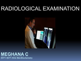
RADIOLOGICAL EXAMINATION
RADIOLOGICAL EXAMINATIONS
I. TYPES OF RADIOLOGIC EQUIPMENT
& ACCESSARIES
II. RADIOLOGIC DIAGNOSTIC
PROCEDURES
III. INTERVENTIONAL RADIOLOGY
TYPES OF RADIOLOGIC EQUIPMENT &
ACCESSARIES
1. Contrast media
2. Radiolucent gasses
3. Radiologic tabel
4. Cassete
5. Fixed x-ray equipment
6. Portable x-ray machine
7. Fluroscope
8. Image intensifier
9. Mobile c-arm image intensifier
10.Computerized digital subtraction processor
RADIOLOGIC DIAGNOSTIC
PROCEDURE
LIST OF RADIOLOGIC DIAGNOSTIC PROCEDURES
CARRIED OUT FOR RADIIOLOGIC EXAMINATIONS
1. Chest x-rays
2. Mammography
3. Steriotactic core breast tissue biopsy
4. Computed tomography
5. Ventriculography
6. Arthrography
7. Angiography & arteriography
8. Bronchography
9. Cholangiography
10. Myelography
11. Git x-ray studies
12. Urography
13. Incidental x-ray films
CHEST X-RAYS
An x-ray study of the chest
may be the part of the
admission procedure to rule
out un suspected pulmonary
diseases that would
contraindicate the use of
inhalational anaesthetic
agents
MOMMOGRAPHY
 A technique for projecting an x-
ray image of soft tissue of the
breast
 mammography is the most
effective screening method for
rear diagnosis of small, palpable
breast tumours
 This procedure may be some
what painfull for the woman ,
because compression of breast
is need for radioloogic imaging.
STERIOTACTIC CORE BREAST TISSUE
BIOPSY
 Imaging equipment is used
to steriotactically isolate
breast lesions that may not
be palpable.
 Percutanious needle
biopsies are performed
with the patient under local
anaesthesia.
VENTRICULOGRAPHY
 Ventricuclogrphy is the study
of the ventricles after
injections of a directly into the
lateral ventricles of the brain.
 If intracranial pressure
Becomes too great after gas
injection, a needle can be
inserted to remove the gas
 It can be used in patients with
signs of increased intra
cranial presure as a result of
Blockade of csf cerebrospinal
fluid circulation.
COMPUTED TOMOGRAPHY
 The x-ray beam moves back & forth across the body to project cross-sectional images this
technique is refered to as computed tomogaphy(ct), computed axial tomography (cat) or simply
scanning.
 It produces a highly contrasted, detailed study of normal & pathologic anatomy
 The exact size & location of lesiion in the brain media stainum & abdominal organs are identified
 Ct scan become an invasive procedure when a radiopaque contrast medium is used to examine
the git.
 Needle aspration can be performed under direct ct visualization
 Ct exposes the patient to ionizing radiations.
 Ct exposes the patient to allergic reactions to contrast medium, if used.
 To ensuure the proper use of this complex equuipment & t protect the patient from
unnecessary/excess radiation this procedure is done under the supervision of the quqlified
radiologist.
ARTHROGRAPHY
 Arthrogaphy is the study of
joint aftr the use of the gas
or cntrast medium into it.
 Through injection of the dye
or gas iinjury to cartilage or
ligaments can be visualized
 Arthrography can beusefull
in knee arthrogram.
ARTHROGRAPHY
ANGIOGRAPHYAND
ARTERIOGRAPHY
 Angiography is the study of the circulatory system after
injectiion of the radiopaque to permit visualization of
the venous blood vessell system.
 These procedure are usefull in the diffferential
diagnosis of arteriovenous malformations,
aneurysms,tumours, vascular accidents, or other
circulatory abnormalities caused either by traumatic
injury or by an acquired structural diseases.
 Angiography is also used at the time of the surgical
procedure to identify the exact location of some types
of lesions in the extremities brain, thoracic &
abdominal cavity .
BRONCHOGRAPHY
 Study of the tracheobronchial tree.
 This is donebyinstallation of a contrast
medium to aid in the diagnosis of the
bronchiectasis, cancer, tuberculosis, and
lung abscessor to detect a foreign body
 The location of a lesion can be determined
and the surgical procedure planned
accordiinngly.
CHOLANGIOGRAPHY
Radiography of the biliary ductus after injection or the administration
of the contrast medium, orally intravenously or percutaniously.
PREOPERATIVE COLANGIOGRAPHY:
In addition to pre operative x-ray diagnostic studies some surgeons
request request radiologic studies in conjunction with cholecystectomy
or cholilithotomy to identify gallstones in the biliary tract.
OPERATIVE CHOLANGIOGRAPHY
Cholangiography performed during a surgical
Procedure on the gall bladder.
Here surgeons include the cholangiography at the time of surgical
procedure in patients in whom they suspect stones might be present in
the bile duct.
The basic difference between pre operative & intra operative
cholangiography is the sit e of administration.
For pre operative cholangiography the contrast medium is injected iv through
percutaneous veinpuncture.
For intra operative cholangiography medium is directaly injection to bile duct.
GASTROINTESTINAL X-RAY
STUDIES
 Studies are performed to identify lesions
in the mucosa of the GI tract, such as an
ulcer, tumour or stricture.
 Barium sulphate is swallowed by the
patients or instilled by enema to be
studied.
MYELOGRAPHY
 Lesions in the spinal canal are studied by
the myelograph.
 It helpfull to localize a filling defects,
spinalcord tumour.
UROGRAPHY
 Urography is the radiologic study of the urinary
tract.
 Urographic studies are described as follows.
 1.cystography
 2.Cystourethrography
 3.Intravenous pyelography (IVP):study of the
structure of the urinary tract and kidney
functions.
 4.Retrogradepyelography: the study of the shape
and position of the kidney & ureter.
 Retrograde pyelography is used to visualize the
renal pelves.
 Voiding cystourethrography:the study of contour
& patiency of the urethra.
INCIDENTAL X-RAY FILMS
 An unanticipated need for an x-ray films
occurs when a sponge, needle, or
instrument is taken during wound closure.
 A x ray film will confirm wether the items is
still in the patient.
INTERVENTIONAL RADIOLOGY
 Invasive procedures performed under radiological control.
Examples:
a) balloon angioplasty
b) Coronary angioplasty stent placement
c) Inferior vena cava fitter placement.
Cardiac catheterization, angioplasty & stent placement are
Performed in a interventional radiology department.
FOLLOWING ARE SOME OF THE
INTERVENTIONAL RADIOLOGY
TECHNIQUES
1. Magnetic Resonance Imaging (MRI)
2. Ultrasonography
3. Plethysmography
4. Endoscopy
5. Nuclear medicine studies
 Radionuclide
 Total body scanning
 PET Scan
 Scintigraphy
 Lymphoscintigraphy (lymph node mapping)
MRI is based on the magnetic properties of the hydrogen
in the body
Unlike CT Scan MRI does not use radiations
The patients lies flat inside a large electromagnet
MRI looks at both the body’s structure & functions.
It distinguishes between fat, muscles, compact bone &
bonemarrow, brain & spinal cord, fluid filled cavities,
ligaments
& tendons and blood vessels.
MAGNETIC RESONANCE IMAGING
(MRI)
THE MAJOR APPLICATIONS OF
MRI ARE AS FOLLOWS
1. Detection of tumours
2. Inflammatory diseases
3. Infections
4. Abscesses
5. Used in the evaluation of the function of
central nervous system, cardiovascular
system, & other organs.
 MRI paramagnetic IV Contrast media, such
as like gadopentetate,are sometimes used to
localize tumours in the central nervous
system.
 MRI paramagnetic IV contrast media
(GADOPENTETATE) do not contain iodine,
and allergic reactions are rare.
 The magnetic field can cause ferromagnetic
components of implants, such as
pacemakers & cochlear implants, to
malfunctions.
 The magnetic field will also disable
metallic device in the area, such as
cardiac monitors, infusions pumps &
wristwatches.
 Some patients feel a sense of
claustrophobia in a conventional MRI
Machine.
 MRI is used during selected neurosurgical
procedures.
 Dedicated MRI Interventional rooms
permit the use of MRI during the actual
ULTRASONOGRAPHY
 Ultrasonography is a technique uses
ehoes of the ultrasound pulses to
delineate objects or areas of different
density in the body.
 Ultrasonography uses sound waves to
produce images on a screen, which allows
medical providers to view internal
structures of the body.
 The basic component of any diagnostic
ultrasound system is its specialized
 The transducer converts electrical
impulses to ultrasonic waves at
afrequency greater than 1 million cycles
per second.
 These ultrasonic frequencies are
transmitted into tissues through a
transducer placed on skin.
 Water soluble gel is placed to the skin to
maintain air tight contact between the skin
and the transducer, because ltrasonic
waves does not travel well through air.
 Ultrasound is not effective in the presence
of bone or gas in the gastrointestinal tract.
 The image can be recorded on video
tapes or printed to provide a permanent
record known as echogram or sonogram.
 Ultrasonogrphy does not expose the
patient to radiation & contrast medium.
ADVANTAGES OF
ULTRASONOGRAPHY
 Whether used as a preoperative or intra-
operative diagnostic technique ultrasonography
is ;
1. Ultrasonography is rapid, painless, non invasive
procedure.
2. Ultrasonography distinguishes between fluid-
filled & solid masses.
3. Ultrasonography is less time consuming
procedure.
4. Ultrasonography requires less tissue
manipulation than do other intraoperative
diagnostic procedure.
5. Ultrasonography does not exposes the patient
USES OF
ULTRASONNOGRAPHY
 Ultrasonography is a useful adjunct in the diagnosis of
the followings;
1. Space occupying lesions in the neonatal brain:- the
ECHOENCEPHALOGRAM will show a shift of the
brain caused by tumour
2. Ultrasound can distinguish between a cystic & a solid
tumour mass in the kidney, pancrea liver ovaries &
testies.
3. Ultrasonography is the best imaging technique in the
patients gallbladder diseases.
Emboli (air, blood, fat)
• Ultrasonography is particularly useful in
the early diagnosis of pulmonary
embolism.
• Ultrasonography is useful to determine
the need for a caesarean section (c-
section) if the head is too large for
vaginal delivery.
• Ultrasonography is also used to
determine the position of the fetes &
 Ultrasonography is used to identify the
gender and to detect foetal abnormalities.
 ECHOCARDIOGRAPHY: (ultrasonic
cardiography).
Cardiac defects like structural defect,
insufficient valvular movement, and blood
flow volumes within the heart chambers &
myocardium can be detected by this
diagnostic technique known as
ECHOCARDIOGRAPHY.
DOPPLER STUDIES
 The Doppler ultrasonic velocitydetector
emits a beam of 5-10 megahertz (mh2)
the is directed through the skin into the
blood stream.
 Doppler originally

Weitere ähnliche Inhalte

Was ist angesagt? (20)

ANES 1502 - M14 PPT: Diagnostic Procedures
ANES 1502 - M14 PPT: Diagnostic ProceduresANES 1502 - M14 PPT: Diagnostic Procedures
ANES 1502 - M14 PPT: Diagnostic Procedures
 
BIOPSY
BIOPSYBIOPSY
BIOPSY
 
Angiography
AngiographyAngiography
Angiography
 
INTRAVENOUS UROGRAM (IVU)
INTRAVENOUS UROGRAM (IVU)INTRAVENOUS UROGRAM (IVU)
INTRAVENOUS UROGRAM (IVU)
 
Intravenous pyelogram
Intravenous pyelogram Intravenous pyelogram
Intravenous pyelogram
 
Ultrasonography
UltrasonographyUltrasonography
Ultrasonography
 
Surgical dressings i
Surgical dressings  iSurgical dressings  i
Surgical dressings i
 
Myelogram
MyelogramMyelogram
Myelogram
 
Ct and mri preparation
Ct and mri preparationCt and mri preparation
Ct and mri preparation
 
SUTURE AND SUTURE MATERIALS IN SURGERY
SUTURE AND SUTURE MATERIALS IN SURGERYSUTURE AND SUTURE MATERIALS IN SURGERY
SUTURE AND SUTURE MATERIALS IN SURGERY
 
BRONCHOSCOPY
BRONCHOSCOPYBRONCHOSCOPY
BRONCHOSCOPY
 
Gastrostomy
GastrostomyGastrostomy
Gastrostomy
 
Basic surgical instrumentation
Basic surgical instrumentationBasic surgical instrumentation
Basic surgical instrumentation
 
Endoscopy
EndoscopyEndoscopy
Endoscopy
 
Surgical draping
Surgical drapingSurgical draping
Surgical draping
 
Cystoscopy /uretroscopy
Cystoscopy /uretroscopyCystoscopy /uretroscopy
Cystoscopy /uretroscopy
 
Surgical Scrubbing Gowning Gloving
Surgical Scrubbing Gowning GlovingSurgical Scrubbing Gowning Gloving
Surgical Scrubbing Gowning Gloving
 
Endoscopy
EndoscopyEndoscopy
Endoscopy
 
Patient preparation for radiological procedure
Patient preparation for radiological procedurePatient preparation for radiological procedure
Patient preparation for radiological procedure
 
BARIUM ENEMA
BARIUM ENEMABARIUM ENEMA
BARIUM ENEMA
 

Ähnlich wie Radiological Examinations

Recent Advances in Interventional Radiologic Proceedures
Recent Advances in Interventional Radiologic ProceeduresRecent Advances in Interventional Radiologic Proceedures
Recent Advances in Interventional Radiologic ProceeduresMUHAMMED SWALIH MP
 
Radiologically guided fnac
Radiologically guided fnacRadiologically guided fnac
Radiologically guided fnacsonali timaniya
 
laboratory tests part 3
laboratory tests part 3laboratory tests part 3
laboratory tests part 3jhonee balmeo
 
Minimal access surgery
Minimal access surgeryMinimal access surgery
Minimal access surgeryAjayKumar4497
 
1) Abdomen & Pelvis Radiography for helminthic diseases.pptx
1) Abdomen & Pelvis Radiography for helminthic diseases.pptx1) Abdomen & Pelvis Radiography for helminthic diseases.pptx
1) Abdomen & Pelvis Radiography for helminthic diseases.pptxIbrahimAboAlasaad
 
Special contract X-ray methods of examination
Special contract X-ray methods of examinationSpecial contract X-ray methods of examination
Special contract X-ray methods of examinationEneutron
 
Ultrasound guided interventional procedure
Ultrasound guided interventional procedureUltrasound guided interventional procedure
Ultrasound guided interventional procedureSyed Yousaf Gilani
 
PRESENTATION. old rad techniques.pptx
PRESENTATION. old rad techniques.pptxPRESENTATION. old rad techniques.pptx
PRESENTATION. old rad techniques.pptxmohamedabushanab1
 
Robotic GI surgery
Robotic GI surgeryRobotic GI surgery
Robotic GI surgeryMahesh Raj
 
Diagnostic radiography in Ghana
Diagnostic radiography in GhanaDiagnostic radiography in Ghana
Diagnostic radiography in GhanaDomlan Mathias
 
medical thoracoscopy2022
medical thoracoscopy2022medical thoracoscopy2022
medical thoracoscopy2022rambhoopal1
 
Dr.CSS_Medical X ray Imaging Equipments in Healthcare
Dr.CSS_Medical X ray Imaging Equipments in HealthcareDr.CSS_Medical X ray Imaging Equipments in Healthcare
Dr.CSS_Medical X ray Imaging Equipments in HealthcareDr.C.Senthamil Selvan Ph.D.
 
White Paper, Esaote VNAV in Endocrinology
White Paper, Esaote VNAV in EndocrinologyWhite Paper, Esaote VNAV in Endocrinology
White Paper, Esaote VNAV in EndocrinologyMIDEAS
 
behgal cancer institute
behgal cancer  institute behgal cancer  institute
behgal cancer institute Dr Behgal K S
 
Radiology_Equipment_Lec-1_Dr. Emad Taleb.pdf
Radiology_Equipment_Lec-1_Dr. Emad Taleb.pdfRadiology_Equipment_Lec-1_Dr. Emad Taleb.pdf
Radiology_Equipment_Lec-1_Dr. Emad Taleb.pdfEmadTaleb1
 

Ähnlich wie Radiological Examinations (20)

Recent Advances in Interventional Radiologic Proceedures
Recent Advances in Interventional Radiologic ProceeduresRecent Advances in Interventional Radiologic Proceedures
Recent Advances in Interventional Radiologic Proceedures
 
Radiologically guided fnac
Radiologically guided fnacRadiologically guided fnac
Radiologically guided fnac
 
Usg 4 surgeons
Usg 4 surgeonsUsg 4 surgeons
Usg 4 surgeons
 
laboratory tests part 3
laboratory tests part 3laboratory tests part 3
laboratory tests part 3
 
Minimal access surgery
Minimal access surgeryMinimal access surgery
Minimal access surgery
 
1) Abdomen & Pelvis Radiography for helminthic diseases.pptx
1) Abdomen & Pelvis Radiography for helminthic diseases.pptx1) Abdomen & Pelvis Radiography for helminthic diseases.pptx
1) Abdomen & Pelvis Radiography for helminthic diseases.pptx
 
Investigations in urology
Investigations in urologyInvestigations in urology
Investigations in urology
 
Biomedical Imaging Informatics
Biomedical Imaging InformaticsBiomedical Imaging Informatics
Biomedical Imaging Informatics
 
Special contract X-ray methods of examination
Special contract X-ray methods of examinationSpecial contract X-ray methods of examination
Special contract X-ray methods of examination
 
Ultrasound guided interventional procedure
Ultrasound guided interventional procedureUltrasound guided interventional procedure
Ultrasound guided interventional procedure
 
Ct urography/ivp
Ct urography/ivpCt urography/ivp
Ct urography/ivp
 
PRESENTATION. old rad techniques.pptx
PRESENTATION. old rad techniques.pptxPRESENTATION. old rad techniques.pptx
PRESENTATION. old rad techniques.pptx
 
Robotic GI surgery
Robotic GI surgeryRobotic GI surgery
Robotic GI surgery
 
Diagnostic radiography in Ghana
Diagnostic radiography in GhanaDiagnostic radiography in Ghana
Diagnostic radiography in Ghana
 
medical thoracoscopy2022
medical thoracoscopy2022medical thoracoscopy2022
medical thoracoscopy2022
 
Dr.CSS_Medical X ray Imaging Equipments in Healthcare
Dr.CSS_Medical X ray Imaging Equipments in HealthcareDr.CSS_Medical X ray Imaging Equipments in Healthcare
Dr.CSS_Medical X ray Imaging Equipments in Healthcare
 
White Paper, Esaote VNAV in Endocrinology
White Paper, Esaote VNAV in EndocrinologyWhite Paper, Esaote VNAV in Endocrinology
White Paper, Esaote VNAV in Endocrinology
 
Radiology
RadiologyRadiology
Radiology
 
behgal cancer institute
behgal cancer  institute behgal cancer  institute
behgal cancer institute
 
Radiology_Equipment_Lec-1_Dr. Emad Taleb.pdf
Radiology_Equipment_Lec-1_Dr. Emad Taleb.pdfRadiology_Equipment_Lec-1_Dr. Emad Taleb.pdf
Radiology_Equipment_Lec-1_Dr. Emad Taleb.pdf
 

Mehr von MEGHANA C

PLASMA PROTEIN (2).pptx
PLASMA PROTEIN (2).pptxPLASMA PROTEIN (2).pptx
PLASMA PROTEIN (2).pptxMEGHANA C
 
AMINO ACID & PROTEIN CHEMISTRY.pptx
AMINO ACID & PROTEIN CHEMISTRY.pptxAMINO ACID & PROTEIN CHEMISTRY.pptx
AMINO ACID & PROTEIN CHEMISTRY.pptxMEGHANA C
 
Pharmacology
PharmacologyPharmacology
PharmacologyMEGHANA C
 
Cerebro vascular accident
Cerebro vascular accidentCerebro vascular accident
Cerebro vascular accidentMEGHANA C
 
Cerebro vascular accident
Cerebro vascular accidentCerebro vascular accident
Cerebro vascular accidentMEGHANA C
 
Pre anesthetic medications by Meghana C
Pre anesthetic medications by Meghana CPre anesthetic medications by Meghana C
Pre anesthetic medications by Meghana CMEGHANA C
 
Intravenous Anestetics by Meghana C
Intravenous Anestetics by Meghana CIntravenous Anestetics by Meghana C
Intravenous Anestetics by Meghana CMEGHANA C
 
Nasogastric tube (ryles tube intubation) by meghana c
Nasogastric tube (ryles tube intubation) by meghana cNasogastric tube (ryles tube intubation) by meghana c
Nasogastric tube (ryles tube intubation) by meghana cMEGHANA C
 
Radioimmunoassay (modified copy)
Radioimmunoassay (modified copy)Radioimmunoassay (modified copy)
Radioimmunoassay (modified copy)MEGHANA C
 

Mehr von MEGHANA C (9)

PLASMA PROTEIN (2).pptx
PLASMA PROTEIN (2).pptxPLASMA PROTEIN (2).pptx
PLASMA PROTEIN (2).pptx
 
AMINO ACID & PROTEIN CHEMISTRY.pptx
AMINO ACID & PROTEIN CHEMISTRY.pptxAMINO ACID & PROTEIN CHEMISTRY.pptx
AMINO ACID & PROTEIN CHEMISTRY.pptx
 
Pharmacology
PharmacologyPharmacology
Pharmacology
 
Cerebro vascular accident
Cerebro vascular accidentCerebro vascular accident
Cerebro vascular accident
 
Cerebro vascular accident
Cerebro vascular accidentCerebro vascular accident
Cerebro vascular accident
 
Pre anesthetic medications by Meghana C
Pre anesthetic medications by Meghana CPre anesthetic medications by Meghana C
Pre anesthetic medications by Meghana C
 
Intravenous Anestetics by Meghana C
Intravenous Anestetics by Meghana CIntravenous Anestetics by Meghana C
Intravenous Anestetics by Meghana C
 
Nasogastric tube (ryles tube intubation) by meghana c
Nasogastric tube (ryles tube intubation) by meghana cNasogastric tube (ryles tube intubation) by meghana c
Nasogastric tube (ryles tube intubation) by meghana c
 
Radioimmunoassay (modified copy)
Radioimmunoassay (modified copy)Radioimmunoassay (modified copy)
Radioimmunoassay (modified copy)
 

Kürzlich hochgeladen

Advanced Views - Calendar View in Odoo 17
Advanced Views - Calendar View in Odoo 17Advanced Views - Calendar View in Odoo 17
Advanced Views - Calendar View in Odoo 17Celine George
 
Disha NEET Physics Guide for classes 11 and 12.pdf
Disha NEET Physics Guide for classes 11 and 12.pdfDisha NEET Physics Guide for classes 11 and 12.pdf
Disha NEET Physics Guide for classes 11 and 12.pdfchloefrazer622
 
Measures of Central Tendency: Mean, Median and Mode
Measures of Central Tendency: Mean, Median and ModeMeasures of Central Tendency: Mean, Median and Mode
Measures of Central Tendency: Mean, Median and ModeThiyagu K
 
Mastering the Unannounced Regulatory Inspection
Mastering the Unannounced Regulatory InspectionMastering the Unannounced Regulatory Inspection
Mastering the Unannounced Regulatory InspectionSafetyChain Software
 
Call Girls in Dwarka Mor Delhi Contact Us 9654467111
Call Girls in Dwarka Mor Delhi Contact Us 9654467111Call Girls in Dwarka Mor Delhi Contact Us 9654467111
Call Girls in Dwarka Mor Delhi Contact Us 9654467111Sapana Sha
 
Web & Social Media Analytics Previous Year Question Paper.pdf
Web & Social Media Analytics Previous Year Question Paper.pdfWeb & Social Media Analytics Previous Year Question Paper.pdf
Web & Social Media Analytics Previous Year Question Paper.pdfJayanti Pande
 
Grant Readiness 101 TechSoup and Remy Consulting
Grant Readiness 101 TechSoup and Remy ConsultingGrant Readiness 101 TechSoup and Remy Consulting
Grant Readiness 101 TechSoup and Remy ConsultingTechSoup
 
Beyond the EU: DORA and NIS 2 Directive's Global Impact
Beyond the EU: DORA and NIS 2 Directive's Global ImpactBeyond the EU: DORA and NIS 2 Directive's Global Impact
Beyond the EU: DORA and NIS 2 Directive's Global ImpactPECB
 
microwave assisted reaction. General introduction
microwave assisted reaction. General introductionmicrowave assisted reaction. General introduction
microwave assisted reaction. General introductionMaksud Ahmed
 
POINT- BIOCHEMISTRY SEM 2 ENZYMES UNIT 5.pptx
POINT- BIOCHEMISTRY SEM 2 ENZYMES UNIT 5.pptxPOINT- BIOCHEMISTRY SEM 2 ENZYMES UNIT 5.pptx
POINT- BIOCHEMISTRY SEM 2 ENZYMES UNIT 5.pptxSayali Powar
 
Q4-W6-Restating Informational Text Grade 3
Q4-W6-Restating Informational Text Grade 3Q4-W6-Restating Informational Text Grade 3
Q4-W6-Restating Informational Text Grade 3JemimahLaneBuaron
 
1029 - Danh muc Sach Giao Khoa 10 . pdf
1029 -  Danh muc Sach Giao Khoa 10 . pdf1029 -  Danh muc Sach Giao Khoa 10 . pdf
1029 - Danh muc Sach Giao Khoa 10 . pdfQucHHunhnh
 
Ecosystem Interactions Class Discussion Presentation in Blue Green Lined Styl...
Ecosystem Interactions Class Discussion Presentation in Blue Green Lined Styl...Ecosystem Interactions Class Discussion Presentation in Blue Green Lined Styl...
Ecosystem Interactions Class Discussion Presentation in Blue Green Lined Styl...fonyou31
 
1029-Danh muc Sach Giao Khoa khoi 6.pdf
1029-Danh muc Sach Giao Khoa khoi  6.pdf1029-Danh muc Sach Giao Khoa khoi  6.pdf
1029-Danh muc Sach Giao Khoa khoi 6.pdfQucHHunhnh
 
JAPAN: ORGANISATION OF PMDA, PHARMACEUTICAL LAWS & REGULATIONS, TYPES OF REGI...
JAPAN: ORGANISATION OF PMDA, PHARMACEUTICAL LAWS & REGULATIONS, TYPES OF REGI...JAPAN: ORGANISATION OF PMDA, PHARMACEUTICAL LAWS & REGULATIONS, TYPES OF REGI...
JAPAN: ORGANISATION OF PMDA, PHARMACEUTICAL LAWS & REGULATIONS, TYPES OF REGI...anjaliyadav012327
 
Software Engineering Methodologies (overview)
Software Engineering Methodologies (overview)Software Engineering Methodologies (overview)
Software Engineering Methodologies (overview)eniolaolutunde
 
Introduction to Nonprofit Accounting: The Basics
Introduction to Nonprofit Accounting: The BasicsIntroduction to Nonprofit Accounting: The Basics
Introduction to Nonprofit Accounting: The BasicsTechSoup
 
Interactive Powerpoint_How to Master effective communication
Interactive Powerpoint_How to Master effective communicationInteractive Powerpoint_How to Master effective communication
Interactive Powerpoint_How to Master effective communicationnomboosow
 

Kürzlich hochgeladen (20)

Advanced Views - Calendar View in Odoo 17
Advanced Views - Calendar View in Odoo 17Advanced Views - Calendar View in Odoo 17
Advanced Views - Calendar View in Odoo 17
 
Disha NEET Physics Guide for classes 11 and 12.pdf
Disha NEET Physics Guide for classes 11 and 12.pdfDisha NEET Physics Guide for classes 11 and 12.pdf
Disha NEET Physics Guide for classes 11 and 12.pdf
 
Measures of Central Tendency: Mean, Median and Mode
Measures of Central Tendency: Mean, Median and ModeMeasures of Central Tendency: Mean, Median and Mode
Measures of Central Tendency: Mean, Median and Mode
 
Mastering the Unannounced Regulatory Inspection
Mastering the Unannounced Regulatory InspectionMastering the Unannounced Regulatory Inspection
Mastering the Unannounced Regulatory Inspection
 
Call Girls in Dwarka Mor Delhi Contact Us 9654467111
Call Girls in Dwarka Mor Delhi Contact Us 9654467111Call Girls in Dwarka Mor Delhi Contact Us 9654467111
Call Girls in Dwarka Mor Delhi Contact Us 9654467111
 
Web & Social Media Analytics Previous Year Question Paper.pdf
Web & Social Media Analytics Previous Year Question Paper.pdfWeb & Social Media Analytics Previous Year Question Paper.pdf
Web & Social Media Analytics Previous Year Question Paper.pdf
 
Código Creativo y Arte de Software | Unidad 1
Código Creativo y Arte de Software | Unidad 1Código Creativo y Arte de Software | Unidad 1
Código Creativo y Arte de Software | Unidad 1
 
Mattingly "AI & Prompt Design: Structured Data, Assistants, & RAG"
Mattingly "AI & Prompt Design: Structured Data, Assistants, & RAG"Mattingly "AI & Prompt Design: Structured Data, Assistants, & RAG"
Mattingly "AI & Prompt Design: Structured Data, Assistants, & RAG"
 
Grant Readiness 101 TechSoup and Remy Consulting
Grant Readiness 101 TechSoup and Remy ConsultingGrant Readiness 101 TechSoup and Remy Consulting
Grant Readiness 101 TechSoup and Remy Consulting
 
Beyond the EU: DORA and NIS 2 Directive's Global Impact
Beyond the EU: DORA and NIS 2 Directive's Global ImpactBeyond the EU: DORA and NIS 2 Directive's Global Impact
Beyond the EU: DORA and NIS 2 Directive's Global Impact
 
microwave assisted reaction. General introduction
microwave assisted reaction. General introductionmicrowave assisted reaction. General introduction
microwave assisted reaction. General introduction
 
POINT- BIOCHEMISTRY SEM 2 ENZYMES UNIT 5.pptx
POINT- BIOCHEMISTRY SEM 2 ENZYMES UNIT 5.pptxPOINT- BIOCHEMISTRY SEM 2 ENZYMES UNIT 5.pptx
POINT- BIOCHEMISTRY SEM 2 ENZYMES UNIT 5.pptx
 
Q4-W6-Restating Informational Text Grade 3
Q4-W6-Restating Informational Text Grade 3Q4-W6-Restating Informational Text Grade 3
Q4-W6-Restating Informational Text Grade 3
 
1029 - Danh muc Sach Giao Khoa 10 . pdf
1029 -  Danh muc Sach Giao Khoa 10 . pdf1029 -  Danh muc Sach Giao Khoa 10 . pdf
1029 - Danh muc Sach Giao Khoa 10 . pdf
 
Ecosystem Interactions Class Discussion Presentation in Blue Green Lined Styl...
Ecosystem Interactions Class Discussion Presentation in Blue Green Lined Styl...Ecosystem Interactions Class Discussion Presentation in Blue Green Lined Styl...
Ecosystem Interactions Class Discussion Presentation in Blue Green Lined Styl...
 
1029-Danh muc Sach Giao Khoa khoi 6.pdf
1029-Danh muc Sach Giao Khoa khoi  6.pdf1029-Danh muc Sach Giao Khoa khoi  6.pdf
1029-Danh muc Sach Giao Khoa khoi 6.pdf
 
JAPAN: ORGANISATION OF PMDA, PHARMACEUTICAL LAWS & REGULATIONS, TYPES OF REGI...
JAPAN: ORGANISATION OF PMDA, PHARMACEUTICAL LAWS & REGULATIONS, TYPES OF REGI...JAPAN: ORGANISATION OF PMDA, PHARMACEUTICAL LAWS & REGULATIONS, TYPES OF REGI...
JAPAN: ORGANISATION OF PMDA, PHARMACEUTICAL LAWS & REGULATIONS, TYPES OF REGI...
 
Software Engineering Methodologies (overview)
Software Engineering Methodologies (overview)Software Engineering Methodologies (overview)
Software Engineering Methodologies (overview)
 
Introduction to Nonprofit Accounting: The Basics
Introduction to Nonprofit Accounting: The BasicsIntroduction to Nonprofit Accounting: The Basics
Introduction to Nonprofit Accounting: The Basics
 
Interactive Powerpoint_How to Master effective communication
Interactive Powerpoint_How to Master effective communicationInteractive Powerpoint_How to Master effective communication
Interactive Powerpoint_How to Master effective communication
 

Radiological Examinations

  • 1. MEGHANA C DOTT, BOTT, M.Sc Med.Biochemistry RADIOLOGICAL EXAMINATION
  • 2. RADIOLOGICAL EXAMINATIONS I. TYPES OF RADIOLOGIC EQUIPMENT & ACCESSARIES II. RADIOLOGIC DIAGNOSTIC PROCEDURES III. INTERVENTIONAL RADIOLOGY
  • 3. TYPES OF RADIOLOGIC EQUIPMENT & ACCESSARIES 1. Contrast media 2. Radiolucent gasses 3. Radiologic tabel 4. Cassete 5. Fixed x-ray equipment 6. Portable x-ray machine 7. Fluroscope 8. Image intensifier 9. Mobile c-arm image intensifier 10.Computerized digital subtraction processor
  • 4. RADIOLOGIC DIAGNOSTIC PROCEDURE LIST OF RADIOLOGIC DIAGNOSTIC PROCEDURES CARRIED OUT FOR RADIIOLOGIC EXAMINATIONS 1. Chest x-rays 2. Mammography 3. Steriotactic core breast tissue biopsy 4. Computed tomography 5. Ventriculography 6. Arthrography 7. Angiography & arteriography 8. Bronchography 9. Cholangiography 10. Myelography 11. Git x-ray studies 12. Urography 13. Incidental x-ray films
  • 5. CHEST X-RAYS An x-ray study of the chest may be the part of the admission procedure to rule out un suspected pulmonary diseases that would contraindicate the use of inhalational anaesthetic agents
  • 6. MOMMOGRAPHY  A technique for projecting an x- ray image of soft tissue of the breast  mammography is the most effective screening method for rear diagnosis of small, palpable breast tumours  This procedure may be some what painfull for the woman , because compression of breast is need for radioloogic imaging.
  • 7. STERIOTACTIC CORE BREAST TISSUE BIOPSY  Imaging equipment is used to steriotactically isolate breast lesions that may not be palpable.  Percutanious needle biopsies are performed with the patient under local anaesthesia.
  • 8. VENTRICULOGRAPHY  Ventricuclogrphy is the study of the ventricles after injections of a directly into the lateral ventricles of the brain.  If intracranial pressure Becomes too great after gas injection, a needle can be inserted to remove the gas  It can be used in patients with signs of increased intra cranial presure as a result of Blockade of csf cerebrospinal fluid circulation.
  • 9. COMPUTED TOMOGRAPHY  The x-ray beam moves back & forth across the body to project cross-sectional images this technique is refered to as computed tomogaphy(ct), computed axial tomography (cat) or simply scanning.  It produces a highly contrasted, detailed study of normal & pathologic anatomy  The exact size & location of lesiion in the brain media stainum & abdominal organs are identified  Ct scan become an invasive procedure when a radiopaque contrast medium is used to examine the git.  Needle aspration can be performed under direct ct visualization  Ct exposes the patient to ionizing radiations.  Ct exposes the patient to allergic reactions to contrast medium, if used.  To ensuure the proper use of this complex equuipment & t protect the patient from unnecessary/excess radiation this procedure is done under the supervision of the quqlified radiologist.
  • 10.
  • 11. ARTHROGRAPHY  Arthrogaphy is the study of joint aftr the use of the gas or cntrast medium into it.  Through injection of the dye or gas iinjury to cartilage or ligaments can be visualized  Arthrography can beusefull in knee arthrogram.
  • 13. ANGIOGRAPHYAND ARTERIOGRAPHY  Angiography is the study of the circulatory system after injectiion of the radiopaque to permit visualization of the venous blood vessell system.  These procedure are usefull in the diffferential diagnosis of arteriovenous malformations, aneurysms,tumours, vascular accidents, or other circulatory abnormalities caused either by traumatic injury or by an acquired structural diseases.  Angiography is also used at the time of the surgical procedure to identify the exact location of some types of lesions in the extremities brain, thoracic & abdominal cavity .
  • 14. BRONCHOGRAPHY  Study of the tracheobronchial tree.  This is donebyinstallation of a contrast medium to aid in the diagnosis of the bronchiectasis, cancer, tuberculosis, and lung abscessor to detect a foreign body  The location of a lesion can be determined and the surgical procedure planned accordiinngly.
  • 15. CHOLANGIOGRAPHY Radiography of the biliary ductus after injection or the administration of the contrast medium, orally intravenously or percutaniously. PREOPERATIVE COLANGIOGRAPHY: In addition to pre operative x-ray diagnostic studies some surgeons request request radiologic studies in conjunction with cholecystectomy or cholilithotomy to identify gallstones in the biliary tract. OPERATIVE CHOLANGIOGRAPHY Cholangiography performed during a surgical Procedure on the gall bladder. Here surgeons include the cholangiography at the time of surgical procedure in patients in whom they suspect stones might be present in the bile duct. The basic difference between pre operative & intra operative cholangiography is the sit e of administration. For pre operative cholangiography the contrast medium is injected iv through percutaneous veinpuncture. For intra operative cholangiography medium is directaly injection to bile duct.
  • 16. GASTROINTESTINAL X-RAY STUDIES  Studies are performed to identify lesions in the mucosa of the GI tract, such as an ulcer, tumour or stricture.  Barium sulphate is swallowed by the patients or instilled by enema to be studied.
  • 17. MYELOGRAPHY  Lesions in the spinal canal are studied by the myelograph.  It helpfull to localize a filling defects, spinalcord tumour.
  • 18. UROGRAPHY  Urography is the radiologic study of the urinary tract.  Urographic studies are described as follows.  1.cystography  2.Cystourethrography  3.Intravenous pyelography (IVP):study of the structure of the urinary tract and kidney functions.  4.Retrogradepyelography: the study of the shape and position of the kidney & ureter.  Retrograde pyelography is used to visualize the renal pelves.  Voiding cystourethrography:the study of contour & patiency of the urethra.
  • 19. INCIDENTAL X-RAY FILMS  An unanticipated need for an x-ray films occurs when a sponge, needle, or instrument is taken during wound closure.  A x ray film will confirm wether the items is still in the patient.
  • 20. INTERVENTIONAL RADIOLOGY  Invasive procedures performed under radiological control. Examples: a) balloon angioplasty b) Coronary angioplasty stent placement c) Inferior vena cava fitter placement. Cardiac catheterization, angioplasty & stent placement are Performed in a interventional radiology department.
  • 21. FOLLOWING ARE SOME OF THE INTERVENTIONAL RADIOLOGY TECHNIQUES 1. Magnetic Resonance Imaging (MRI) 2. Ultrasonography 3. Plethysmography 4. Endoscopy 5. Nuclear medicine studies  Radionuclide  Total body scanning  PET Scan  Scintigraphy  Lymphoscintigraphy (lymph node mapping)
  • 22. MRI is based on the magnetic properties of the hydrogen in the body Unlike CT Scan MRI does not use radiations The patients lies flat inside a large electromagnet MRI looks at both the body’s structure & functions. It distinguishes between fat, muscles, compact bone & bonemarrow, brain & spinal cord, fluid filled cavities, ligaments & tendons and blood vessels. MAGNETIC RESONANCE IMAGING (MRI)
  • 23. THE MAJOR APPLICATIONS OF MRI ARE AS FOLLOWS 1. Detection of tumours 2. Inflammatory diseases 3. Infections 4. Abscesses 5. Used in the evaluation of the function of central nervous system, cardiovascular system, & other organs.
  • 24.  MRI paramagnetic IV Contrast media, such as like gadopentetate,are sometimes used to localize tumours in the central nervous system.  MRI paramagnetic IV contrast media (GADOPENTETATE) do not contain iodine, and allergic reactions are rare.  The magnetic field can cause ferromagnetic components of implants, such as pacemakers & cochlear implants, to malfunctions.
  • 25.  The magnetic field will also disable metallic device in the area, such as cardiac monitors, infusions pumps & wristwatches.  Some patients feel a sense of claustrophobia in a conventional MRI Machine.  MRI is used during selected neurosurgical procedures.  Dedicated MRI Interventional rooms permit the use of MRI during the actual
  • 26. ULTRASONOGRAPHY  Ultrasonography is a technique uses ehoes of the ultrasound pulses to delineate objects or areas of different density in the body.  Ultrasonography uses sound waves to produce images on a screen, which allows medical providers to view internal structures of the body.  The basic component of any diagnostic ultrasound system is its specialized
  • 27.  The transducer converts electrical impulses to ultrasonic waves at afrequency greater than 1 million cycles per second.  These ultrasonic frequencies are transmitted into tissues through a transducer placed on skin.  Water soluble gel is placed to the skin to maintain air tight contact between the skin and the transducer, because ltrasonic waves does not travel well through air.
  • 28.  Ultrasound is not effective in the presence of bone or gas in the gastrointestinal tract.  The image can be recorded on video tapes or printed to provide a permanent record known as echogram or sonogram.  Ultrasonogrphy does not expose the patient to radiation & contrast medium.
  • 29. ADVANTAGES OF ULTRASONOGRAPHY  Whether used as a preoperative or intra- operative diagnostic technique ultrasonography is ; 1. Ultrasonography is rapid, painless, non invasive procedure. 2. Ultrasonography distinguishes between fluid- filled & solid masses. 3. Ultrasonography is less time consuming procedure. 4. Ultrasonography requires less tissue manipulation than do other intraoperative diagnostic procedure. 5. Ultrasonography does not exposes the patient
  • 30. USES OF ULTRASONNOGRAPHY  Ultrasonography is a useful adjunct in the diagnosis of the followings; 1. Space occupying lesions in the neonatal brain:- the ECHOENCEPHALOGRAM will show a shift of the brain caused by tumour 2. Ultrasound can distinguish between a cystic & a solid tumour mass in the kidney, pancrea liver ovaries & testies. 3. Ultrasonography is the best imaging technique in the patients gallbladder diseases.
  • 31. Emboli (air, blood, fat) • Ultrasonography is particularly useful in the early diagnosis of pulmonary embolism. • Ultrasonography is useful to determine the need for a caesarean section (c- section) if the head is too large for vaginal delivery. • Ultrasonography is also used to determine the position of the fetes &
  • 32.  Ultrasonography is used to identify the gender and to detect foetal abnormalities.  ECHOCARDIOGRAPHY: (ultrasonic cardiography). Cardiac defects like structural defect, insufficient valvular movement, and blood flow volumes within the heart chambers & myocardium can be detected by this diagnostic technique known as ECHOCARDIOGRAPHY.
  • 33. DOPPLER STUDIES  The Doppler ultrasonic velocitydetector emits a beam of 5-10 megahertz (mh2) the is directed through the skin into the blood stream.  Doppler originally