
Empfohlen
Weitere ähnliche Inhalte
Was ist angesagt?
Was ist angesagt? (20)
Andere mochten auch
Andere mochten auch (20)
Ähnlich wie Patellofemoral Pain Syndrome (Final)
Ähnlich wie Patellofemoral Pain Syndrome (Final) (20)
Patellofemoral Pain Syndrome (Final)
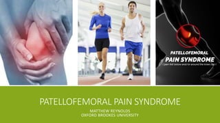
- 1. PATELLOFEMORAL PAIN SYNDROME MATTHEW REYNOLDS OXFORD BROOKES UNIVERSITY
- 2. WHY PFPS? • Two patients with PFPS • Initial unsuccessful treatments • Very little knowledge of condition • Vast/ unspecific understanding of causes • Two podcasts Got me interested!
- 3. CASE STUDY INTRODUCTION – HOW DID THEY PRESENT? ▪ 34 y/o lady ▪ Referred with 4 year history of left knee pain huge walk ▪ MRI = ? Patellar tendon impingement + Minor chondral degeneration ▪ Body Chart: 8/10 Lateroinferior Left Knee Pain (No neural involvement) ▪ Aggs: Hiking, Running, Prolonged Sitting, Driving ▪ Eases: Rest ▪ SH: Very active! Hiking 2/7, Walking, Therapeutic Care Worker ▪ Obs: TOP infrapatellar fat pad, patella maltracking laterally ▪ ROM: Knee = Full / PFJ = Reduced medial glide, P1 ▪ Joint: Intact ligaments and meniscus, +ve Hoffa’s test ▪ Strength: 4/5 Knee Ext, P1 EOR
- 4. WHAT I DID INITIALLY? ▪ Clinical Impression: ▪ Anterior Knee Pain secondary to Infrapatellar Fat Pad Impingement ▪ Treatment: ▪ Initial: ▪ Advice on pacing ▪ Advice on activity choice (cycling, swimming) ▪ HEP = Wall Squat
- 5. • Umbrella term for all peripatellar or retropatellar pain • Also referred to as: • Runners Knee • Patellofemoral Joint Syndrome • Chondromalacia Patellae • Anterior Knee Pain • Numerous PFJ structures are susceptible to overload: • Peripatellar Synovitis • Lateral Retinaculum • Infrapatellar Fat Pad • Medial Patellofemoral Ligament • May predispose to the development of Patellofemoral Osteoarthritis WHAT IS PATELLOFEMORAL PAIN SYNDROME (PFPS)? = potent sources of noxious input (Dye, 2005; Fithian, Powers, and Khan, 2010; Luhmann et al., 2008; Post, 2016)
- 6. PFPS PRESENTATION • Accounts for 11-17% of all knee pain presentations to GP • Typically physically active young adults <40 years • Adolescences = Periods of rapid growth • Older Adults = Degenerative changes in PFJ • Women > Men • Vicious Cycle: • Pain • Inactivity • Weight Gain • Increased PFJ Loading • Increased Pain ▪ Common Symptoms: • Gradual onset diffuse anterior knee pain • Associated with increased frequency or duration of PFJ loading activities: • Squatting • Ascending/ Descending Stairs • Prolonged Sitting (‘Movie Gowers Knee’) • Hiking • Running • Rarely pain when PFJ is unloaded: • Sleeping • Standing • Resting • Crepitus • NO locking or giving way, minimal swelling • Stiffness, but FULL ROM (Crossley, Callaghan, and van Linschoten, 2015; Duncan et al., 2006; Hinman et al., 2014)
- 7. DIAGNOSIS ▪ Primarily based on symptom identification ▪ No definitive clinical test! ▪ Best option = Anterior Knee Pain elicited during a functional squat ▪ TOP of patellar edges ▪ Limited evidence for imaging ▪ Differential Diagnosis: ▪ Patellar Tendinopathy ▪ Osgood Schlatter disease ▪ Ligament Sprains/ Ruptures ▪ Patellar Dislocation ▪ Meniscal Tears ▪ Patellar Subluxation or Instability ▪ RA (Nunes et al., 2013)
- 8. FUNCTIONAL ANATOMY • Full Extension = Patella sits lateral to trochlea • During Flexion = Patella moves medially, when laterally again • Actively controlled by: o VMO o Vastus Lateralis • Increasing knee flexion = Greater articular surface contact area • PFJ Loads: • Level Walking = 1.5 times body weight • Ascending Stairs = 3.3 times body weight • Running = 5.6 times body weight • Squatting = 7.5 times body weight • ABNORMAL ALIGNMENT AND TRACKING = INCREASED OR UNACUSTOMED LOADS = PATELLOFEMORAL PAIN! (Baheti and Jamati , 2016; Brukner and Khan, 2012; Fulkerson et al., 2004; Wung, 2009)
- 10. PAIN PREDISPOSING FACTORS AND IDENTIFICATION Increased or Unaccustomed PFJ Loads Abnormal Alignment or Tracking Extrinsic Factors LOCAL Intrinsic Factors REMOTE Intrinsic Factors Ground Force Reaction Increase in Flexion Activities Increased Femoral IR Increased Apparent Knee Valgus Increased Tibial Rotation Pronated Foot Type Inadequate Flexibility Vasti Neuromuscular Control Patellar Position Soft Tissue Contributions PFPS
- 11. REMOTE INTRINSIC FACTORS Increased Femoral IR Structural Femoral Anteversion Inadequate Strength Hip ERs and Abds Altered Neuromotor Control Hip ERs and Abds ROM Deficits Hip (Boling et al., 2009; Plastaras et al., 2015)
- 12. REMOTE INTRINSIC FACTORS Increased Knee Valgus Structural - Genu, Tibial, Coxa Valgum - Increased Q Angle Inadequate Strength - Hip ERs and Abds - Quads + Hammy’s Altered Neuromotor Control - Hip ERs and Abds - Lumbopelvic ROM Deficits Hip (Barton et al., 2009; Esculier et al., 2015; Messier et al., 1991; Myer et al., 2010) - Increased Q Angle - Increased Hip Adduction - Increased Knee Abduction
- 13. REMOTE INTRINSIC FACTORS Increased Tibial Rotation Femoral Rotations Subtalar Motion Not in isolation. Rather caused by these:
- 14. REMOTE INTRINSIC FACTORS Pronated Foot Type (Boling et al., 2009)
- 15. REMOTE INTRINSIC FACTORS Inadequate Flexibility Quadriceps - Rectus Femoris Hamstrings TFL / ITB Gastrocnemius (Patil et al., 2010; Witvrouw et al., 2000)
- 16. LOCAL INTRINSIC FACTORS Patellar Position Lateral Displacement Lateral Tilt Rotation Posterior Tilt
- 17. LOCAL INTRINSIC FACTORS Soft Tissue Contributions Tight Lateral Structures Overall Hypermobility Compliant Medial Structures (Witvrouw et al., 2000)
- 18. LOCAL INTRINSIC FACTORS Vasti Neuromuscular Control Reduced Activity of Quads Delayed Onset of VMO Altered Reflex Response Delayed Onset of VL (Van Tiggelen et al., 2009; Witvrouw et al., 2000)
- 19. SUMMARY RISK FACTORS ▪ Increased Femoral IR ▪ Increased Knee Abduction Moment ▪ Pronated Foot Type ▪ Decreased Quads Flexibility ▪ Patellar Hypermobility ▪ Tight Lateral Structures (ITB and Lateral Retinaculum) ▪ Decreased VMO Strength ▪ Decreased VMO / VL Synchronisation NOT RISK FACTORS ▪ Q Angle ▪ Increased Hip Adduction (alone) ▪ Increased Tibial Rotation (alone) ▪ Decreased Hamstring Flexibility
- 20. REHABILITATION GOALS Acute: 1. Immediate symptom reduction Rehabilitation: 2. Identify the cause: ▪ Extrinsic ▪ Intrinsic 3. Rehab the cause 4. Gradually ‘reload’ PFJ and return to normal function Gradual ‘Reloading’ of PFJ and Return to Normal Function Identify and Rehab Cause Reduction of Symptoms (Collins et al., 2013; Witvrouw et al., 2014)
- 21. 1. IMMEDIATE SYMPTOM REDUCTION ▪ True rest ▪ Pacing ▪ Pain Relief – Ice / Heat / Medication ▪ Only masks further damage! ▪ Taping ▪ Mobilisations McConnell’s technique Medial Tilt technique (Aminaka and Gribble, 2005; Chang et al., 2015; Crossley et al., 2015; Wilson et al., 2003)
- 22. 2-3. IDENTIFY AND REHAB THE CAUSE ▪ Identification = ▪ Risk Factor Understanding ▪ Subjective/ Objective Assessment ▪ Multimodal Physio Rehab Program: ▪ Training Error ▪ Muscle Weakness ▪ Poor Motor Control ▪ Decreased Flexibility ▪ Abnormal Biomechanics (Collins et al., 2012)
- 23. TRAINING ERROR ▪ Extrinsic factors: ▪ Changes in: ▪ Body Mass ▪ Training/ Activity Workload: ▪ Speed of Gait ▪ Surfaces ▪ Frequency ▪ Quality ▪ Footwear ▪ Graded return as rehab progresses Extrinsic Factors Ground Force Reaction Increase in Flexion Activities
- 24. MUSCLE WEAKNESS ▪ Biggest evidence base! ▪ (Pain Free) Strengthening of: ▪ Knee Extensors – Quads, VMO ▪ Hip External Rotators and Abductors – Glute Med ▪ Closed Chain = Open Chain ▪ Closed Chain = 0-45 degrees range ▪ Open Chain = 90-50 degrees + 20-0 degrees ranges ▪ Open Chain: ▪ Quads = Static Quads, IRQ, SLR ▪ Glute Med = Side Lying Abduction, Side Plank Abduction, Clam ▪ Closed Chain: ▪ Single-Leg Squat, Shallow Knee Dip, Lateral Band Walks ▪ Hamstrings and Quads = POWER ▪ 10 reps / 3 sets / Large Resistance / 2 min Rest Periods ▪ Glutes = ENDURANCE ▪ 15-25 reps / 3-5 sets / Large Resistance / 1-2 min Rest Periods (Alba-Martín et al., 2015; American College of Sports Medicine, 2009; Boren et al., 2011; Clijsen et al., 2014; Distefano et al., 2009; McBeth et al., 2012; Santos et al., 2015; van der Heijden et al., 2015)
- 25. POOR MOVEMENT CONTROL ▪ Hip Function and Vasti Retraining ▪ Taping Techniques ▪ Single Leg Stand (10-20 secs, 10-15 sets): ▪ SLS ▪ SLS Eyes Closed ▪ SLS with Bilateral Trunk Side Flexion ▪ SLS with Forward/ Backward Leaning ▪ SLS with Balance Boards/ Cushions ▪ Single Leg Dip (10-15 reps, Good Quality Movements): ▪ Shallow (30-40 degrees) Single Leg Dip ▪ Deep Single Leg Dip ▪ Single Leg Dip with Trunk Flexion ▪ Single Leg Dip with Weights ▪ Single Leg Dip with Balance Boards/ Cushions ▪ Single Leg Dip (Bum Tap) (Cowan et al., 2002; Crossley et al., 2002; Davis and Powers, 2010; Goom, 2012a; Goom, 2012b))
- 26. REDUCED FLEXIBILITY ▪ Goal = Improving compliance ▪ Stretches and Foam Roller: ▪ Hip Flexors ▪ Quads ▪ ITB ▪ Hamstrings ▪ Gastrocnemius
- 27. ABNORMAL BIOMECHANICS ▪ Goal = Reducing the PFJ load ▪ Orthotics ▪ Taping ▪ Strengthening ▪ Motor Control ▪ Flexibility (Collins et al., 2009; Smith et al., 2015)
- 28. 4. GRADUAL ‘RELOADING’ AND RETURN TO FUNCTION (Dye, 2005; Goom, 2012c) Normal Injured Post Rehab Envelope of Function = ‘the function of a mechanical transmission, defined by the torque that can be safely withstood and transmitted by that system without damage
- 29. What have I learnt? What would I do differently? SELF REFLECTION What have I learnt? • What causes a more likely • What structures are likely producing pain • How to structure my rehab • Best treatment choices = Multimodal! • The importance of explanation! What would I do differently? • Structure my objective assessment to identify cause • Focus first on pain relief and explaining importance of unloading the PFJ • Use correct taping technique early to aid offloading • Use mobilisations to affect local extrinsic factors • Focus on VMO and Glute Med Rehab, Motor Control • Using Open Chain if Closed Chain produces pain Working in range of least PFJ loading
- 30. ANY QUESTIONS
- 31. THANK YOU!
- 32. REFERENCES ▪ Alba-Martín, P., Gallego-Izquierdo, T., Plaza-Manzano, G., Romero-Franco, N., Núñez-Nagy, S. and Pecos-Martín, D. (2015) ‘Effectiveness of therapeutic physical exercise in the treatment of patellofemoral pain syndrome: A systematic review’, Journal of Physical Therapy Science, 27(7), pp. 2387–2390. doi: 10.1589/jpts.27.2387. ▪ American College of Sports Medicine (2009) ‘Progression models in resistance training for healthy adults’, Medicine and Science in Sports and Exercise, 41(3), pp. 687–708. doi: 10.1249/MSS.0b013e3181915670. ▪ Aminaka, N. and Gribble, P. (2005) ‘A systematic review of the effects of therapeutic taping on patellofemoral pain syndrome’, Journal of Athletic Training, 40(4), pp. 341–351. ▪ Baheti, N.D. and Jamati , M.K. (eds.) (2016) Physical Therapy: Treatment of Common Orthopedic Conditions. 1st edn. India: Jaypee Brothers Medical Publishers. ▪ Barton, C.J., Levinger, P., Menz, H.B. and Webster, K.E. (2009) ‘Kinematic gait characteristics associated with patellofemoral pain syndrome: A systematic review’, Gait & Posture, 30(4), pp. 405–416. doi: 10.1016/j.gaitpost.2009.07.109. ▪ Boling, M.C., Padua, D.A., Marshall, S.W., Guskiewicz, K., Pyne, S. and Beutler, A. (2009) ‘A prospective investigation of biomechanical risk factors for patellofemoral pain syndrome: The joint undertaking to monitor and prevent ACL injury (JUMP-ACL) cohort’, The American Journal of Sports Medicine, 37(11), pp. 2108–2116. doi: 10.1177/0363546509337934. ▪ Boren, K., Conrey, C., Coguic, J.L., Paprocki, L., Voight, M. and Robinson, K.T. (2011) ‘Electromyographic analysis of gluteus medius and gluteus maximus during rehabilitation exercises’, The International Journal of Sports Physical Therapy, 6(3), pp. 206–223. ▪ Brukner, P. and Khan, K.A.A. (2012) Clinical Sports Medicine. 4th edn. United States: McGraw-Hill Education Pty Ltd. ▪ Chang, W.-D., Chen, F.-C., Lee, C.-L., Lin, H.-Y. and Lai, P.-T. (2015) ‘Effects of Kinesio Taping versus McConnell Taping for Patellofemoral Pain Syndrome: A Systematic Review and Meta-Analysis’, Evidence-Based Complementary and Alternative Medicine, 2015, pp. 1–11. doi: 10.1155/2015/471208.
- 33. REFERENCES ▪ Clijsen, R., Fuchs, J. and Taeymans, J. (2014) ‘Effectiveness of exercise therapy in treatment of patients with Patellofemoral pain syndrome: Systematic review and Meta-Analysis’, Physical Therapy Journal, 94(12), pp. 1697–1708. doi: 10.2522/ptj.20130310. ▪ Collins, N., Crossley, K., Beller, E., Darnell, R., McPoil, T. and Vicenzino, B. (2009) ‘Foot orthoses and physiotherapy in the treatment of patellofemoral pain syndrome: Randomised clinical trial’, British Journal of Sports Medicine, 43(3), pp. 163–168. doi: 10.1136/bmj.a1735. ▪ Collins, N.J., Bierma-Zeinstra, S.M.A., Crossley, K.M., van Linschoten, R.L., Vicenzino, B. and van Middelkoop, M. (2013) ‘Prognostic factors for patellofemoral pain: A multicentre observational analysis’, British Journal of Sports Medicine, 47(4), pp. 227–233. doi: 10.1136/bjsports-2012-091696. ▪ Collins, N.J., Bisset, L.M., Crossley, K.M. and Vicenzino, B. (2012) ‘Efficacy of nonsurgical interventions for anterior knee pain: systematic review and meta-analysis of randomised trials’, Sports Medicine, 42(1), pp. 31–49. doi: 10.2165/11594460-000000000-00000. ▪ Cowan, S.M., Bennell, K.L. and Hodges, P.W. (2002) ‘Therapeutic Patellar taping changes the timing of Vasti muscle activation in people with Patellofemoral pain syndrome’, Clinical Journal of Sport Medicine, 12(6), pp. 339–347. doi: 10.1097/00042752-200211000-00004. ▪ Crossley, K., Bennell, K., Green, S., Cowan, S. and McConnell, J. (2002) ‘Physical therapy for patellofemoral pain: A randomized, double-blinded, placebo- controlled trial’, The American Journal of Sports Medicine, 30(6), pp. 857–65. ▪ Crossley, K.M., Callaghan, M.J. and van Linschoten, R. (2015) ‘Patellofemoral pain’, BMJ, 351. doi: 10.1136/bmj.h3939. ▪ Davis, I.S. and Powers, C. (2010) ‘Patellofemoral pain syndrome: Proximal, distal, and local Factors—International research retreat’, Journal of Orthopaedic and Sports Physical Therapy, 40(3), pp. A1–48. doi: 10.2519/jospt.2010.0302. ▪ Distefano, L.J., Blackburn, J.T., Marshall, S.W. and Padua, D.A. (2009) ‘Gluteal muscle activation during common therapeutic exercises’, Journal of Orthopaedic and Sports Physical Therapy, 39(7), pp. 532–540. doi: 10.2519/jospt.2009.2796.
- 34. REFERENCES ▪ Duncan, R.C., Hay, E.M., Saklatvala, J. and Croft, P.R. (2006) ‘Prevalence of radiographic osteoarthritis--it all depends on your point of view’, Rheumatology, 45(6), pp. 757–760. doi: 10.1093/rheumatology/kei270. ▪ Dye, S.F. (2005) ‘The Pathophysiology of Patellofemoral Pain: A Tissue Homeostasis Perspective’, Clinical Orthopaedics and Related Research, 436, pp. 100–110. doi: 10.1097/01.blo.0000172303.74414.7d. ▪ Esculier, J.-F., Roy, J.-S. and Bouyer, L.J. (2015) ‘Lower limb control and strength in runners with and without patellofemoral pain syndrome’, Gait & Posture, 41(3), pp. 813–819. doi: 10.1016/j.gaitpost.2015.02.020. ▪ Fithian, D.C., Powers, C.M. and Khan, N. (2010) ‘Rehabilitation of the knee after Medial Patellofemoral ligament reconstruction’, Clinics in Sports Medicine, 29(2), pp. 283–290. doi: 10.1016/j.csm.2009.12.008. ▪ Fulkerson, J.P., Dye, S.F., Farr, J., Post, W.R. and Buuck, D.A. (2004) Disorders of the Patellofemoral Joint: Principles and Practice. 4th edn. United States: Lippincott Williams and Wilkins. ▪ Goom, T. (2012a) ‘Assessment and rehab of movement control and balance’, 28 May. Available at: http://www.running-physio.com/balance/ (Accessed: 30 May 2016). ▪ Goom, T. (2012b) ‘Patellofemoral pain syndrome – PFPS – part 1’, 2 July. Available at: http://www.running-physio.com/pfps1/ (Accessed: 30 May 2016). ▪ Goom, T. (2012c) ‘Patellofemoral pain syndrome – PFPS – part 2’, 5 July. Available at: http://www.running-physio.com/pfps2/ (Accessed: 30 May 2016). ▪ van der Heijden, R.A., Lankhorst, N.E., van Linschoten, R., Bierma-Zeinstra, S.M.A. and van Middelkoop, M. (2015) ‘Exercise for treating patellofemoral pain syndrome’, Cochrane Database of Systematic Reviews, 1. doi: 10.1002/14651858.CD010387.pub2. ▪ Hinman, R.S., Lentzos, J., Vicenzino, B. and Crossley, K.M. (2014) ‘Is Patellofemoral osteoarthritis common in middle-aged people with chronic Patellofemoral pain?’, Arthritis Care & Research, 66(8), pp. 1252–1257. doi: 10.1002/acr.22274. ▪ Luhmann, S.J., Schoenecker, P.L., Dobbs, M.B. and Eric Gordon, J. (2008) ‘Adolescent patellofemoral pain: Implicating the medial patellofemoral ligament as the main pain generator’, Journal of Children’s Orthopaedics, 2(4), pp. 269–277. doi: 10.1007/s11832-008-0104-2.
- 35. REFERENCES ▪ McBeth, J.M., Earl-Boehm, J.E., Cobb, S.C. and Huddleston, W.E. (2012) ‘Hip muscle activity during 3 side-lying hip-strengthening exercises in distance runners’, Journal of Athletic Training, 47(1), pp. 15–23. ▪ Messier, S.P., Davis, S.E., Curl, W.W., Lowery, R.B. and Pack, R.J. (1991) ‘Etiologic factors associated with patellofemoral pain in runners’, Medicine and Science in Sports and Exercise, 23(9), pp. 1008–1015. doi: 10.1249/00005768-199109000-00003. ▪ Mostamand, J., Bader, D.L. and Hudson, Z. (2009) ‘The effect of patellar taping on joint reaction forces during squatting in subjects with Patellofemoral Pain Syndrome (PFPS)’, Journal of Bodywork and Movement Therapies, 14(4), pp. 375–381. doi: 10.1016/j.jbmt.2009.07.003. ▪ Myer, G.D., Ford, K.R., Barber Foss, K.D., Goodman, A., Ceasar , A., Rauh, M.J., Divine, J.G. and Hewett , T.E. (2010) ‘The Incidence and Potential Pathomechanics of Patellofemoral Pain in Female Athletes’, Clinical Biomechanics, 25(7), pp. 700–707. doi: 10.1016/j.clinbiomech.2010.04.001. ▪ Nunes, G.S., Stapait, E.L., Kirsten, M.H., de Noronha, M. and Santos, G.M. (2013) ‘Clinical test for diagnosis of patellofemoral pain syndrome: Systematic review with meta-analysis’, Physical Therapy in Sport, 14(1), pp. 54–59. doi: 10.1016/j.ptsp.2012.11.003. ▪ Patil, S., White, L., Jones, A. and Hui, A.C.W. (2010) ‘Idiopathic anterior knee pain in the young: A prospective controlled study’, Acta Orthopaedica Belgica, 76, pp. 356–359. ▪ Plastaras, C., McCormick, Z., Nguyen, C., Rho, M., Nack, S.H., Roth, D., Casey, E., Carneiro, K., Cucchiara, A., Press, J., McLean, J. and Caldera, F. (2015) ‘Is Hip Abduction Strength Asymmetry Present in Female Runners in the Early Stages of Patellofemoral Pain Syndrome?’, The American Journal of Sports Medicine, 44(1), pp. 105–112. doi: 10.1177/0363546515611632. ▪ Post, W.R. (2016) ‘Patellofemoral syndrome--a term to be avoided: Letter to the editor’, The American Journal of Sports Medicine, 44(5), pp. NP22–NP22. doi: 10.1177/0363546516643489.
- 36. REFERENCES ▪ Santos, T.R.T., Oliveira, B.A., Ocarino, J.M., Holt, K.G. and Fonseca, S.T. (2015) ‘Effectiveness of hip muscle strengthening in patellofemoral pain syndrome patients: A systematic review’, Brazilian Journal of Physical Therapy, 19(3), pp. 167–176. doi: 10.1590/bjpt-rbf.2014.0089. ▪ Smith, T.O., Drew, B.T., Meek, T.H. and Clark, A.B. (2015) ‘Knee orthoses for treating patellofemoral pain syndrome’, Cochrane Database of Systematic Reviews, (12). doi: 10.1002/14651858.cd010513.pub2. ▪ Van Tiggelen, D., Cowan, S., Coorevits, P., Duvigneaud, N. and Witvrouw, E. (2009) ‘Delayed vastus medialis obliquus to vastus lateralis onset timing contributes to the development of patellofemoral pain in previously healthy men’, The American Journal of Sports Medicine, 37(6), pp. 1099–1105. doi: 10.1177/0363546508331135. ▪ Wilson, T., Carter, N. and Thomas, G. (2003) ‘A multicenter, single-masked study of medial, neutral, and lateral patellar taping in individuals with patellofemoral pain syndrome’, Journal of Orthopaedic and Sports Physical Therapy, 33(8), pp. 437–448. doi: 10.2519/jospt.2003.33.8.437. ▪ Witvrouw, E., Callaghan, M.J., Stefanik, J.J., Noehren, B., Bazett-Jones, D.M., Willson, J.D., Earl-Boehm, J.E., Davis, I.S., Powers, C.M., McConnell, J. and Crossley, K.M. (2014) ‘Patellofemoral pain: Consensus statement from the 3rd international Patellofemoral pain research retreat held in Vancouver, September 2013’, British Journal of Sports Medicine, 48(6), pp. 411–414. doi: 10.1136/bjsports-2014-093450. ▪ Witvrouw, E., Lysens, R., Bellemans, J., Cambier, D. and Vanderstraeten, G. (2000) ‘Intrinsic Risk Factors For the Development of Anterior Knee Pain in an Athletic Population’, The American Journal of Sports Medicine, 28(4), pp. 480–489. ▪ Wung, R. (2009) Patellofemoral joint motion and patellar tracking. Available at: https://www.youtube.com/watch?v=Q-80Qi5cx9o (Accessed: 29 May 2016).
Hinweis der Redaktion
- So why PFPS, well…… I saw two patients early on referred with anterior knee pain My initial treatments saw only small improvements, which left me frustrated. During early reading on the condition I quickly realised a gap in my knowledge, and that the condition was more complex than I first thought, with a number of potential causes and treatment options. I then listened to two podcasts on the condition, which grabbed my interest further, one because of my 2 patients, and two, because listening to a few specialists talking about the condition directed me towards some new literature which helped improve my basic understand of the condition, and left me wanting to find out more!
- So, with that in mind, I’ve included a brief case summary of one of those two patients, to track how my treatment of the condition improved with greater understanding, and highlight exactly what I’d do different next time round. 34 y/o lady Referred with 4 year history of left knee pain after huge walk from coast to coast in Spain. Body Chart: Lateroinferior Left Knee Pain Aggs: Hiking, Running, Prolonged Sitting, Driving Eases: Rest SH: Very active! Hiking 2/7, Walking, Therapeutic Care Worker Obs: TOP infrapatellar fat pad, patella maltracked laterally during a squat ROM: Knee = Full / PFJ = Reduced medial glide, P1 Joint: +ve Hoffa’s test Strength: 4/5 quad strength, P1 EOR
- Clinical Impression: Anterior Knee Pain secondary to Infrapatellar Fat Pad Impingement Treatment: Initial: Advice on pacing Advice on activity choice (cycling, swimming) HEP = Wall Squat
- PFPS is the current preferred term used to describe pain in and around the patella: Peripatellar – ‘Around’ kneecap Retropatellar – ‘Back’ of kneecap It has previously been incorrectly referred to as Chondromalacia Patellae, which suggests damage to cartilage of the patellar. This was deemed misleading as the general consensus around the condition is that it refers to patients with pain, but WITHOUT cartilage damage. In the last month, it has even been argued that the current term is outdated and should be avoided, in an article by William Post, of the International Patellofemoral Study Group. The group argued that going forward the term ‘anterior knee pain’ should be used, as the phrase ‘syndrome’ refers to a well-defined group of signs and symptoms, which they propose are not consistently present with the condition. Numerous PFJ structures are susceptible to overload, and premeditate the painful symptoms: The articular cartilage of the joint cannot directly be a source of nociception, as it’s avascular and aneural. However, a cartilage lesion may lead to chemical or mechanical synovial irritation, edema, or erosion – all of which can result in pain. Peripatellar Synovitis, in the absence of obvious cartilage damage, is considered one of the main causes of PF pain. One brave researcher, Scott Dye, highlighted this by having an arthroscopic probe moved around inside his knee while he was awake in 2005! He described in his paper that when the underside of his patella was probed he felt no pain, but when the synovium was probed, he had excruciating pain! Other soft tissues such as the lateral retinaculum, infrapatellar fat pad, and the medial patellofemoral ligament have been implicated as a potent source of noxious input. All are highly innervated, and intimately related to the mechanically and chemically sensitive synovium. Medial Patellofemoral Ligament: It originates on the superomedial aspect of the patella and inserts in the space between the adductor tubercle and the medial femoral epicondyle. Its main function is to prevent lateral displacement of the patella. Recent studies suggest the medial patellofemoral ligament may play a significant role in medial knee pain associated with the condition (Luhmann et al., 2008). This ligament is located 2cm proximal and parallel to the medial joint line. Indeed, medial patellofemoral ligament reconstruction has been described as a pain-relieving surgery in some (Fithian, Powers, and Khan, 2010). The condition may also predispose to the development of Patellofemoral Osteoarthritis, highlighting the importance of early management before it’s too late.
- PF pain is common, accounting for up to 17% of all knee pain presentations in general practice. Common symptoms to look out for include: Gradual onset of anterior knee pain A history with an association to increased frequency or duration of PF loaded activities, such as squatting, stairs, hiking, prolonged sitting, or running. Each of which double up as the main aggravating factors. Pain is also often completely relieved when the PFJ is unloaded, such as when sleeping, standing, or resting in general. Crepitus is also common in all age groups with the condition, not just with older adults. But locking, giving way and swelling are all rare. And finally stiffness may be described, but full ROM is generally always maintained on assessment. It typically occurs in physically active young adults <40 years of age. But it can also present in adolescences, especially during periods of rapid growth; and older adults, where degenerative changes to the PFJ may be present as well, and be accompanied by joint stiffness and crepitus. Indeed, radiographic signs of OA were found to be evident in approximately 70% of people with PF pain aged >40. The condition is more frequently seen in women than men. And it can persist for long periods, affecting ADLs, QoL, and Physical Activity levels; which together can cause a vicious cycle to ensue as inactivity results in weight gain, which subsequently increases the load on the PFJ, and in turn causes further pain.
- Patellofemoral pain is a clinical diagnosis, primarily based on identifying the symptoms on the previous slide. There is no definitive clinical test to diagnose the condition, however the best available test has been found to be anterior knee pain elicited during a functional squatting manoeuver. A study found that PF pain was evident in 80% of people who were positive for this test. Most other tests have limited evidence, however the same study found PF pain to be evident in 75% of people with tenderness on palpation of the patellar edges. There is limited evidence for the need of imaging to diagnose the condition. However it can be used to identify potential sources of nociception if required. Differential Diagnosis: (are varied, and need to be considered and disregarded before a definitive diagnosis of PFPS can be made) Localised pain around the inferior patellar pole suggest patellar tendinopathy. Localised tenderness and swelling around the tibial tuberosity in adolescences points to Osgood Schlatter’s disease. A history of acute knee trauma and swelling (with or without locking) usually suggests ligament sprains or ruptures, patellar dislocation, or acute meniscal tears. Sensations of the patella ‘moving’ or ‘slipping’, or ‘popping out’ suggest patellar subluxation or instability. While prolonged morning stiffness >30 mins, involving multiple joints or tendons, and joint swelling, may point towards Rheumatoid Arthritis.
- Functional Anatomy In full extension the patella sits lateral to the trochlea. During flexion, it moves medially and comes to lie within the intercondylar notch until 130 degrees of flexion, when it starts to move laterally again. This mediolateral movement is actively controlled by the quadriceps, particularly VMO and Vastus Lateralis. PLAY VIDEO! With increasing knee flexion, a greater area of the patellar articular surface comes into contact with the femur. Patellofemoral joint loads change dramatically with different loaded knee flexion activities. During level walking, the forces through the joint are around 1.5 times body weight, whereas when squatting, the forces are believed to be 7.5 times body weight. This all highlights the importance of avoiding activities that increase these loads, or produce unaccustomed loads onto, or into the PFJ. As such, any change to this balance may alter the magnitude and distribution of PFJ stresses.
- So, what factors can predispose to PFP… Well, as previously mentioned, the condition is likely initiated by increased or unaccustomed PFJ loads. And the various potential risk factors for this can be broken down into being either intrinsic or extrinsic factors. Extrinsic load is influenced by the ground reaction force exerted on the body when in contact with it. As such, it can be affected by body mass, speed of gait, surfaces, and footwear. In addition, the number and frequency of loading cycles on the joint can increase PFJ loads, particularly during knee flexion weight-bearing activities. As a result, any increase in activity that intensifies PFJ loads may cause overloading of the PFJ structures sufficiently to initiate pain. Intrinsic factors can be either remote or local, and affect both the PFJ loads AND alignment, or tracking. I will now talk briefly about the different intrinsic factors backed by evidence, and their possible mechanisms to help aid identification… GRF = the force exerted by the ground on a body in contact with it.
- Increased femoral internal rotation has been found to contribute to the development of PFPS, and was backed by a huge 1600 person prospective cohort study by Boling et al. (2009). It presents in clinical observation as ‘squinting patellae’, where the patellae both face medially. Some potential mechanisms include: Femoral Anteversion structural deficit Hip ROM deficits And altered neuromotor control, and/ or, inadequate strength of the hip external rotators and abductors During gait analysis (walking and running), step down and/or single leg squat tests, it can appear more prominent, and can be visualised as apparent knee valgus. However, a recent study in the American Journal of Sports Medicine found that hip asymmetry was not present in a study of 21 female runners with early unilateral PFPS, raising doubts over whether it may contribute to it’s development. But of course, the transferability of the study is limited to only the female athletic population with the condition, and must be considered.
- Increased apparent knee valgus has been associated with the development of PF pain, and is believed to be caused by a weakness of glute med. But the various mechanisms of cause are backed by different levels of evidence. An increased Q angle as the mechanism is backed only by a weak case-control study from the 1990’s (Messier et al., 1991). Q-Angle - It is measured in clinic using a long-armed goniometer, to measure the knee angle between the quadriceps muscles (AIIS) and the patella tendon. Normal for men is considered to be around 14 degrees, while for women it’s 3 degrees more due to their naturally wider pelvis. An increased Q angle predisposes the patella to a lateral deviation during quadriceps contractions, causing it to press abnormally into the lateral femoral condyle. An increased dynamic hip adduction mechanism was similarly found to be backed only by retrospective studies in a systematic review by Barton et al. (2009). Another study into the affects of hip adduction (Esculier et al., 2015) also found that where increased hip adduction was the only link between runners with and without PFPS, further sub-analysis found there to be decreased glute med activation patterns, particularly in those with the condition who adopted a natural rearfoot strike pattern. However, an increased knee abduction moment as the mechanism of cause was backed in a cohort study by Myer et al. (2010). As with the previous slide, genu valgum, can be observed in standing, and is often exaggerated during gait midstance, and even further when completing a step down or single leg squat test.
- Increased structural or functional tibial rotations can affect PFJ loads directly, and also through transferred rotations to the femur, and subtalar joint motions. However, there is little data that examines the role of tibial rotation in isolation. As such, it is often assessed and treated by addressing the functional deficits associated with femoral and subtalar rotations.
- A pronated foot type has been associated to the development of PFPS, and was backed by the same cohort study by Boling et al. (2009) that highlighted the role of femoral internal rotation. Subtalar pronation can also be observed in standing and during gait.
- Inadequate flexibility in any muscle that affects knee movement may directly cause increased PFJ loads. Evidence for reduced hamstring length as a cause is limited only to a grade 5 case control study by Patil et al. (2010). But perhaps the biggest evidence base comes in support of reduced quadriceps flexibility as the main cause of the increased loads. Numerous studies have gone on to confirm the link highlighted initially by the 2-year prospective study by Witvrouw et al. (2000).
- The first of the local intrinsic factors contributing to PF pain is Patellar Position. It can be affected in a number of ways, and all of its passive and active movements should be assessed to identify abnormalities. Lateral displacement and tilt is caused by increased patellar contact area with the lateral femoral condyle. Displacement may present as a restricted medial glide, while the same glide would cause a lateral tilt to increase if present. A posterior tilt would present as a ‘dimple’ in the infrapatellar fat pad, as the inferior patella pole is displaced posteriorly into the fat pad. While a rotational displacement would present with the long axis of the patella not lining up parallel to the long axis of the femur.
- The second local intrinsic factor comes in the form of superficial and deep soft tissue abnormalities, which can directly influence the position of the patellar. A hypermobile patellar in all directions can leave the PFJ susceptible to increased loads during flexion weight-bearing activities, as highlighted in the Witvrouw et al. (2000) study. Tight lateral structures, such as the ITB and the lateral retinaculum, would cause the patellar to displace or tilt laterally, and may also be identifiable through reduced compliance on palpation. While compliant medial structures, such as the medial retinaculum, VMO, and the medial patellofemoral ligament, would have a similar affect on patella position, and appear over compliant on palpation.
- Finally, altered neuromuscular control of the VMO and Vastus Lateralis muscles provide the last known intrinsic factor predisposing PF pain. Both should be assessed in a number of positions using EMG biofeedback, if accessible, including those functionally relevant to the patient. Biofeedback can help identify changes to the synchronisation and reflex response times of the vasti muscles, which is particularly important with VMO, as it has been proposed as a direct indicator of PFPS (Van Tiggelen et al., 2009; Witvrouw et al., 2000). Muscle bulk should also be observed to identify obvious muscle wasting and weakness, but should not be used as a single entity, as normal muscle bulk does not guarantee normal function.
- So to summarise, the main risk factors are here…….. And the factors not backed by evidence as risks are here, and each is talked about in more detail in the PowerPoints notes. So, with all that in mind, how do we rehab the condition!!!
- Well, referral to physiotherapy should be made as soon as possible, since greater severity and longer duration of PF pain predicts poorer prognosis (Collins et al., 2013). For best ‘results and patient adherence’, an accurate diagnosis with a clear explanation of the condition and the reasoning behind intervention choices is advocated, and shown to improve prognosis (Witvrouw et al., 2014). Benefits can be expected between 6-12 weeks after starting a physiotherapy program. And such a program should follow a simple rule of offload, rehab, and reload. In the early stages the goal is to provide immediate reduction is pain. Once achieved, the goal should switch to identifying, and then rehabbing the cause, before gradually ‘reloading’ the PFJ to aid return to normal function.
- Here the aim is to reduce load, by modifying or reducing aggravating movements/ activities. Rest from such activities, in particular kneeling, is usually suffice. But we all know that not everyone understands the phrase REST! If they’re commonly required to kneel, advice on gel knee pads and taking regular breaks is recommended. So if not, advice on pacing regarding the aggravating activity can be beneficial, if they’re not too irritable already. This can be done by modifying speed, distance, frequency, footwear, and stride length. While some patients may require advice on pain relief if unable to avoid aggravating movements, but this isn’t ideal, and is only masking the continued irritation of the joint, which must be stressed. Taping should have an immediate pain-relieving effect in itself, and if it does, strongly suggests the diagnosis of PFPS. Various studies have highlighted these affects, particularly with use of both the McConnell medial glide technique, and the medial tilt technique seen here (Aminaka and Gribble, 2005; Wilson et al., 2003). Other studies have looked at the differences between Kinesio and normal taping techniques, and found that both can relieve pain, but only normal taping techniques can alter the patellar alignment (Chang et al., 2015). In addition, if deficits have been found in the PFJ’s ROM, mobilisations can be used to correct this. The medial glide mobilisation is frequently highlighted as beneficial in the presentation of PFPS, to increase compliance of the lateral soft tissues (Crossley et al., 2015).
- Identification of the cause has been discussed already, and would utilise an understanding of the known risk factors, alongside a thorough subjective and objective examination. As a result of the wide range of potential risk factors, and the evidence that supports them, the general consensus is that any physiotherapy rehabilitation program should be multimodal, and aim to assist with the following problems, each of which will be discussed: (Collins et al., 2012) Training Error Muscle Weakness Poor Motor Control Decreased Flexibility Abnormal Biomechanics
- It’s important to address and remove the negative affects any extrinsic factors may be having on the PFJ’s load. As previously mentioned these may include body mass, and any recent changes to training workload, such as speed of gait, surfaces, frequency, quality, and footwear. As rehabilitation progresses, these changes can be gradually reintroduced to avoid placing excessive loads on the PFJ.
- By far the biggest evidence base for successful treatment of the condition comes in the form of muscle strengthening, in particular strengthening within pain free ranges of the knee extensors, hip external rotators, and hip abductors (Alba-Martín et al., 2015; Clijsen et al., 2014; Santos et al., 2015; van der Heijden et al., 2015). The focus has been placed on VMO and glute med in particular, for their influences on PFJ load. Numerous studies have highlighted that there is no benefit of using closed rather than open chain exercises, or visa versa, in muscle activation. As such, open chain exercises can be used initially if a patients pain is too limiting, before progressing to more functional weight-bearing exercises as pain reduces. Due to the contact areas of the PFJ during knee flexion, the following ranges have been advised as the best to work within for respective pain free strengthening. With that in mind, studies have found that Static Quads, IRQ, and SLR are all beneficial when strengthening the quads. While side-lying abduction has been found to offer the best exercise for activation of glute med (Boren et al., 2011; Distefano et al., 2009; McBeth et al., 2012). Others have found value in side plank abduction and clams, in activating the same muscle. If a patient is having difficulty reducing pain levels, the taping techniques previously discussed can be used as an adjunct treatment to aid their strengthening program. Due to the role of the respective muscle groups, it is advised to strengthen the power of the muscles working on the knee, using less reps, and larger weights. Whereas the glutes endurance should be the focus, and use more reps, more sets, and reduced rest periods for best functional effects (American College of Sports Medicine, 2009).
- Poor single leg balance and control of a single leg knee dip are common, with patients often rotating at the hip or knee, as discussed previously. A number of EMG studies have shown changes in the timing of muscle contractions (millisecond difference), which are believed to have an affect on these deficiencies. Glute med has been found to contract later, as has VMO when compared against vastus lateralis (Cowan et al., 2002). By retraining the control of these movements, evidence has shown that we can influence the timing of their contractions, as well as reducing pain (Crossley et al., 2002; Davis and Powers, 2010). With this is mind, experts in the field have proposed repetitive practice of the functional activities patients are having difficulty with, and progressing them when appropriate. It has also been found that the taping techniques previously discussed can be used to help facilitate VMO contraction timings, and can be used as an adjunct to the above treatments (Cowan et al., 2002). But there is no evidence to support its use in aiding the magnitude or strength of a muscle contraction. If we look at two activities people with the condition struggle with, we can propose methods for progressing their difficulty, as seen on the slide. But it must be stressed that these are not backed by evidence, and rather expert clinical experience (Goom, 2012a; Goom, 2012b). It may also prove beneficial to use a mirror to provide some visual feedback to help aid the patient with their movement control.
- There is limited evidence currently regarding the specific management of reduced flexibility in PFPS. So for now it’s treated in much the same way as in any other condition, stretches and the use of a foam roller. I’m not going to go into detail about how to stretch each of these, as you all no better than me! But some I’ve come across during my research are included on the slide for good measure!
- There are few specific treatments for changing the abnormal biomechanics seen in the condition. Instead they are predominantly influenced via the methods already mentioned, making your initial assessment essential for identifying the biggest threats. But there is some evidence regarding the use of orthotics. A recent systematic review by Smith et al. (2015) into the role of knee orthotics found a lack of evidence to support its affects. And another study found that VMO strengthening reduces PFJ loads more than orthotics themselves (Collins et al., 2009). However, the same study found foot orthoses improved patients pain levels with the condition (Collins et al., 2009). But it also questioned their importance, as it found no significant differences between foot orthoses and physiotherapy, or between physiotherapy and physiotherapy plus orthoses. If foot orthoses were to be used, an orthosis that supports or alters the patients medial arch, or foot pronation, is advised.
- The final stage of rehabilitation comes in the form of a program of gradual ‘reloading’ and return to the previously aggravating functional activities. The work by Scott Dye (2005) has been hugely helpful in the treatment of PFPS, and describes the ‘envelope of function’ as a useful way of looking into, and explaining the role of gradual reloading to a patient He describes the envelope of function as ‘the function of a mechanical transmission, defined by the torque that can be safely withstood and transmitted by that system without damage’. Its important to stress to the patient exactly what their knee is capable of, and if they push above this limit they’ll cause further pain and inflammation. But if they work within it, they can gradually increase the level of manageable loads the knee can tolerate. Tom Goom’s (2012c) easy to understand diagrams have been include as a useful tool to help explain this quickly and clearly to a patient. In a nutshell, they show a manageable area that represents a patients ‘envelope of function’. And an excessive area that represents PFJ loads that will exceed the patients tissue tolerance and cause damage. It also shows how low loads can still be excessive if they have a high frequency; and visa versa. The first graph represents the patients normal tissue capacity. The second and third highlight the changes PFPS can have on functional activities and ADLs. And the final graph demonstrates the affects a controlled rehab program will have if gradual reloading is adhered to.
- What have I learnt? What causes a more likely So the risk factors I’ve discussed already. And I now feel more comfortable in being able to identify each of these, to help start forming a picture as to what structures may be being aggravated. What structures are likely producing pain Peripatellar Synovium, Infrapatellar Fat Pad, Medial PF Ligament, Lat/ Med Retinaculum. How to structure my rehab Offload, Rehab, Reload principles. Best treatment choices Multimodal! No best, or go to treatment currently, keep mind open and cover each of the topics I discussed, and not focus on just one. But that strengthening Glute Med and improving VMO synchronisation is likely beneficial in a lot of cases. The importance of explanation! How beneficial explaining the ideal of an envelope of function can aid patients understand, and stress importance of unloading the joint. What would I do differently? Structure my objective assessment to identify cause Considering hip and foot, as well as knee, particularly glute med and VMO Assess motor control with single leg dip/ single leg stand, identify the potential cause. Focusing first on pain relief and explaining importance of unloading the PFJ Use correct taping technique early to aid offloading Depending on the structures causing nociception, infrapatellar fat pad technique, or McConnell’s technique if patella maltracking is the cause? Use mobilisations to affect local extrinsic factors In the case of this lady, a medial glide, backed my the evidence, would likely have aided the offloading of her PFJ by increasing the compliance of the lateral structures of the joint. Focus on VMO and Glute Med Rehab, Motor Control Using Open Chain exs if Closed Chain exs produces pain Working in range of least PFJ loading…. Side Lying Abduction is best open chain exercise for activating glute med, single leg dip for improving motor control.