1. Template provided by: “posters4research.com”
c d
A 55-year old man was diagnosed with
acute renal failure secondary to rhabdomyolysis
after 3 hours in the exaggerated lithotomy
position during perineal urethroplasty,
overlapping sphincteroplasty, and tube
cystostomy. LDH, CK-Total, CK-MM, and
creatinine were all increased. He was initially
managed with volume replacement and diuresis.
Acute hemodialysis was initiated when his
creatinine progressively increased. Complete
recovery was noted after six dialysis sessions.
To date, this is the first reported case in the
Philippines of acute renal failure secondary to
rhabdomyolysis after extreme surgical
positioning.
Rhabdomyolysis has been described as the
dissolution of sarcolemma of muscle and the
release of potentially toxic intracellular
components into the systemic circulation and the
attendant consequences. It has the potential to
cause myoglobinuric acute renal failure (ARF).
Postoperative ARF secondary to
intraoperative rhabdomyolysis is a rare surgical
complication, seen most commonly, with urologic
surgical procedures. The extended (high)
lithotomy position is associated with case reports
of postoperative rhabdomyolysis and subsequent
renal dysfunction.
Although relatively uncommon, we present
a case of acute myoglobinuric renal failure after
uretheroplasty in the exaggerated lithotomy
position, to date, the first reported case in the
Philippines.
Five years PTA, a 55-year old man
sustained perineal injury secondary to a goring
accident. His levator ani muscle, sphincter
urethrae muscle, and pudendal and perineal
nerves were all injured, thereby severing all his
sphincteric functions. He subsequently underwent
transverse loop colostomy and suprapubic tube
cystostomy in a local hospital.
Two years PTA, he noted passage of urine
at his anus. He was diagnosed to have a
urethrorectal fistula and was advised surgery. The
patient had a BMI of 31 kg/m2, rendering him
Obese Class II. PE was unremarkable. Initial pre-
operative laboratory examinations were all within
the normal range, except for a slightly elevated
creatinine of 128 umol/L.
The complete operation done was revision
of colostomy, perineal urethroplasty, overlapping
sphincteroplasty, and bilateral suprapubic tube
cystostomy under GETA/CLEA. Total skin-to-skin
operative time was 6 hrs and 33 mins, with a
total exaggerated lithotomy time (flexion < 90
degrees) (Fig 1) of 3 hrs and 16 mins. He was
transfused with 1 unit of PRBC. A total of 3 liters
of PNSS and 1 liter of D5NR was given during the
operation.
Rhabdomyolysis is a potentially life-
threatening syndrome characterized by the
breakdown of skeletal muscle resulting in the
subsequent release of intracellular contents into
the circulatory system. The classic laboratory
finding is an elevated serum CK of at least five
times the normal value, where CK-MM
predominates.
In the post-operative period,
rhabdomyolysis is most likely secondary to
surgical positioning. Urological surgeons must
often exercise strategic positions in order to
access retroperitoneal and pelvic organs.
Aside from the exaggerated lithotomy, other
reported positions associated with rhabdomyolysis
occur in the high lithotomy, the tilted lithotomy, the
lateral decubitus position, and the flank position.
Figure 2 compares these positions.
The risk of rhabdomyolysis is directly
proportional to the duration of the position.
Procedures of less than 5 hours in duration have
minimal risk. There are conflicting reports on the
relationship of the patient’s BMI with the incidence
of complications, but more studies presently favor
that BMI is also directly related to complications.
Other risk factors that have yet to be established
are sex, interface pressures, height, and type of
stirrups.
There is lack of Level I evidence from which
the best management plans for rhabdomyolysis.
No RCTs of treatment have been conducted, and
most evidence is based on retrospective clinical
studies, case reports and animal models.
Nevertheless, adequate fluid resuscitation is the
cornerstone of therapy.
On review of surgical records at the UP-
PGH from the year 2000 until the present, there
has been no case of rhabdomyolysis after any
urologic surgery. We also asked all of our
consultant nephrologists and urologists and they
said that they have not dealt with such a case
during their practice. As far as we know, this is the
first reported case in the Philippines, and we feel
the need to let the other practitioners know of this
possible complication.
This case will be reported to the Philippine
Society of Nephrology with a recommendation to
create a database for these patients so that
information and evidence could be collected that
will form the basis for the treatment and
prevention of this rare complication. //
We have presented an obese 55-year old
man who was diagnosed with rhabdomyolysis and
acute renal failure after the exaggerated lithotomy
position. After only 6 dialysis sessions, the
patient’s creatinine was already normal. To our
knowledge, this is the first reported case in the
Philippines of ARF secondary to rhabdomyolysis
from extreme surgical positioning.
BSTRACTA
NTRODUCTIONI
At the post-anesthesia care unit, the patient’s
urine output was only 32.4 cc/hr. On the first post-
operative day, urine output was <10 cc/hr. Hydration
was increased to 6 hours. Post-operative labs
revealed a two-fold rise in creatinine at 224 umol/L.
On the 2nd post-operative day, the patient was
referred to Nephrology for a four-fold rise in creatinine
(499 umol/L). He complained of back and bilateral
thigh pain. The assessment for the patient was Acute
Renal Failure, Rifle F, on top of Possible Chronic
Kidney Disease secondary to Hypertensive
Nephrosclerosis, to consider rhabdomyolysis.
The next day, the diagnosis of rhabdomyolysis
was confirmed when the urinalysis tested positive for
myoglobin and markers for muscle damage were all
markedly elevated: LDH 896 mmol/L (7x elevated),
CK-Total 27.66 ukat/L (5x elevated), CK-MM 26
ukat/L (7x elevated). Creatinine also was
progressively increasing at 810 umol/L. The patient
underwent acute hemodialysis and was discharged
after 2 HD sessions. He underwent four more HD
sessions at an OPD basis, and on his 3rd month post-
op, creatinine was already 110 umol/L. Dialysis was
terminated.
ISCUSSIOND
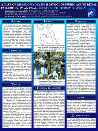
Fig 2. The other surgical positions related to
rhabdomyolysis and subsequent ARF. (a)
high lithotomy (b) flank (c) lateral decubitus
and (d) tilted lithotomy.
a b
c d
Fig 1. This is the exaggerated lithotomy position in
which the patient was operated on. Note the
angle of the trunk with the thighs at less than
90 degrees (almost 45 degrees).
IGURESF
HE CASET
UMMARYS
ECOMMENDATIONSR
A CASE OF RHABDOMYOLYSIS & MYOGLOBINURIC ACUTE RENAL
FAILURE FROM AN EXAGGERATED LITHOTOMY POSITION
Mary Ondinee U. Manalo, M.D., Resident, Department of Medicine, UP-PGH
Rommel Bataclan, M.D., Fellow, Section of Nephrology, Department of Medicine, UP-PGH
Roy Lascano, M.D. , Resident, Section of Urology, Department of Surgery, UP-PGH
Rey Jaime M. Tan, M.D., FPCP, Consultant, Section of Nephrology, Department of Medicine, UP-PGH
OURSE & IAGNOSTICSC D