SlideShare ist ein Scribd-Unternehmen logo
1 von 106
Slipped capital femoral
epiphysis
Presenter- Dr.Madhukar
INTRODUCTION
• The capital femoral
epiphysis is unique.
• It is one of the few
epiphysis in the body
that is inside the joint
capsule.
Definition
• SCFE term refers to slippage of the overlying
epiphysis of proximal femur posteriorly and
inferiorly due to weakness of the growth plate in
relation to metaphysis.
• Although this disorder is termed slipped capital
femoral epiphysis(SCFE), this is a misnomer. The
femoral epiphysis maintains its normal
relationship within the acetabulum, and it is the
femoral neck and the shaft that displace upwards
and anteriorly relative to the femoral epiphysis
and acetabulum.
Epidemiology
• Incidence is 2-3 per 100,000 population.
• Most common in adolescent period with rapid growth
plate (boys 10-16yrs; girls 12-14yrs)
• Very early onset <10yrs and very late onset >16yrs
should be evaluated for endocrine disorders.
• Males have 2.4 times the risk as females.
• Obesity is a risk factor because it places more shear
forces around the proximal growth plate in the hip.
• Bilateral slippage is common of which 2nd slip is about
12-18 months later to the 1st (left is effected 1st before
right)
Etiology
• Local trauma
• Obesity
• Endocrine disorders (like primary or secondary
hupoparthyroidism, adiposogenital
dystrophy(hypogonodal male)
• Growth hormone deficiency
• Atypical SCFE is associated with renal failure,
radiation therapy.
Pathology
• The epiphysis slowly displaces inferiorly and
posteriorly with the femoral neck shifting upward and
rotating anteriorly to the anterverted position thus
resulting in varus deformity, adduction and external
rotation of the femur.
• The displacement takes place through the layer of
hypertrophied mature cartilage cells adjacent to the
calcified cartilage zone
• The interval produced by separation becomes filled
with fibrous tissue, embryonic cartilage and callus
particularly at the posterior and inferior angle
Abduction and internal rotation of the hip are
greatly limited by 2 factors
- large hump formed by the fibrous cartilage
overgrowth about the anterior exposed portion
of the neck impinges against the anterior and
superior margins of the acetabulum
-The capital epiphysis is fixed posterior and inferior
where further outward movement is limited by
its impingement against capsule
Classification
• Based on symptoms
-acute
-chronic
-acute on chronic
• Functional classification
-stable
-unstable
• Depending on head shaft angle
-mild
-moderate
-severe
• Acute slip: sudden onset of usually severe
symptoms that have been present for less
than 2 weeks.x-ray shows displacement
without any bone healing
• Chronic slips –gradual onset and symptoms
of more than 2weeks duration some bony
healing and remodeling along the medial
femoral neck usually seen on x -rays
• Acute on chronic—symptoms lasting longer
than one month and a recent sudden
exacerbation of pain after a relatively trivial
injury
• Pre-slip– essentially an x-ray finding
manifested by irregularity widening and
indistinctness of the epiphyseal plate
• Depending on head shaft angle
Mild slipping – exist when the neck is displaced less than
1/3 rd of the diameter of the femoral head or when the
head shaft angle deviates from normal by 30 degrees
or less on both a,p and lateral views
Moderate slipping—when neck is displaced between 1/3
rd and ½ the diameter of femoral head or angle
deviation between30-60 degrees
Severe slipping:
when the neck is displaced more than half the
diameter of the head or deviation of the head shaft
angle of more than 60 degrees than normal
Loders classification
stable unstable
• Wt bearing possible with or
without crutches
• Less severe slips
• Effusion absent
• Good prognosis
• Less chances for avn
• Wt bearing is not possible
even with crutches
• Slip is more severe
• Effusion is almost always
present
• Bad or worst prognosis
• More chances for avn
Clinical features
• The onset is insidious
• Course is slowly progressive
• Symptoms:-
• Pain in the groin and around the hip
• Antalgic limp
• Sortening of the affected limb
• Limb may be in external rotation
• Flexion, abduction, internal rotation are limited.
• External rotation, adduction are increased.
• Axis deviation(pathognomic) – when hip is flexed,
the limb goes into external rotaion.
Hip flexion and external rotation are limited and with
flexion of the affected hip, the limb rotates externally.
• Preslipping stage :
• At first slight discomfort develops about the
groin usually after activity and it subsides by
rest this may be associated with slight
stiffness and an occasional limp
• The pain may radiate along antero-medial
thigh and to the inner aspect of the knee
• Chronic slipping stage:
• An antalagic limp becomes pronounced and
persistent .
• The findings include tenderness about the hip
and limitation of motion abduction and internal
rotation
• the limb gradually develops an adduction and
external rotation deformity
• when hip is flexed the external rotation deformity
is accentuated
• Stage of fixed deformity
-Pain and muscle spasm disappear but the
limp,external rotation, adduction and
shortening persists
• Signs:
-The patient is likely to be a boy between age of
10-17 years or a girl between 11 and 13 in
whom menstruation has not at started
• The radiographic abnormalities that may be
seen in SCFE depend on the degree of
displacement of the capital epiphysis.
• The AP radiograph of the hip, supplemented
by a frog-lateral view, is usually sufficient to
make a correct diagnosis.
• Several diagnostic indicators of SCFE have
been identified on the anteroposterior
projection of the hip
• The TRIANGLE SIGN OF CAPENER may
be of value in recognizing early SCFE.
• On a plain film of the normal adolescent hip,
an intracapsular area at the medial aspect of
the femoral neck is seen overlapping the
posterior wall of the acetabulum, creating a
dense triangular shadow-triangle of capener
• In most cases of SCFE, this triangle is lost.
• In a later stage, periarticular osteoporosis
becomes apparent, as do widening and
blurring of the physis and a decrease in height
of the epiphysis
• Moreover, as the disease progresses, slippage
of the capital epiphysis can be identified by
the absence of an intersection of the epiphysis
with a line drawn tangent to the lateral cortex
of the femoral neck
Metaphyseal blanch sign of steel
• In Ap view- crescent shaped area of increased
density overlying the metaphysis adjacent to
the physis.
• This increased density is due to the
overlapping of the femoral neck and the
posteriorly displaced capital epiphysis.
• Chronic stages of this disorder exhibit reactive
bone formation along the superolateral aspect
of the femoral neck, along with remodeling;
this creates a protuberance and broadening of
the femoral neck, which gives it a “pistol-grip”
appearance known as a HERNDON HUMP.
Radiological appearances
- Preslipping stage-indicated by the absence of the
normal shoulder on the upper aspect of the neck and
head [TRETHOWAN’S SIGN] in which a line called KLEINS
LINE ,drawn along the superior surface of the neck
will pass above the femoral head rather than through
the head
-The head is more or less sickle shaped instead of
hemispherical and its height is diminished
-The epiphyseal is widened and rarefaction or even
streaks of sclerosis may be seen underneath it
• Early stage
-The head of the femur lies in the acetabulum
but it is rotated so that its lower and laterally.
-The head is slightly displaced in relation to the
neck
• Advanced stage:
-The femoral head is atrophic especially in its
projecting lower half and it has now become
so rotated and displaced that only a small
anterior portion is actually in the acetabulum
-The projecting lower edge of the head is now
curved laterally and upwards and is in contact
with the lower border of the neck
-The head is often completely separated from
the neck and lies loose in the acetabulum
except for the displacement, the contour of
the bones is normal but the joint margins and
the joint space may be hazy from the
extravasation of blood.
• CT SCANS
Are valuable in visualizing early slipping.CT
provides the most accurate measurement of
the extent of epiphyseal displacement and
angulation
Diagnosis
• Diagnosis is a combination of clinical suspicion plus
radiological investigations.
• 20-50% cases of scfe are misdiagnosed on their 1st
presentation to a medical facility.
• This is because the common symptom is knee pain.
This is a referred pain from the hip.
• The symptom complex consist of-
• A limp of spontaneous onset(without apparent cause)
• Pain referred to the knee in obese adolescent.
• Characteristic radiographic appearance.
Differential diagnosis
1. Tuberculosis of hip
-Attitude- adducted and medially roated
-Movements-restricted in all directions atrophy
is greater
-Hip is more sensitive and painful even at rest
radiographs show extensive demineralization
of the femoral head and acetabulum without
any epiphyseal displacement
2. Perthes disease
-History : limitations of movement slight atrophy
and shortening are identical with those of mild
epiphyseal coxa vara the chief points of difference
-age: perthes diseases rarely begins after the
tenth year while slipped capital femoral epiphysis
begins before it
-X-ray: in perthes the head is not displaced
but actually deformed
Difference between SCFE and
PERTHE’S
SCFE PERSTHE’S
age- 10-14 yrs
Late onset 14-16 yrs
4-7 yrs
Late onset 7-10 yrs
Thin and tall adolscent or short
obese individuals
Occurs in normal child
Presents as pain with slippage
and limping nated later satge
Initially child limps and then at
later stages complaints of pain
Limb may externally roated but
no fixed flexion deformity
Fixed flexion deformity is noted
3. Congenital dislocation of hip
-Usually present since birth.
-Head of the femur may be palpated outside
the acetabulum.
-Telescopy present in majority of the case.
Prognosis
The second hip becomes affected in about 1 in 4
cases
• The end result depends to a certain extent on
the degree of displacement of the head
• In early case where there is minimal slipping
the end result is completely satisfied
• Good results can be achieved with good
reduction provided avascular necrosis avoided
• Good results can be achieved even with up to
50% displacement of the head if good
reduction is followed by corrective osteotomy
• Some patients with a displaced epiphysis after
treatment develop avascular necrosis cartilage
necrosis and arthritic changes during adult life
Management
• Conservative management- rest and traction
• Closed manupulative reduction
• Operative management
insitu pinning -open instiu pinning
-Bone peg Epiphysiodesis
-Osteotomies
-
Closed reduction
• Manipulative reduction has been reported for
acute and acute on chronic slips with
moderate and sever displacement.
• An association of avascular necrosis with
manipulative reduction has been reported.
-The development of avascular necrosis may be
related to severity of the slip rather than to
the manipulative reduction provided that only
a very gentle reduction is performed. This is
performed with the patient on a fracture table
and internal rotation alone usually is sufficient
to obtain adequate reduction
-Longitudinal traction is applied to counteract
the muscle spasm straight longitudinal
traction especially when combined with
medial rotation will effectively reduce the
displacement of an acute slip and this can be
followed by internal fixation
The aim of surgery are
• To prevent further slip
• To reduce the displacement
• To effect early closure of the epiphyseal plate
Internal fixation
• In situ pin or screw fixation
Percutaneous in situ pinning is currently the
most often used treatment for mild moderate
and some severe acute on chronic slipped
capital femoral epiphysis
Open in situ pinning may be indicated for more
severe acute or acute on chronic slipping
Percutaneous in-situ pinning
• Although earlier reports indicated that two or
three pins were necessary for stability and
results of multiple pinning generally have
been satisfactory
• Reports now recommend the use of a single
larger diameter central pin or screw because
single pin insertion is technically simpler than
insertion of multiple pins but this is still
surgeon dependent
• Whether pins or cannulated hip screws are
used, they should not be removed for at least
12 months or until the physis closes.
• Persistent pin penetration has been the most
serious disadvantage of in situ pinning.
• Adverse effects attributed to unrecognized pin
penetration include joint stiffness.
• The pins should be placed in the femoral neck
and into the centre of the head in the safe
zone to avoid pin penetration.
• Lehman et al and Shaw recommended the use
of cannulated hip screw with injection of radio
opaque dye to detect pin penetration.
• The pin tip is to be advanced to 8mm or1/3rd
of the radius of the femoral head from the sub
chondral bone.
• This places the actual tip 7 to 18 mm from the
subchondral bone, leaving a safe margin.
Open in situ pinning
• Technique (Morrissy):
• Place the patient on the fracture table with
the affected leg abducted 10 to 15 degrees
and internally rotated as for as possible
without force. This brings the femoral neck as
close as possible parallel to the floor to assist
in obtaining true anterior and lateral image
views after standard preparation and draping
under image control insert a k-wire
• Through the antero-lateral area of the thigh
down to the femoral neck, adjusting the guide
wire on the antero-posterior projection to
determine the axis of the femoral neck and
obtain a lateral view to determine the amount
of posterior inclination needed.
• When the starting point and the posterior
inclination have been estimated, insert the
guide through a small puncture wound.
• Advance the guide wire to the physis and
confirm placement in the central axis of the
femoral head by intensification. If the position
is correct advance the guide assembly across
the plate until the proper depth is reached
determine the correct screw length by
passing a guide wire of identical length
advance correct length screw over the guide
pin and then remove the pin.
• Use both anteroposterior and lateral views to
conform that the screws do not penetrate the
joint and close the stab wound with a single
subcuticular suture .
• If two screws are deemed necessary for an
acute slip the first screw should lie in the
central axis of the femoral head and the
second below it avoiding the superolateral
quadrant .
• The second screw should stop at least 8mm
from the subchondral bone
• After treatment
• Range of motion exercises are to begin the day
after surgery .
• Partial weight bearing is allowed for most of the
patients on first day using crutches.
• Crutches are used until all signs of synovitis are
gone and motion is free and painless .all vigorous
activities are forbidden until the physis have
closed .
• The screws are removed after physeal closure has
been demonstrated.
• Prophylactic pinning of the contra-lateral hip
is performed in rare instances such as In high
risk non complaint patients or patients with
epiphysio-lysis from irradiation therapy or
renal failure .
Bone peg epiphysiodesis
- Described by Ferguson and Howorth in 1931
-Reported complications after pin or screw
penentration into the joint increased its
popularity
-There is rapid physeal closure and low
incidence of complications especially in
chondro-lysis
• Disadvantages
Graft insufficency leading to increase in severity
of slip and failure of physeal fusion
• Longer opreating time
• Increased blood loss
• Longer hospitalization
• Longer rehabilitation
• Spica cast immobilization may be necessary to
present further slip
• Post operative complication
• -avascularnecrosis
• -chondro-lysis
• -infection
• -lengthy immobilization
• -heterotopic ossification
Technique of bone peg epiphysiodesis
through anterolateral apporach(wenier
etal)
-Make a mid lateral incision beginning 4 to 5
inches below the level of the greater
trochanter in the lateral mid line of the upper
portion of the thigh,proceeding proximally to
the greater trochanter and then angling
obliquely to the antero-superior iliac spine
- Muscles are split and retracted capsule of the hip
joint is exposed and opened in an H shape
fashion
- If bony hump present on the antero-lateral meta-
physeal region resulting from slip remove it with
an osteotome
- Make a square or rectangular window in the
anterior surface of femoral neck insert a large
hollow drill through the window and drill it across
the physis into the epiphysis under image
intensifier
-Remove a cylindrical core consisting of
metaphyseal bone physis and a portion of the
epiphyseal bone there by guaranting passage
of the drill across the physis and into the
epiphysis enlarge the cylindrical tunnel with a
curret and remove more of the physis remove
sections of cortico-cancellous bone from the
outer layer of ilium and sand witch them
together and drive this composite bone peg
across the physis into epiphysis
After treatment
Spica cast applied in acute slips for six weeks
and then touch down wait bearing is allowed
Chronic slips are ambulated on crutches after 48
to 96 hrs of bed rest
Weight bearing can be started at approximately
10 weeks
-In an effort to simplify bone peg epiphysiodes schmidt et
al developed a percutaneuos fluoroscopically guided
technique using cortical allograft inserted through a
hole drilled in the lateral femoral cortex passed
through the femoral neck and across the physis
-Procedure is similar to the percutaneous pin fixation
except that rather than cannulated screw a 10mm
reamer is inserted over the guide pin and a tunnel is
reamed
-Reamed tunnels is filled with a cortical strut allograft.
-operative time the and morbedity similar to single screw
fixiation
Osteotomy
-Chronic slips with moderate or severe
displacement produce perment irregularities
in the femoral head and acetabulum and
some form of reconstructive procedure often
is indicated the goals of any osteotomy are-
Restoration of the normal relationship of
the femoral head and neck
Dealy of the onset of degenerative joint
disease
There are two basic types of osteotomy
• Closing wedge osteotomy through the femoral
neck usually near the physis to correct the
deformity and
• Compensetory osteotomy through the
trochanteric region to the produce deformity
in the opposite direction
Four femoral neck osteotomies are described
• The technique of fish
• The technique of dunn just distal to the
slip(subcapital).
• The base of neck technique of kramar et al.
• The technique of abraham et al.
• The itertrochantric osteotomy by southwick.
Compensatory osteotomies in the trochanteric
region as well as partial cheilectomy to reduce
deformity
Cuneiform osteotomy of the femoral
neck(fish)
-Reduction by a cuneiform osteotomy made just
distal to the physis and internal fixation may
be necessary for a severe chronic or acute on
chronic slip.
-The reported incidence of avascular necrosis
and chondrolysis after this procedure have
been exceptionally high
• Technique
-Place the patient supine make an anterolateral approach
to the hip
-Open the capsule longitudinally determine the size of
the wedge to be removed by noting the degree of slip
and the position of the epiphysis make the base of the
wedge anteriorly and superiorly for correct positioning
of the epiphsysis after removing sufficient bone
-Reduce the epiphsysis by flexion abduction and internal
rotation of the limb after reduction fix the epiphysis to
the neck with 3 or 4 pins
Cuneiform osteotomy of femoral neck
{fish}
After treatment
-The patient should be free in bed with the
involved limb supported on pillows
-When comfortable he is allowed out of bed
using crutches with only touch down weight
bearing of the involved limb
-Full weight bearing is permitted after x-rays
show the osteotomy to be completely healed
at approximately 5 months pins are removed
full activity allowed 2 months later
Cuneiform osteotomy of femoral neck
(dunn)
-Dunn described an osteotomy for severe
chronic slips in chidern with open physis he
emphasized that this procedure should not be
done if the physis is closed
• The slip of the femoral head strips the
periosteum from the back of the femoral neck
and beak of new bone is laid down beneath it
• The main retincular blood supply runs of the
femoral neck a lateral apporach allows
stripping of the periosteum and its contained
vessels under direct vision to avoid damaging
the blood supply to the femoral head
• Technique
-Through a lateral approach make an incision in the
periosteum and elevate the posterior vascular
covering of the femoral neck make to osteotomy
cuts one in the long axis of the neck to remove
the bony beak and the second at right angles to
the neck to shorten it by 3 to 4 mm. appose the
surface of the osteotomy and insert three
threaded pins up the femoral neck to its cut
surface.
-Reduce the deformity and conform position on
x ray. In the lateral view the head should
appear to sit squarely on the neck but in AP
view the head should be tilted into about 20
degrees of valgus when reduction is
satisfactory drive the pins into the femoral
head close the wound in layers
• After treatment
-A cast is applied from the nipple line to the toes on
the affected side holding the extremity in the
nuetral roatatoin and abduction and to above
knee on the opposite side.
-Cast is removed at 4 weeeks and active and passive
motions are begun after two weeks crutch
walking is allowed at 3 to 4 months after surgery
partial weight bearing is permitted full weight
-Bearing permitted only after x-ray evidence of
union of osteotomy
Compensatory basilar osteotomy of
femoral neck
-Kramer et al described a compensatory
osteotomy of the base of the femoral neck
that corrects the varus and retoversion
components of moderate or severe chronic
slipped capital femoral epiphysis
-They suggested that it is safer than an
Osteotomy made near or at the physis
because the line of the osteotomy is distal to
the major bloood supply in the posterior
retinacuium.
-Threaded pains are used for fixation of the
osteotomy and the epiphysis not only is the
anatomical relationship of the proximal femur
restored but also futher slipping is prevented
• Technique
-Determine preoperatively the size of the wedge
to be removed by measuring the head-neck
angle on AP radiograph and confirmed at
surgery by measuring the width of the callus
by direct inspection.
-Part of wedge will be in line with the widest part of the
slip in the anterior and superior aspects of the neck
make the more distal osteotomy cut first perpendicular
to the femoral neck and following the anterior
intertrochanteric line from proximal to distal extended
this osteotomy
-Cut to the posterior cortex but leave this cortex intact
(this will permit the greenstick fracture in the posterior
cortex when closed)make the second osteotomy cut
directed obliquely so that its cutting edge stays distal to
the posterior retinacular blood supply.
-Drill one or two 5mm threaded steinmann pins
into the femoral neck proximally to ensure
that the proximal portion of the femur is kept
under control before completing the
osteotomy
-Complete the osteotomy by medial rotation nd
abduction of the distal segment.
-Advance the threaded steinmann across the
osteotomy site and physis to prevent for the
slipping
After treatment
Bed rest for 2 to 3 weeks followed by non weight
bearing
partial weight bearing is allowed depending on
the stability of the osteotomy
stienmenn pins should be removed only after
physis has fused
Extra capsular base of neck osteotomy
• Extra capsular base of neck osteotomy
• Abraham et al recommended this osteotomy as
safe and effective in preventing furthur slipping
and improving hip range of motion in pts with
severe chronic slip
• They noted that with severe slips the amount of
correction of varus and posterior tilt of the
femoral head is limited and complete restoration
of normal head shaft angle may not be possible
• Removal of a wedge larger than 20 mm
compromises femoral neck length and may
greately increase femoral antversion
• Also pinning across the osteotomy site becomes
more difficult when correction of more than 55
degrees of varus or valgus is attempted
• These same restrictions also are applicable to
intra capsular base of neck osteotomies and
southwick procedure
• Before surgery the head shaft angle is
measured on lateral films by measuring the
angle formed by epiphyseal line and the
femoral shaft in the affected limb and
comparing it with normal limb
• The head shaft angle for posterior tilt or
retroversion is measured on frog lateral view
and compared with normal side
• The difference between normal and abnormal
angles are used to determine the size of
wedge removed during osteotomy
• Make a standard anterolateral approach mark
the anterior joint line or intertrochantric line
• Delineate a triangle on the anterior surface of
the femoral neck to indicate the two plane
wedge osteotomy
• Locate the proximal cut by placing a 3cm long
k wire on the anterior surface of the femur
from the lesser to greater trochanter at the
base of the neck along the edge of the capsule
confirm position by c-arm
• Externally rotate the leg and drill a second K wire on
the AP plane just distal to the guide wire
• Begin the second osteotomy line from the lesser
trochanter to the growth plate of the greater trochanter
• Make a single osteotomy along the posterior cortex and
completely remove the wedge and by maintaining
traction internally rotate or abduct the leg to close the
osteotomy.
• Use cannulated screws to hold the osteotomy, use only
one screw to span the physis of the femoral head
avoiding superolateral quadrant .
Southwick’s intertrochantric
osteotomy
 This is measured biplane intertrochanteric
osteotomy in which degree of correction is
accurately pre determined before surgery.
 Osteotomy through the trochanter is most useful
when the head has slipped from 30-70d is
considered minimum and is fixed insitu with
threaded pins
 Initially the limitation of joint movement due to
muscle is overcomed by longitudinal traction in
mild abduction and an internal rotation strap for
few days.
• The X-ray measurements are made on both AP
and frog leg lateral views.
• AP view gives the degree of varus deformity.
• Frog leg lateral view gives the measure of
posterior tilting.
• Measurements are made by template models
on X-ray prior to surgery.
Complications
1.Avascular necrosis :
• More common with moderate and severe slips and less
common with mild slips.
• Avn is common following femoral neck osteotomy33%,Open
reduction with knowles pin- 27%, less common with in situ
fixation – 1.5% and after trochanteric osteotomy – 10%.
• AVN probably results from interruption of retrograde blood
supply and is apparently influenced by the original injury.
• Forceful repetitive manipulatations, open reductions,
osteotomies of femoral neck and superolateral placement of
pins.
2. Chondrolysis:
• Sometimes termed as ‘acute cartilaginous necrosis’ is
an affection of the hip joint characterized
pathologically by rapid loss of articular cartilage and
clinically by development of continuous pain and
severe restriction of motion.
• A joint space that measured less than 3mm width
and decreased range of motion are required for
the diagnosis.
• Occurs more often in females and black.
• More common following trochanteric osteotomy-
59%, open reduction -55%. Femoral neck
osteotomy – 37%, pin protruding into the joint –
51%. It is usually diagnosed within the first 6-9
months of observation
• The joint space is rapidly lost and severe limitation of
movement is present.
• Subsequently the width of the joint space usually increases
slowly and range of motion also increases slightly.
• If severe joint space narrowing persists with limitation of joint
motion arthrodesis or arthroplasty should be considered.
• Conservative treatment by bed rest , traction.
• Usually the joint goes to ankylosis
3. Osteorthritis :
• Normally pressure forces of weight bearing are widely
distributed over the femoral head.
• In coxa vara pressure forces are concentrated over a smaller
area causing degeneration of cartilage and incongruous joint
surfaces.
• The time required for these changes to develop was long and
varied from 18 months to 4 years after onset.
4. Fractures:
• Fractures can occur just distal to the knowles pins. So the pins
should be removed after the epiphyseal plate has fused.
• Over reaming of the femoral neck also cause a stress fracture.
5. Continued slipping:
• In case of conservative treatment or in which pins were not
placed far enough proximally or removed before the physis
had completely fused.
THANK YOU

Weitere ähnliche Inhalte

Was ist angesagt?

Osteotomies around hip by dr rohit kumar
Osteotomies around hip by dr rohit kumarOsteotomies around hip by dr rohit kumar
Osteotomies around hip by dr rohit kumar
Dr Rohit Kumar
 
Blood supply of femoral head at various ages
Blood supply of femoral head at various agesBlood supply of femoral head at various ages
Blood supply of femoral head at various ages
songao
 

Was ist angesagt? (20)

Scaphoid fracture and nonunion
Scaphoid fracture and nonunion Scaphoid fracture and nonunion
Scaphoid fracture and nonunion
 
Non Union
Non UnionNon Union
Non Union
 
Osteotomies around hip by dr rohit kumar
Osteotomies around hip by dr rohit kumarOsteotomies around hip by dr rohit kumar
Osteotomies around hip by dr rohit kumar
 
Fracture disease
Fracture diseaseFracture disease
Fracture disease
 
Physeal injuries
Physeal injuriesPhyseal injuries
Physeal injuries
 
Recurrent shoulder dislocation
Recurrent shoulder dislocationRecurrent shoulder dislocation
Recurrent shoulder dislocation
 
Blood supply of femoral head at various ages
Blood supply of femoral head at various agesBlood supply of femoral head at various ages
Blood supply of femoral head at various ages
 
Cubitus varus deformity
Cubitus varus deformityCubitus varus deformity
Cubitus varus deformity
 
Tuberculosis of hip joint
Tuberculosis of hip jointTuberculosis of hip joint
Tuberculosis of hip joint
 
Supracondylar Fractures
Supracondylar FracturesSupracondylar Fractures
Supracondylar Fractures
 
Ctev.ppt by krr
Ctev.ppt by krrCtev.ppt by krr
Ctev.ppt by krr
 
Clinical Examination of Hip
Clinical Examination of HipClinical Examination of Hip
Clinical Examination of Hip
 
Congenital talipes equinovarus (club foot/ctev) ppt by Dr Pratik
Congenital talipes equinovarus (club foot/ctev) ppt by Dr PratikCongenital talipes equinovarus (club foot/ctev) ppt by Dr Pratik
Congenital talipes equinovarus (club foot/ctev) ppt by Dr Pratik
 
Hallux valgus.pptx
Hallux valgus.pptxHallux valgus.pptx
Hallux valgus.pptx
 
Surgical approaches to hip joint
Surgical approaches to hip jointSurgical approaches to hip joint
Surgical approaches to hip joint
 
Devlopmental dysplasia of hip(DDH) by DR.NAVEEN RATHOR
Devlopmental dysplasia of hip(DDH) by DR.NAVEEN RATHORDevlopmental dysplasia of hip(DDH) by DR.NAVEEN RATHOR
Devlopmental dysplasia of hip(DDH) by DR.NAVEEN RATHOR
 
Scaphoid fractures
Scaphoid fracturesScaphoid fractures
Scaphoid fractures
 
Shoulder Dislocations
Shoulder DislocationsShoulder Dislocations
Shoulder Dislocations
 
Lisfranc injury
Lisfranc injuryLisfranc injury
Lisfranc injury
 
Intertrochanteric fractures of the femur
Intertrochanteric fractures of the femurIntertrochanteric fractures of the femur
Intertrochanteric fractures of the femur
 

Ähnlich wie Slipped capital femoral epiphysis

Slipped capital femoral epiphysis vamshi kiran feb 6/2013
Slipped capital femoral epiphysis vamshi kiran feb 6/2013Slipped capital femoral epiphysis vamshi kiran feb 6/2013
Slipped capital femoral epiphysis vamshi kiran feb 6/2013
badamvamshikiran
 
Osteochondritis of different bones
Osteochondritis of different bonesOsteochondritis of different bones
Osteochondritis of different bones
Pramod Govindraj
 
Paediatric Orthopaedic
Paediatric OrthopaedicPaediatric Orthopaedic
Paediatric Orthopaedic
Fara Dyba
 

Ähnlich wie Slipped capital femoral epiphysis (20)

Scfe seminar
Scfe seminarScfe seminar
Scfe seminar
 
SCFE / slipped capital femoral epiphysis
SCFE / slipped capital femoral epiphysis SCFE / slipped capital femoral epiphysis
SCFE / slipped capital femoral epiphysis
 
Slipped capital femoral epiphysis
Slipped capital femoral epiphysisSlipped capital femoral epiphysis
Slipped capital femoral epiphysis
 
Spondylolisthesis
SpondylolisthesisSpondylolisthesis
Spondylolisthesis
 
Slipped capital femoral epiphysis
Slipped capital femoral epiphysisSlipped capital femoral epiphysis
Slipped capital femoral epiphysis
 
Congenital hip disease
Congenital hip disease Congenital hip disease
Congenital hip disease
 
Slipped capital femoral epiphysis vamshi kiran feb 6/2013
Slipped capital femoral epiphysis vamshi kiran feb 6/2013Slipped capital femoral epiphysis vamshi kiran feb 6/2013
Slipped capital femoral epiphysis vamshi kiran feb 6/2013
 
Legg-Calve-Perthes Disease.pptx
Legg-Calve-Perthes Disease.pptxLegg-Calve-Perthes Disease.pptx
Legg-Calve-Perthes Disease.pptx
 
Osteochondritis of different bones
Osteochondritis of different bonesOsteochondritis of different bones
Osteochondritis of different bones
 
Slipped Capital Femoral Epiphysis (SCFE)
Slipped Capital Femoral Epiphysis (SCFE)Slipped Capital Femoral Epiphysis (SCFE)
Slipped Capital Femoral Epiphysis (SCFE)
 
Perthes disease
Perthes diseasePerthes disease
Perthes disease
 
Paediatric Orthopaedic
Paediatric OrthopaedicPaediatric Orthopaedic
Paediatric Orthopaedic
 
Slipped Upper Femoral Epiphysis (SUFE)
Slipped Upper Femoral Epiphysis (SUFE)Slipped Upper Femoral Epiphysis (SUFE)
Slipped Upper Femoral Epiphysis (SUFE)
 
Irritable hip and perthe's disease
Irritable hip and perthe's diseaseIrritable hip and perthe's disease
Irritable hip and perthe's disease
 
Case discussion of perthes disease-Dr. Siddharth Deshwal PG Orthopaedics
Case discussion of perthes disease-Dr. Siddharth Deshwal PG OrthopaedicsCase discussion of perthes disease-Dr. Siddharth Deshwal PG Orthopaedics
Case discussion of perthes disease-Dr. Siddharth Deshwal PG Orthopaedics
 
Perthes disease
Perthes diseasePerthes disease
Perthes disease
 
Legg Calve Perthes disease
Legg Calve Perthes diseaseLegg Calve Perthes disease
Legg Calve Perthes disease
 
SHEUERMANN+DISEASE.pptx
SHEUERMANN+DISEASE.pptxSHEUERMANN+DISEASE.pptx
SHEUERMANN+DISEASE.pptx
 
SCFE
SCFESCFE
SCFE
 
paralytic and postural scoliosis
paralytic and postural scoliosisparalytic and postural scoliosis
paralytic and postural scoliosis
 

Kürzlich hochgeladen

Call Girls Bhubaneswar Just Call 9907093804 Top Class Call Girl Service Avail...
Call Girls Bhubaneswar Just Call 9907093804 Top Class Call Girl Service Avail...Call Girls Bhubaneswar Just Call 9907093804 Top Class Call Girl Service Avail...
Call Girls Bhubaneswar Just Call 9907093804 Top Class Call Girl Service Avail...
Dipal Arora
 
Russian Escorts Girls Nehru Place ZINATHI 🔝9711199012 ☪ 24/7 Call Girls Delhi
Russian Escorts Girls  Nehru Place ZINATHI 🔝9711199012 ☪ 24/7 Call Girls DelhiRussian Escorts Girls  Nehru Place ZINATHI 🔝9711199012 ☪ 24/7 Call Girls Delhi
Russian Escorts Girls Nehru Place ZINATHI 🔝9711199012 ☪ 24/7 Call Girls Delhi
AlinaDevecerski
 
College Call Girls in Haridwar 9667172968 Short 4000 Night 10000 Best call gi...
College Call Girls in Haridwar 9667172968 Short 4000 Night 10000 Best call gi...College Call Girls in Haridwar 9667172968 Short 4000 Night 10000 Best call gi...
College Call Girls in Haridwar 9667172968 Short 4000 Night 10000 Best call gi...
perfect solution
 

Kürzlich hochgeladen (20)

Call Girls Bhubaneswar Just Call 9907093804 Top Class Call Girl Service Avail...
Call Girls Bhubaneswar Just Call 9907093804 Top Class Call Girl Service Avail...Call Girls Bhubaneswar Just Call 9907093804 Top Class Call Girl Service Avail...
Call Girls Bhubaneswar Just Call 9907093804 Top Class Call Girl Service Avail...
 
Call Girls Dehradun Just Call 9907093804 Top Class Call Girl Service Available
Call Girls Dehradun Just Call 9907093804 Top Class Call Girl Service AvailableCall Girls Dehradun Just Call 9907093804 Top Class Call Girl Service Available
Call Girls Dehradun Just Call 9907093804 Top Class Call Girl Service Available
 
Call Girls Ludhiana Just Call 9907093804 Top Class Call Girl Service Available
Call Girls Ludhiana Just Call 9907093804 Top Class Call Girl Service AvailableCall Girls Ludhiana Just Call 9907093804 Top Class Call Girl Service Available
Call Girls Ludhiana Just Call 9907093804 Top Class Call Girl Service Available
 
Call Girls Bangalore Just Call 8250077686 Top Class Call Girl Service Available
Call Girls Bangalore Just Call 8250077686 Top Class Call Girl Service AvailableCall Girls Bangalore Just Call 8250077686 Top Class Call Girl Service Available
Call Girls Bangalore Just Call 8250077686 Top Class Call Girl Service Available
 
Top Rated Bangalore Call Girls Ramamurthy Nagar ⟟ 9332606886 ⟟ Call Me For G...
Top Rated Bangalore Call Girls Ramamurthy Nagar ⟟  9332606886 ⟟ Call Me For G...Top Rated Bangalore Call Girls Ramamurthy Nagar ⟟  9332606886 ⟟ Call Me For G...
Top Rated Bangalore Call Girls Ramamurthy Nagar ⟟ 9332606886 ⟟ Call Me For G...
 
Bangalore Call Girls Nelamangala Number 9332606886 Meetin With Bangalore Esc...
Bangalore Call Girls Nelamangala Number 9332606886  Meetin With Bangalore Esc...Bangalore Call Girls Nelamangala Number 9332606886  Meetin With Bangalore Esc...
Bangalore Call Girls Nelamangala Number 9332606886 Meetin With Bangalore Esc...
 
Russian Escorts Girls Nehru Place ZINATHI 🔝9711199012 ☪ 24/7 Call Girls Delhi
Russian Escorts Girls  Nehru Place ZINATHI 🔝9711199012 ☪ 24/7 Call Girls DelhiRussian Escorts Girls  Nehru Place ZINATHI 🔝9711199012 ☪ 24/7 Call Girls Delhi
Russian Escorts Girls Nehru Place ZINATHI 🔝9711199012 ☪ 24/7 Call Girls Delhi
 
Call Girls Faridabad Just Call 9907093804 Top Class Call Girl Service Available
Call Girls Faridabad Just Call 9907093804 Top Class Call Girl Service AvailableCall Girls Faridabad Just Call 9907093804 Top Class Call Girl Service Available
Call Girls Faridabad Just Call 9907093804 Top Class Call Girl Service Available
 
Call Girls Haridwar Just Call 8250077686 Top Class Call Girl Service Available
Call Girls Haridwar Just Call 8250077686 Top Class Call Girl Service AvailableCall Girls Haridwar Just Call 8250077686 Top Class Call Girl Service Available
Call Girls Haridwar Just Call 8250077686 Top Class Call Girl Service Available
 
Best Rate (Patna ) Call Girls Patna ⟟ 8617370543 ⟟ High Class Call Girl In 5 ...
Best Rate (Patna ) Call Girls Patna ⟟ 8617370543 ⟟ High Class Call Girl In 5 ...Best Rate (Patna ) Call Girls Patna ⟟ 8617370543 ⟟ High Class Call Girl In 5 ...
Best Rate (Patna ) Call Girls Patna ⟟ 8617370543 ⟟ High Class Call Girl In 5 ...
 
Premium Bangalore Call Girls Jigani Dail 6378878445 Escort Service For Hot Ma...
Premium Bangalore Call Girls Jigani Dail 6378878445 Escort Service For Hot Ma...Premium Bangalore Call Girls Jigani Dail 6378878445 Escort Service For Hot Ma...
Premium Bangalore Call Girls Jigani Dail 6378878445 Escort Service For Hot Ma...
 
Premium Call Girls Cottonpet Whatsapp 7001035870 Independent Escort Service
Premium Call Girls Cottonpet Whatsapp 7001035870 Independent Escort ServicePremium Call Girls Cottonpet Whatsapp 7001035870 Independent Escort Service
Premium Call Girls Cottonpet Whatsapp 7001035870 Independent Escort Service
 
(👑VVIP ISHAAN ) Russian Call Girls Service Navi Mumbai🖕9920874524🖕Independent...
(👑VVIP ISHAAN ) Russian Call Girls Service Navi Mumbai🖕9920874524🖕Independent...(👑VVIP ISHAAN ) Russian Call Girls Service Navi Mumbai🖕9920874524🖕Independent...
(👑VVIP ISHAAN ) Russian Call Girls Service Navi Mumbai🖕9920874524🖕Independent...
 
Call Girls Coimbatore Just Call 9907093804 Top Class Call Girl Service Available
Call Girls Coimbatore Just Call 9907093804 Top Class Call Girl Service AvailableCall Girls Coimbatore Just Call 9907093804 Top Class Call Girl Service Available
Call Girls Coimbatore Just Call 9907093804 Top Class Call Girl Service Available
 
Pondicherry Call Girls Book Now 9630942363 Top Class Pondicherry Escort Servi...
Pondicherry Call Girls Book Now 9630942363 Top Class Pondicherry Escort Servi...Pondicherry Call Girls Book Now 9630942363 Top Class Pondicherry Escort Servi...
Pondicherry Call Girls Book Now 9630942363 Top Class Pondicherry Escort Servi...
 
Top Rated Bangalore Call Girls Richmond Circle ⟟ 9332606886 ⟟ Call Me For Ge...
Top Rated Bangalore Call Girls Richmond Circle ⟟  9332606886 ⟟ Call Me For Ge...Top Rated Bangalore Call Girls Richmond Circle ⟟  9332606886 ⟟ Call Me For Ge...
Top Rated Bangalore Call Girls Richmond Circle ⟟ 9332606886 ⟟ Call Me For Ge...
 
Call Girls Nagpur Just Call 9907093804 Top Class Call Girl Service Available
Call Girls Nagpur Just Call 9907093804 Top Class Call Girl Service AvailableCall Girls Nagpur Just Call 9907093804 Top Class Call Girl Service Available
Call Girls Nagpur Just Call 9907093804 Top Class Call Girl Service Available
 
Night 7k to 12k Chennai City Center Call Girls 👉👉 7427069034⭐⭐ 100% Genuine E...
Night 7k to 12k Chennai City Center Call Girls 👉👉 7427069034⭐⭐ 100% Genuine E...Night 7k to 12k Chennai City Center Call Girls 👉👉 7427069034⭐⭐ 100% Genuine E...
Night 7k to 12k Chennai City Center Call Girls 👉👉 7427069034⭐⭐ 100% Genuine E...
 
College Call Girls in Haridwar 9667172968 Short 4000 Night 10000 Best call gi...
College Call Girls in Haridwar 9667172968 Short 4000 Night 10000 Best call gi...College Call Girls in Haridwar 9667172968 Short 4000 Night 10000 Best call gi...
College Call Girls in Haridwar 9667172968 Short 4000 Night 10000 Best call gi...
 
VIP Hyderabad Call Girls Bahadurpally 7877925207 ₹5000 To 25K With AC Room 💚😋
VIP Hyderabad Call Girls Bahadurpally 7877925207 ₹5000 To 25K With AC Room 💚😋VIP Hyderabad Call Girls Bahadurpally 7877925207 ₹5000 To 25K With AC Room 💚😋
VIP Hyderabad Call Girls Bahadurpally 7877925207 ₹5000 To 25K With AC Room 💚😋
 

Slipped capital femoral epiphysis

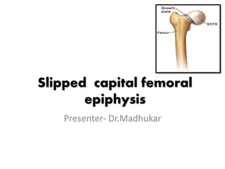
  • 2. INTRODUCTION • The capital femoral epiphysis is unique. • It is one of the few epiphysis in the body that is inside the joint capsule.
  • 3. Definition • SCFE term refers to slippage of the overlying epiphysis of proximal femur posteriorly and inferiorly due to weakness of the growth plate in relation to metaphysis. • Although this disorder is termed slipped capital femoral epiphysis(SCFE), this is a misnomer. The femoral epiphysis maintains its normal relationship within the acetabulum, and it is the femoral neck and the shaft that displace upwards and anteriorly relative to the femoral epiphysis and acetabulum.
  • 4. Epidemiology • Incidence is 2-3 per 100,000 population. • Most common in adolescent period with rapid growth plate (boys 10-16yrs; girls 12-14yrs) • Very early onset <10yrs and very late onset >16yrs should be evaluated for endocrine disorders. • Males have 2.4 times the risk as females. • Obesity is a risk factor because it places more shear forces around the proximal growth plate in the hip. • Bilateral slippage is common of which 2nd slip is about 12-18 months later to the 1st (left is effected 1st before right)
  • 5. Etiology • Local trauma • Obesity • Endocrine disorders (like primary or secondary hupoparthyroidism, adiposogenital dystrophy(hypogonodal male) • Growth hormone deficiency • Atypical SCFE is associated with renal failure, radiation therapy.
  • 6. Pathology • The epiphysis slowly displaces inferiorly and posteriorly with the femoral neck shifting upward and rotating anteriorly to the anterverted position thus resulting in varus deformity, adduction and external rotation of the femur. • The displacement takes place through the layer of hypertrophied mature cartilage cells adjacent to the calcified cartilage zone • The interval produced by separation becomes filled with fibrous tissue, embryonic cartilage and callus particularly at the posterior and inferior angle
  • 7. Abduction and internal rotation of the hip are greatly limited by 2 factors - large hump formed by the fibrous cartilage overgrowth about the anterior exposed portion of the neck impinges against the anterior and superior margins of the acetabulum -The capital epiphysis is fixed posterior and inferior where further outward movement is limited by its impingement against capsule
  • 8. Classification • Based on symptoms -acute -chronic -acute on chronic • Functional classification -stable -unstable • Depending on head shaft angle -mild -moderate -severe
  • 9. • Acute slip: sudden onset of usually severe symptoms that have been present for less than 2 weeks.x-ray shows displacement without any bone healing • Chronic slips –gradual onset and symptoms of more than 2weeks duration some bony healing and remodeling along the medial femoral neck usually seen on x -rays
  • 10. • Acute on chronic—symptoms lasting longer than one month and a recent sudden exacerbation of pain after a relatively trivial injury • Pre-slip– essentially an x-ray finding manifested by irregularity widening and indistinctness of the epiphyseal plate
  • 11. • Depending on head shaft angle
  • 12.
  • 13. Mild slipping – exist when the neck is displaced less than 1/3 rd of the diameter of the femoral head or when the head shaft angle deviates from normal by 30 degrees or less on both a,p and lateral views Moderate slipping—when neck is displaced between 1/3 rd and ½ the diameter of femoral head or angle deviation between30-60 degrees Severe slipping: when the neck is displaced more than half the diameter of the head or deviation of the head shaft angle of more than 60 degrees than normal
  • 14.
  • 15. Loders classification stable unstable • Wt bearing possible with or without crutches • Less severe slips • Effusion absent • Good prognosis • Less chances for avn • Wt bearing is not possible even with crutches • Slip is more severe • Effusion is almost always present • Bad or worst prognosis • More chances for avn
  • 16.
  • 17. Clinical features • The onset is insidious • Course is slowly progressive • Symptoms:- • Pain in the groin and around the hip • Antalgic limp • Sortening of the affected limb • Limb may be in external rotation • Flexion, abduction, internal rotation are limited. • External rotation, adduction are increased. • Axis deviation(pathognomic) – when hip is flexed, the limb goes into external rotaion.
  • 18. Hip flexion and external rotation are limited and with flexion of the affected hip, the limb rotates externally.
  • 19. • Preslipping stage : • At first slight discomfort develops about the groin usually after activity and it subsides by rest this may be associated with slight stiffness and an occasional limp • The pain may radiate along antero-medial thigh and to the inner aspect of the knee
  • 20. • Chronic slipping stage: • An antalagic limp becomes pronounced and persistent . • The findings include tenderness about the hip and limitation of motion abduction and internal rotation • the limb gradually develops an adduction and external rotation deformity • when hip is flexed the external rotation deformity is accentuated
  • 21. • Stage of fixed deformity -Pain and muscle spasm disappear but the limp,external rotation, adduction and shortening persists • Signs: -The patient is likely to be a boy between age of 10-17 years or a girl between 11 and 13 in whom menstruation has not at started
  • 22. • The radiographic abnormalities that may be seen in SCFE depend on the degree of displacement of the capital epiphysis. • The AP radiograph of the hip, supplemented by a frog-lateral view, is usually sufficient to make a correct diagnosis. • Several diagnostic indicators of SCFE have been identified on the anteroposterior projection of the hip
  • 23. • The TRIANGLE SIGN OF CAPENER may be of value in recognizing early SCFE. • On a plain film of the normal adolescent hip, an intracapsular area at the medial aspect of the femoral neck is seen overlapping the posterior wall of the acetabulum, creating a dense triangular shadow-triangle of capener • In most cases of SCFE, this triangle is lost.
  • 24.
  • 25. • In a later stage, periarticular osteoporosis becomes apparent, as do widening and blurring of the physis and a decrease in height of the epiphysis • Moreover, as the disease progresses, slippage of the capital epiphysis can be identified by the absence of an intersection of the epiphysis with a line drawn tangent to the lateral cortex of the femoral neck
  • 26.
  • 27. Metaphyseal blanch sign of steel • In Ap view- crescent shaped area of increased density overlying the metaphysis adjacent to the physis. • This increased density is due to the overlapping of the femoral neck and the posteriorly displaced capital epiphysis.
  • 28.
  • 29. • Chronic stages of this disorder exhibit reactive bone formation along the superolateral aspect of the femoral neck, along with remodeling; this creates a protuberance and broadening of the femoral neck, which gives it a “pistol-grip” appearance known as a HERNDON HUMP.
  • 30.
  • 31.
  • 32. Radiological appearances - Preslipping stage-indicated by the absence of the normal shoulder on the upper aspect of the neck and head [TRETHOWAN’S SIGN] in which a line called KLEINS LINE ,drawn along the superior surface of the neck will pass above the femoral head rather than through the head -The head is more or less sickle shaped instead of hemispherical and its height is diminished -The epiphyseal is widened and rarefaction or even streaks of sclerosis may be seen underneath it
  • 33.
  • 34. • Early stage -The head of the femur lies in the acetabulum but it is rotated so that its lower and laterally. -The head is slightly displaced in relation to the neck
  • 35. • Advanced stage: -The femoral head is atrophic especially in its projecting lower half and it has now become so rotated and displaced that only a small anterior portion is actually in the acetabulum -The projecting lower edge of the head is now curved laterally and upwards and is in contact with the lower border of the neck
  • 36. -The head is often completely separated from the neck and lies loose in the acetabulum except for the displacement, the contour of the bones is normal but the joint margins and the joint space may be hazy from the extravasation of blood.
  • 37. • CT SCANS Are valuable in visualizing early slipping.CT provides the most accurate measurement of the extent of epiphyseal displacement and angulation
  • 38. Diagnosis • Diagnosis is a combination of clinical suspicion plus radiological investigations. • 20-50% cases of scfe are misdiagnosed on their 1st presentation to a medical facility. • This is because the common symptom is knee pain. This is a referred pain from the hip. • The symptom complex consist of- • A limp of spontaneous onset(without apparent cause) • Pain referred to the knee in obese adolescent. • Characteristic radiographic appearance.
  • 39. Differential diagnosis 1. Tuberculosis of hip -Attitude- adducted and medially roated -Movements-restricted in all directions atrophy is greater -Hip is more sensitive and painful even at rest radiographs show extensive demineralization of the femoral head and acetabulum without any epiphyseal displacement
  • 40. 2. Perthes disease -History : limitations of movement slight atrophy and shortening are identical with those of mild epiphyseal coxa vara the chief points of difference -age: perthes diseases rarely begins after the tenth year while slipped capital femoral epiphysis begins before it -X-ray: in perthes the head is not displaced but actually deformed
  • 41. Difference between SCFE and PERTHE’S SCFE PERSTHE’S age- 10-14 yrs Late onset 14-16 yrs 4-7 yrs Late onset 7-10 yrs Thin and tall adolscent or short obese individuals Occurs in normal child Presents as pain with slippage and limping nated later satge Initially child limps and then at later stages complaints of pain Limb may externally roated but no fixed flexion deformity Fixed flexion deformity is noted
  • 42. 3. Congenital dislocation of hip -Usually present since birth. -Head of the femur may be palpated outside the acetabulum. -Telescopy present in majority of the case.
  • 43. Prognosis The second hip becomes affected in about 1 in 4 cases • The end result depends to a certain extent on the degree of displacement of the head • In early case where there is minimal slipping the end result is completely satisfied • Good results can be achieved with good reduction provided avascular necrosis avoided
  • 44. • Good results can be achieved even with up to 50% displacement of the head if good reduction is followed by corrective osteotomy • Some patients with a displaced epiphysis after treatment develop avascular necrosis cartilage necrosis and arthritic changes during adult life
  • 45. Management • Conservative management- rest and traction • Closed manupulative reduction • Operative management insitu pinning -open instiu pinning -Bone peg Epiphysiodesis -Osteotomies -
  • 46. Closed reduction • Manipulative reduction has been reported for acute and acute on chronic slips with moderate and sever displacement. • An association of avascular necrosis with manipulative reduction has been reported.
  • 47. -The development of avascular necrosis may be related to severity of the slip rather than to the manipulative reduction provided that only a very gentle reduction is performed. This is performed with the patient on a fracture table and internal rotation alone usually is sufficient to obtain adequate reduction
  • 48. -Longitudinal traction is applied to counteract the muscle spasm straight longitudinal traction especially when combined with medial rotation will effectively reduce the displacement of an acute slip and this can be followed by internal fixation
  • 49. The aim of surgery are • To prevent further slip • To reduce the displacement • To effect early closure of the epiphyseal plate
  • 50. Internal fixation • In situ pin or screw fixation Percutaneous in situ pinning is currently the most often used treatment for mild moderate and some severe acute on chronic slipped capital femoral epiphysis Open in situ pinning may be indicated for more severe acute or acute on chronic slipping
  • 51. Percutaneous in-situ pinning • Although earlier reports indicated that two or three pins were necessary for stability and results of multiple pinning generally have been satisfactory • Reports now recommend the use of a single larger diameter central pin or screw because single pin insertion is technically simpler than insertion of multiple pins but this is still surgeon dependent
  • 52. • Whether pins or cannulated hip screws are used, they should not be removed for at least 12 months or until the physis closes. • Persistent pin penetration has been the most serious disadvantage of in situ pinning. • Adverse effects attributed to unrecognized pin penetration include joint stiffness.
  • 53. • The pins should be placed in the femoral neck and into the centre of the head in the safe zone to avoid pin penetration. • Lehman et al and Shaw recommended the use of cannulated hip screw with injection of radio opaque dye to detect pin penetration. • The pin tip is to be advanced to 8mm or1/3rd of the radius of the femoral head from the sub chondral bone. • This places the actual tip 7 to 18 mm from the subchondral bone, leaving a safe margin.
  • 54.
  • 55. Open in situ pinning • Technique (Morrissy): • Place the patient on the fracture table with the affected leg abducted 10 to 15 degrees and internally rotated as for as possible without force. This brings the femoral neck as close as possible parallel to the floor to assist in obtaining true anterior and lateral image views after standard preparation and draping under image control insert a k-wire
  • 56. • Through the antero-lateral area of the thigh down to the femoral neck, adjusting the guide wire on the antero-posterior projection to determine the axis of the femoral neck and obtain a lateral view to determine the amount of posterior inclination needed. • When the starting point and the posterior inclination have been estimated, insert the guide through a small puncture wound.
  • 57. • Advance the guide wire to the physis and confirm placement in the central axis of the femoral head by intensification. If the position is correct advance the guide assembly across the plate until the proper depth is reached determine the correct screw length by passing a guide wire of identical length advance correct length screw over the guide pin and then remove the pin.
  • 58. • Use both anteroposterior and lateral views to conform that the screws do not penetrate the joint and close the stab wound with a single subcuticular suture . • If two screws are deemed necessary for an acute slip the first screw should lie in the central axis of the femoral head and the second below it avoiding the superolateral quadrant . • The second screw should stop at least 8mm from the subchondral bone
  • 59. • After treatment • Range of motion exercises are to begin the day after surgery . • Partial weight bearing is allowed for most of the patients on first day using crutches. • Crutches are used until all signs of synovitis are gone and motion is free and painless .all vigorous activities are forbidden until the physis have closed . • The screws are removed after physeal closure has been demonstrated.
  • 60. • Prophylactic pinning of the contra-lateral hip is performed in rare instances such as In high risk non complaint patients or patients with epiphysio-lysis from irradiation therapy or renal failure .
  • 61. Bone peg epiphysiodesis - Described by Ferguson and Howorth in 1931 -Reported complications after pin or screw penentration into the joint increased its popularity -There is rapid physeal closure and low incidence of complications especially in chondro-lysis
  • 62. • Disadvantages Graft insufficency leading to increase in severity of slip and failure of physeal fusion • Longer opreating time • Increased blood loss • Longer hospitalization • Longer rehabilitation
  • 63. • Spica cast immobilization may be necessary to present further slip • Post operative complication • -avascularnecrosis • -chondro-lysis • -infection • -lengthy immobilization • -heterotopic ossification
  • 64. Technique of bone peg epiphysiodesis through anterolateral apporach(wenier etal) -Make a mid lateral incision beginning 4 to 5 inches below the level of the greater trochanter in the lateral mid line of the upper portion of the thigh,proceeding proximally to the greater trochanter and then angling obliquely to the antero-superior iliac spine
  • 65.
  • 66. - Muscles are split and retracted capsule of the hip joint is exposed and opened in an H shape fashion - If bony hump present on the antero-lateral meta- physeal region resulting from slip remove it with an osteotome - Make a square or rectangular window in the anterior surface of femoral neck insert a large hollow drill through the window and drill it across the physis into the epiphysis under image intensifier
  • 67. -Remove a cylindrical core consisting of metaphyseal bone physis and a portion of the epiphyseal bone there by guaranting passage of the drill across the physis and into the epiphysis enlarge the cylindrical tunnel with a curret and remove more of the physis remove sections of cortico-cancellous bone from the outer layer of ilium and sand witch them together and drive this composite bone peg across the physis into epiphysis
  • 68. After treatment Spica cast applied in acute slips for six weeks and then touch down wait bearing is allowed Chronic slips are ambulated on crutches after 48 to 96 hrs of bed rest Weight bearing can be started at approximately 10 weeks
  • 69. -In an effort to simplify bone peg epiphysiodes schmidt et al developed a percutaneuos fluoroscopically guided technique using cortical allograft inserted through a hole drilled in the lateral femoral cortex passed through the femoral neck and across the physis -Procedure is similar to the percutaneous pin fixation except that rather than cannulated screw a 10mm reamer is inserted over the guide pin and a tunnel is reamed -Reamed tunnels is filled with a cortical strut allograft. -operative time the and morbedity similar to single screw fixiation
  • 70. Osteotomy -Chronic slips with moderate or severe displacement produce perment irregularities in the femoral head and acetabulum and some form of reconstructive procedure often is indicated the goals of any osteotomy are- Restoration of the normal relationship of the femoral head and neck Dealy of the onset of degenerative joint disease
  • 71. There are two basic types of osteotomy • Closing wedge osteotomy through the femoral neck usually near the physis to correct the deformity and • Compensetory osteotomy through the trochanteric region to the produce deformity in the opposite direction
  • 72. Four femoral neck osteotomies are described • The technique of fish • The technique of dunn just distal to the slip(subcapital). • The base of neck technique of kramar et al. • The technique of abraham et al. • The itertrochantric osteotomy by southwick. Compensatory osteotomies in the trochanteric region as well as partial cheilectomy to reduce deformity
  • 73.
  • 74. Cuneiform osteotomy of the femoral neck(fish) -Reduction by a cuneiform osteotomy made just distal to the physis and internal fixation may be necessary for a severe chronic or acute on chronic slip. -The reported incidence of avascular necrosis and chondrolysis after this procedure have been exceptionally high
  • 75. • Technique -Place the patient supine make an anterolateral approach to the hip -Open the capsule longitudinally determine the size of the wedge to be removed by noting the degree of slip and the position of the epiphysis make the base of the wedge anteriorly and superiorly for correct positioning of the epiphsysis after removing sufficient bone -Reduce the epiphsysis by flexion abduction and internal rotation of the limb after reduction fix the epiphysis to the neck with 3 or 4 pins
  • 76. Cuneiform osteotomy of femoral neck {fish}
  • 77. After treatment -The patient should be free in bed with the involved limb supported on pillows -When comfortable he is allowed out of bed using crutches with only touch down weight bearing of the involved limb -Full weight bearing is permitted after x-rays show the osteotomy to be completely healed at approximately 5 months pins are removed full activity allowed 2 months later
  • 78. Cuneiform osteotomy of femoral neck (dunn) -Dunn described an osteotomy for severe chronic slips in chidern with open physis he emphasized that this procedure should not be done if the physis is closed
  • 79.
  • 80. • The slip of the femoral head strips the periosteum from the back of the femoral neck and beak of new bone is laid down beneath it • The main retincular blood supply runs of the femoral neck a lateral apporach allows stripping of the periosteum and its contained vessels under direct vision to avoid damaging the blood supply to the femoral head
  • 81. • Technique -Through a lateral approach make an incision in the periosteum and elevate the posterior vascular covering of the femoral neck make to osteotomy cuts one in the long axis of the neck to remove the bony beak and the second at right angles to the neck to shorten it by 3 to 4 mm. appose the surface of the osteotomy and insert three threaded pins up the femoral neck to its cut surface.
  • 82. -Reduce the deformity and conform position on x ray. In the lateral view the head should appear to sit squarely on the neck but in AP view the head should be tilted into about 20 degrees of valgus when reduction is satisfactory drive the pins into the femoral head close the wound in layers
  • 83. • After treatment -A cast is applied from the nipple line to the toes on the affected side holding the extremity in the nuetral roatatoin and abduction and to above knee on the opposite side. -Cast is removed at 4 weeeks and active and passive motions are begun after two weeks crutch walking is allowed at 3 to 4 months after surgery partial weight bearing is permitted full weight -Bearing permitted only after x-ray evidence of union of osteotomy
  • 84. Compensatory basilar osteotomy of femoral neck -Kramer et al described a compensatory osteotomy of the base of the femoral neck that corrects the varus and retoversion components of moderate or severe chronic slipped capital femoral epiphysis
  • 85. -They suggested that it is safer than an Osteotomy made near or at the physis because the line of the osteotomy is distal to the major bloood supply in the posterior retinacuium. -Threaded pains are used for fixation of the osteotomy and the epiphysis not only is the anatomical relationship of the proximal femur restored but also futher slipping is prevented
  • 86. • Technique -Determine preoperatively the size of the wedge to be removed by measuring the head-neck angle on AP radiograph and confirmed at surgery by measuring the width of the callus by direct inspection.
  • 87. -Part of wedge will be in line with the widest part of the slip in the anterior and superior aspects of the neck make the more distal osteotomy cut first perpendicular to the femoral neck and following the anterior intertrochanteric line from proximal to distal extended this osteotomy -Cut to the posterior cortex but leave this cortex intact (this will permit the greenstick fracture in the posterior cortex when closed)make the second osteotomy cut directed obliquely so that its cutting edge stays distal to the posterior retinacular blood supply.
  • 88. -Drill one or two 5mm threaded steinmann pins into the femoral neck proximally to ensure that the proximal portion of the femur is kept under control before completing the osteotomy -Complete the osteotomy by medial rotation nd abduction of the distal segment. -Advance the threaded steinmann across the osteotomy site and physis to prevent for the slipping
  • 89.
  • 90. After treatment Bed rest for 2 to 3 weeks followed by non weight bearing partial weight bearing is allowed depending on the stability of the osteotomy stienmenn pins should be removed only after physis has fused
  • 91. Extra capsular base of neck osteotomy • Extra capsular base of neck osteotomy • Abraham et al recommended this osteotomy as safe and effective in preventing furthur slipping and improving hip range of motion in pts with severe chronic slip • They noted that with severe slips the amount of correction of varus and posterior tilt of the femoral head is limited and complete restoration of normal head shaft angle may not be possible
  • 92. • Removal of a wedge larger than 20 mm compromises femoral neck length and may greately increase femoral antversion • Also pinning across the osteotomy site becomes more difficult when correction of more than 55 degrees of varus or valgus is attempted • These same restrictions also are applicable to intra capsular base of neck osteotomies and southwick procedure
  • 93. • Before surgery the head shaft angle is measured on lateral films by measuring the angle formed by epiphyseal line and the femoral shaft in the affected limb and comparing it with normal limb • The head shaft angle for posterior tilt or retroversion is measured on frog lateral view and compared with normal side • The difference between normal and abnormal angles are used to determine the size of wedge removed during osteotomy
  • 94. • Make a standard anterolateral approach mark the anterior joint line or intertrochantric line • Delineate a triangle on the anterior surface of the femoral neck to indicate the two plane wedge osteotomy • Locate the proximal cut by placing a 3cm long k wire on the anterior surface of the femur from the lesser to greater trochanter at the base of the neck along the edge of the capsule confirm position by c-arm
  • 95. • Externally rotate the leg and drill a second K wire on the AP plane just distal to the guide wire • Begin the second osteotomy line from the lesser trochanter to the growth plate of the greater trochanter • Make a single osteotomy along the posterior cortex and completely remove the wedge and by maintaining traction internally rotate or abduct the leg to close the osteotomy. • Use cannulated screws to hold the osteotomy, use only one screw to span the physis of the femoral head avoiding superolateral quadrant .
  • 96.
  • 97. Southwick’s intertrochantric osteotomy  This is measured biplane intertrochanteric osteotomy in which degree of correction is accurately pre determined before surgery.  Osteotomy through the trochanter is most useful when the head has slipped from 30-70d is considered minimum and is fixed insitu with threaded pins  Initially the limitation of joint movement due to muscle is overcomed by longitudinal traction in mild abduction and an internal rotation strap for few days.
  • 98.
  • 99. • The X-ray measurements are made on both AP and frog leg lateral views. • AP view gives the degree of varus deformity. • Frog leg lateral view gives the measure of posterior tilting. • Measurements are made by template models on X-ray prior to surgery.
  • 100. Complications 1.Avascular necrosis : • More common with moderate and severe slips and less common with mild slips. • Avn is common following femoral neck osteotomy33%,Open reduction with knowles pin- 27%, less common with in situ fixation – 1.5% and after trochanteric osteotomy – 10%. • AVN probably results from interruption of retrograde blood supply and is apparently influenced by the original injury. • Forceful repetitive manipulatations, open reductions, osteotomies of femoral neck and superolateral placement of pins.
  • 101. 2. Chondrolysis: • Sometimes termed as ‘acute cartilaginous necrosis’ is an affection of the hip joint characterized pathologically by rapid loss of articular cartilage and clinically by development of continuous pain and severe restriction of motion.
  • 102. • A joint space that measured less than 3mm width and decreased range of motion are required for the diagnosis. • Occurs more often in females and black. • More common following trochanteric osteotomy- 59%, open reduction -55%. Femoral neck osteotomy – 37%, pin protruding into the joint – 51%. It is usually diagnosed within the first 6-9 months of observation
  • 103. • The joint space is rapidly lost and severe limitation of movement is present. • Subsequently the width of the joint space usually increases slowly and range of motion also increases slightly. • If severe joint space narrowing persists with limitation of joint motion arthrodesis or arthroplasty should be considered. • Conservative treatment by bed rest , traction. • Usually the joint goes to ankylosis
  • 104. 3. Osteorthritis : • Normally pressure forces of weight bearing are widely distributed over the femoral head. • In coxa vara pressure forces are concentrated over a smaller area causing degeneration of cartilage and incongruous joint surfaces. • The time required for these changes to develop was long and varied from 18 months to 4 years after onset.
  • 105. 4. Fractures: • Fractures can occur just distal to the knowles pins. So the pins should be removed after the epiphyseal plate has fused. • Over reaming of the femoral neck also cause a stress fracture. 5. Continued slipping: • In case of conservative treatment or in which pins were not placed far enough proximally or removed before the physis had completely fused.

Hinweis der Redaktion

  1. er