Presentation.pptx
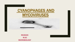
- 2. CYANOPHAGES ■ Cyanophages are the viruses that attack on cyanobacteria that is members of the blue –green algae in general. ■ Cyanophages uses cyanobacteria for their replication and multiplication. ■ Cyanophage is classified in a bacteriophage family that is myoviridae, podoviridae and siphoviridae. ■ For the first time Safferman and Morris (1963) isolated a virus from the waste stabilization pond of Indiana University (U. S. A.) that attacked and destroyed the three genera: Lyngbya, Plectonema and Phormidium. ■ Therefore, they named the virus by using the first letter of the three genera as LPP. ■ There after, several serological strains of LPP were isolated and named as LPP-1, LPP-2, LPP-3, LPP-4 and LPP-5.The viruses are commonly called as blue-green algal viruses and cyanophages. ■ They screened 78 host orgaisms and found the cyanophages only in 11 filamentous cyanobacteria.
- 3. ■ After the discovery of LPP-1, a large number of cyanophages was discovered by the other workers including R. N. Singh and coworkers from Banaras Hindu University. ■ Padan and Shilo (1973) reviewed different types of cyanophages.
- 4. Morphology of cyanophages ■ Morphology of LPP -1 has been studied in detail as compared to the other cyanophages. ■ The cyanophages differ morphologically as well as in physico – chemical properties. ■ The LPP-1 group of cyanophages has an icosahedral head and a tail and are similar to T3 and T7 bacteriophages, whereas the N-1 group resembles with T2 and T4 phages. Like T-even phages the tail may be contractile or non-contractile. ■ The AS-1 group has the largest cyanophages. ■ The group G-III and D-1 are serologically related but do not show any relationship with T-phages.
- 5. Physio – chemical and morphological characteristics of some cyanophages Characters LPP 1 LPP2 N1 SM1 AS1 MORPHOLOG Y icosahedral icosahedral Icosahedral icosahedral hexagonal TAIL, SIZE (nm ) Short 20*15 Short 20*15 Long 110*10 Absent …………… Long 243*22 NATURE OF TAIL Non contractile Non contractile Contractile Absent Contractile G + C CONTENT(%) 53 ………… 37 66-67 53-54 pH RANGE 5-11 5-11 ……………… 5-11 4-10 TEMPRATURE 4-40 C 4-40 C …………… 4-40 C ………… LATENT PERIOD(HOUR) 7 6 7 32 mins 8
- 6. Growth cycle of cyanophages ■ The replication of genetic material of cyanophages has been reviewed by Padan and Shilo (1973) , Sherman and Brown(1978). ■ Like bacteriophage, cyanophages too follow the same one step growth curve. The growth cycle of cyanophages resembles with that of T4 phage however the latent and rise period is different. ■ The growth cycle of LPP -1 has been studied in greater detail, which is carried out in following manner. 1. Adsorption- LPP 1 is adsorbed or attached on the surface of host cell. 2. Injection – After attachment, the DNA is injected into the host cell leaving the protein coat outside. How does the DNA is injected, it’s mechanism is still unknown.
- 7. 3. Reduce protein synthesis – Soon after injection of the genome the rate of protein synthesis is reduced and gradually blocked at the 5 hours of injection. 4.Multiplication- The cyanophages start to multiply in the invaginated photosynthetic lamellae or in virogenic storma. 5.Assembly- The viral nucleic acid and protein coat starts to assemble and form a procapsid. 6.Release- At the end, after maturation and assembly, the progeny cyanophages are released from the host cell by cell lysis. ■ After injection, the following three types of formed: (I) Earliest protein- These proteins are formed immediately after genome enters. (II) Early proteins – These proteins are formed after 2 hours of entry of genome till lysis of the cell. (III) Late proteins – They are formed after 4 hours of injection until the cell lysis.
- 9. ■ After 3 hours of infection, degradation of the host DNA begins and by the end of 7th hour it is converted into acid soluble material. ■ However, complete degradation of host DNA does not occur. Sufficient amount of degraded DNA is used up in building of viral DNA. ■ The latent period differs in different viruses, for example 7 hours in LPP-1 and N-1.Thereafter these starts the rise period which also varies with the viral types. ■ At the end, after maturation and assembly, the progeny cyanophages are released fromz each cell leaving aside the lysed cell. ■ The burst size for different cyanophages follows the following sequence : 350 plaque forming units(pfu) in LPP -1, 100 pfu in N-1, 50 pfu in AS-1, 100 pfu in SM-1. ■ The plaques can be observed either on algal lawn growing on nutrient agar or in broth cultures. ■ After infection, several physiological processes are disturbed such as respiration, photosynthesis, host DNA metabolism and nitrogen fixation.
- 10. Ecological importance of cyanophages ■ Water stabilization ponds,eutrophic lakes and polluted water support the growth luxuriant growth of cyanobacteria. ■ These can be obnoxious bloom in water reservoirs like lakes and result in fish mortality. ■ Therefore, the cyanophages can play a significant role in control of blooms. ■ So far the problems with then are that they are specific to genus and difficult to isolate.
- 12. What are mycoviruses? ■ Mycoviruses or mycophages are viruses that infect fungi. ■ Most of the mycoviruses are latent but some induce symptom. ■ The majority of mycoviruses have double stranded RNA genomes and isometric particles but 30% have a single stranded RNA (+ sense). ■ So far at least 5,000 fungal species are known to contain viruses. ■ Most of the species of Penicillium and Aspergillus are found to be infected with the virus. ■ The existence of mycoviruses appear to be intracellular. ■ The study of mycoviruses is known as mycovirology.
- 13. History ■ During 1950s, several disorders in fungi were described and some authors suspected for the involvement of viruses. ■ For the first time Hollings(1962) gave the conclusive evidence of viruses that infected the cultivated mushrooms, Agaricus bisporus causing the die back disease. ■ The most characteristics and consistent features of mushroom virus disease are the loss of crop and the degeneration of myceliun in the compost.
- 14. Types of mycoviruses ■ So far very few mycoviruses have been fully characterized and most are only the virus like particles(VLPs). ■ Some of the mycoviruses are isometric particles( 105-110 nm diameter) Cassidy is roughly spherical polyhedron. ■ The mycoviruses have a heterogeneous property with a diameter ranging from 25-50 nm. ■ Most of the mycoviruses have single capsid protein but different molecular weight. Some of the viruses have more than one capsids. ■ Mycoviruses are classified into following groups: 1. ds RNA mycoviruses 2. (+)ss RNA mycoviruses 3. (+)ss RNA mycoviruses with RT 4. (+)ss DNA mycoviruses
- 17. CLASSIFICATION OF MYCOVIRUS ■ ICTV classifies mycoviruses into 2 groups as per their taxonomy: 1. Penicillium chrysogenum virus group 2. Penicillium stoloniferum virus group ■ Mycoviruses have been categorised into 4 types as per their particle morphology for their taxonomic affinity : 1. Rod-shpaed particles 2. Filament particle 3. Isometric particles 4. Bacilli form particles
- 19. Replication of mycoviruses ■ Buck (1979,1980) has reviewed the replication of mycoviruses inside the fungal cells. ■ He has reported some host enzymes capable of transcribing the ssRNA and dsRNA in laboratory conditions and probably dsRNA in vivo. ■ Some dsRNA mycoviruses code RNA polymerases necessary for effective in vivo transcription and replication of dsRNA. ■ Mycoviruses are found in fungal spores and it is believed that they are transmitted through the spores. The presence of viral RNA in the fungal cells does not appear to affect any cellular properties such as antibiotic production.
- 21. Examples of mycoviruses ■ Mycovirus of mushroom 1. At least 6 viruses and VLP have been reported from the cultivated mushrooms, A. bisporus nearly from all countries where it is grown widely. 2.The mycoviruses occur in a mixture of cells and are extremely hard to separate 3.In some laboratories it could be demonstrated that the presence of viruses in sporophores of mushrooms resulted in reduction in Crop yield and decrease in mycelial growth.
- 22. Mycoviruses in plant pathogenic fungi ■ Due to the presence of mycoviruses in pathogenic fungi the virulence of pathogens gradually declines resulting in even death of fungi. ■ A highly pathogenic isolate of G. graminis from wheat roots gradually lost the virulence over a period of 17 months in culture. ■ In virulent isolates no viruses could be detected. ■ After a few months, 35 nm virions and later on 26 nm virions were observed in increasing quantities resulting in gradual loss in pathogenicity of the fungus.
- 23. REFERENCE ■ Dubey, R. C. and Maheshwari D. K. A Textbook of Microbiology ,3rd edition, S. Chand and Co, Ram Nagar, New Delhi ■ https://www. sciencedirect .com>cyanophages ■ https://www.frontiers.org ■ https://www.sciencedirect.com>mycoviruses