1. Food processing methods such as pasteurisation,
concentration by evaporation, spray drying and the
addition of chemical and sugar additives are
becoming less favored by consumers due to the
desire of more nutritious, chemical free and higher
quality foods. New technologies are now required
to inactivate deleterious or pathogenic micro
organisms without leaving chemical residues in the
food or destroying healthy compounds within the
food. UV light irradiation holds considerable
promise in food processing as an alternative to
traditional thermal processing. It is widely accepted
that the disinfection efficiency of Pulsed UV (PUV)
technology takes a much shorter time than
continuous UV light technology due to higher
intensities.
Treatment : During phase logarithm, each bacteria
was subjected to PUV treatment (Control = no
treatment, 20P, 40P, 60P, 80P, 120P and 160P). 100
µl of the treated bacteria was plated following serial
dilutions at 37oC for 24hours and Total Viable
Count (TVC) performed.
Protein analysis : Immediately post treatment, 1 ml
of the each treated bacteria was also transferred to
a sterile Eppendorf tube which was centrifuged at
8,000rpm for 10min for protein and DNA analysis.
The BCA Protein Assay was used to determine the
loss of internal microbial proteins against PUV
treatment. Standards created from known
concentrations of BSA (Bovine Serum Albumin)
(Fig. 1.2.) were used to determine the protein
content of the supernatant with the aid of the
colorimetric assay.
Figure 1.2. Standard curve of BSA at 562nm.
DNA analysis : DNA extraction was performed
using a DNA purification kit – High Pure PCR
Template Preparation Kit. The kit is composed of a
cell lysis buffer, proteinase K enzyme, binding
buffer, elution wash buffers, a pre warmed final
elution buffer and filter tubes. Quantitative PCR (q
PCR) was then performed using the Fast Start
Essential DNA Probes Master. A primer specific for
a region – 16S rRNA of all Bacillus spp. was used
to amplify genomic DNA sequences of Bacillus
megaterium and Bacillus cereus in this study .
Cycle threshold (Ct) values of known bacteria
numbers were used to determine the level of DNA
damage in the samples.
Table 1.1. DNA quantification standards.
qPCR was also performed in conjunction with
Propidium Monazide (PMA) dye prior to DNA
extraction and purification. The PMA works in
conjunction with PCR methods in identification of
viable only bacterial cells.
Black, J. G., 2008. Microbiology. 7th ed. Asia: John Wiley and Sons Inc.
Farrell, H.P., Garvey, M. Cormican, M., Laffey, J.G., Rowan, N.J., 2009.
Investigation of critical inter related factor affecting the efficacy of pulsed light
for inactivation of clinically relevant bacterial pathogens. Journal Applied
Microbiology, 91 (2009) pp 1494 – 1508.
Fox, C., 2011. Studies on the variation in strain susceptibility of
Staphylococcus aureus to high intensity pulsed UV light irradiation.
Garvey, M. (2009) Pulsed UV light inactivation of Cryptosporidium Parvum
and other microbial species in drinking water supplies in Ireland.
Garvey, M., Farrell, H., Cormican, M., Rowan, N., 2014. Investigations of the
relationship between use of in vitro cell culture quantitative PCR and a
mouse based bioassay for evaluating critical factors affecting the disinfection
performance of pulsed UV light for treating Cryptosporidium parvum oocysts
in saline. Journal of Microbiology Methods. 80 (3) pp 267 – 273.
Patrasa, A. Bruntona, N.P O’Donnell, C. Tiwari, B.K. 2010. Effect of thermal
processing on anthocyanin stability in foods; mechanisms and kinetics of
degradation, trends in food science and technology. 21 (2010) pp. 3 – 11.
Awuah, G.B. Ramaswamy, H.S. Economides A. 2006. Thermal processing
and quality principles and overview, chemical engineering and food
processing. 46 (2007) pp. 584 – 602.
Goosen, N., Moolenaar, G.E., 2008. Repair of UV damage in bacteria. DNA
repair. 7 (2008) pp 353 – 379.
Special acknowledgment to Prof. Neil Rowan, AIT, Dr. Alessia Stocca, AIT,
Dr. Damien Brady, AIT, Dr. Sile O Flaherty, AIT and class colleagues and
friends.
The effectiveness of PUV in this study is
demonstrated in Fig. 1.2. PUV inflicts 4 types of
cellular damage – photo hydration, photo splitting,
photo crossing and photo dimerization, all of which
prevent DNA replication. 100 % reduction in both
Bacillus spp. was seen at 120P and 160P. Bacillus
megaterium was deemed the more sensitive of the
two bacteria as total reduction of the bacteria was
also seen at 80P (Fig. 1.4.). Increase in protein
concentration as a result of internal cellular protein
damage (Fig 1.5.) also demonstrates the
effectiveness of PUV. Less protein was released by
Bacillus megaterium as a result of PUV.
The measurement of increase in protein as a result
of intracellular damage does not correlate the
results of TVC. Therefore the increase in protein
demonstrates protein leakage of the cell rather than
bacterial inactivation. As the DNA analysis
performed in this study was not successful due to a
non functioning primer, replication of the study is
required to determine the relationship between
protein leakage and DNA damage following PUV
treatment. Further research may also be warranted
which determines the effect of PUV on the
pathogenic toxins released by both Bacillus spp.
Project By: Lorraine Hannon. BSc. (Hons) in Applied Biosciences
Supervisor: Prof. Neil Rowan, Athlone Institute of Technology.
Bacillus spp. are often involved in issues related to
food poisoning and food spoilage characteristics.
Widely found in soil and water, Bacillus spp., are
rod shaped, endospore forming, gram positive
bacteria. They can be either facultative anaerobic or
obligate aerobic and are also a highly resistant
genera due to the production of endospores from
vegetative cells enabling the bacterium to resist
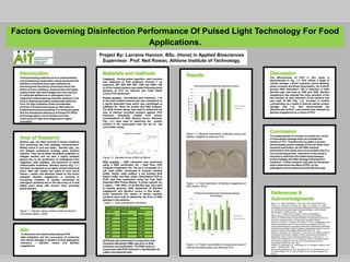
unfavourable conditions. Bacillus cereus (Fig. 1.1.)
has been recognized as an agent of food poisoning
since 1955 and causes two types of food borne
illness – emetic and diarrheal based on the toxins
released. Bacillus megaterium is involved in
increasing numbers of studies examining its
pathogenic potential due to the discovery of a heat
stable toxin along with several toxin encoding
genes present.
Figure 1.1. Bacillus cereus viewed under fluorescent
microscope (Black, 2008). .
Factors Governing Disinfection Performance Of Pulsed Light Technology For Food
Applications.
Figure 1.3. Bacterial Inactivation of Bacillus cereus and
Bacillus megaterium following PUV.
Figure 1.4. Total inactivation of Bacillus megaterium at
80P (Author, 2015).
Figure 1.5. Protein concentration increase as a result of
internal microbial protein loss following PUV.
To determine the relationship between PUV
light irradiation and the occurrence of molecular
and cellular damage in bacteria of food pathogenic
relevance – Bacillus cereus and Bacillus
megaterium.
Table 1.1. Ct Values for Standards
Bacteria
Numbers
Bacillus
megaterium
Bacillus
cereus
1 x 10⁹ 13.543 16.929
1 x 10⁸ 14.465 20.728
1 x 10⁷ 16.987 24.461
1 x 10⁶ 18.486 27.732
1 x 10⁵ 21.432 30.399
1 x 10⁴ 22.543 32.105
Hinweis der Redaktion
Copyright Colin Purrington (http://colinpurrington.com/tips/academic/posterdesign).