Restoration of enododontically treated teeth
•
7 gefällt mir•1,872 views

A rational approach to assessing root filled teeth in order to select an appropriate long term restorative option

Empfohlen
Empfohlen
Weitere ähnliche Inhalte
Was ist angesagt?
Was ist angesagt? (20)
Andere mochten auch
Andere mochten auch (10)
Ähnlich wie Restoration of enododontically treated teeth
Ähnlich wie Restoration of enododontically treated teeth (20)
Kürzlich hochgeladen
Kürzlich hochgeladen (20)
Restoration of enododontically treated teeth
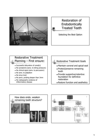
- 1. 1 Restoration of Endodontically Treated Teeth Selecting the Best Option Restorative Treatment Planning – First ensure: Successful obturation of canal(s) No symptoms (sens. to biting pressure) No clinical signs (sens. to percussion) No sens. to palpation No sinus tract No perio. probing deeper than 3mm No radiographic evidence of inflammatory disease Restorative Treatment Goals Maintain coronal and apical seal Protect/preserve remaining tooth Provide supportive/retentive foundation for definitive restoration Restore function and aesthetics How does endo. weaken remaining tooth structure? 1 Fracture In Endo Treated Teeth Iatrogenic Causes Non- Iatrogenic Causes Tooth Structure Loss Intra- canal Medicaments Restorative Procedures Primary Causes Secondary Causes History of Recurrent Pathology Anatomical Position of tooth Ageing of Dental Tissues 3
- 2. 2 Inorganic Fraction Carbonated Apatite •Stiffness •Ultimate Compressive strength Water (free & bound) Viscoelasticity Stress absorbtion Plasticizing & toughening Distribution of stress/strain Organic Fraction (Type 1 collagen) Resistance to crack propagation Toughness Ultimate tensile strength Dentine The role of different constituents on the mechanical integrity of dentine 3 Please see reference 3. For a thorough discussion of factors in play in the predisposition of endo. treated teeth to fracture Endodontic Factors Tooth stiffness Access cavity preparation Removal of roof of pulp chamber Canal preparation Medicaments Heavy obturation forces? (Lateral condensation) 5% reduction in tooth stiffness3 How does previous restorative ₮ weaken teeth? Isolates cusps Undermines cusps Broken cusps – too weak to withstand occlusal forces Sub-gingival margins2 Restorative Factors Tooth Stiffness Occlusal cavity prep. – 20% reduction Loss of marginal ridge integrity MOD cavity prep. – 63% reduction 3 Planning Treatment
- 3. 3 Consider 1. Amount of remaining tooth structure 2. Anatomic position 3. Occlusal/Para-function forces 4. Restorative purpose/ requirements 5. Aesthetic requirements 1. Remaining Tooth Structure More tooth structure – better prognosis E.g Crown prep. with even 1 mm dentine above gingival margin Double the fracture resistance of preps finishing flat & level with gingival margin Ideally a ferrule effect2 Assessment of Remaining Tooth Structure 1 2. Anatomic Position Canines – Canine Guidance – Sufficient natural dentine to resist lateral forces Group function – Canine/Pre-molar guidance 1 3. Occlusal / Para-functional Forces Evidence of heavy bruxism Thin weak mesio- & disto-buccal cusps Early silver- reinforced GIC base Cut back and tooth prepared for full gold inlay/onlay (partial crown) 1 4. Restorative purpose? Single stand alone restoration? Bridge abutment? RPD Overdenture abutment?
- 4. 4 Stand-alone 1 Bridge abutment 1 Crown lengthening to obtain sufficient tooth structure for ferrule Crown/root ratio? Over-denture Root Filled Anterior teeth Assessment of Remaining Structure De-vitalized by Trauma Otherwise intact Restore the access cavity only Aesthetics? Minimally restored- The other proximal surface is intact Restore with composite Small proximal rest. Small proximal-incisal rest.
- 5. 5 Large mesial and distal cavities plus access cavity Restore with post-core and full coverage crown Heavily restored Structurally compromised tooth Long crown – insufficient remaining stucture Reduce tooth and Crown lengthening procedure for the distal & facial – ferrule Post? Root Filled Posterior Teeth Assessment of Remaining Structure Marginal ridges intact 1. 1 Restore access cavity only 1 Moderately sized cavity Remove all restoration – any cracks?????? 1 Marginal ridge undermined?
- 6. 6 Restoration only Marginal ridge intact 1 Thin, weak or undermined cusps Restore with overlay restoration Cusp reduction External bevel Cast metal overlay Ceramic and pre-processed resin also possible 1 Core Build-ups Avoid posts wherever possible Preps. for partial crowns Grooves for added resistance and retention Core paste Core paste Using the pulp chamber to retain the core •Shoulder for ceramic or pre-processed resin •chamfer for cast metal 1
- 7. 7 Margins to finish on sound tooth Partial crown preparation to finish on sound tooth 1 Nayyar core- for full crown 2mm 2mm Undercut Weak sections trimmed down Ferrule Crown restoration Core paste 4 Undercuts in the pulp chamber provide retention and resistance for the core Use the pulp chamber Core paste 1 Posts? Insufficient tooth to retain the core Insufficient core length to retain crown Post Core paste2 2 Varying amounts of loss of tooth structure ***** *** ? Prognosis
- 8. 8 2 mm of remaining coronal tooth allows for preparation creating ferrule effect4 Types of Posts Pre-fabricated and Cast Prefabricated(*) and Cast/Custom Posts(#) Uniformly distributed through cement layer Little or noneSimilar to parallel, serrated #Cast post – parallel, serrated Wedging effectLittle or noneLow#Cast post – smooth tapered Wedging effect at the tapered end Little or noneSimilar to parallel serrated *Parallel, serrated - tapered end Relatively low – distributed by individual threads Low after counter rotation Highest*Parallel threaded High stresses - accentuating installation stress Very high – wedging stress Intermediate*Tapered self-tapping Uniformly distributed through cement layer Little or noneHigher *Parallel serrated (cemented vented) Wedging effectLittle or noneLow*Tapered smooth Functional StressInstallation stressRetentionType 5 Post Materials Pre-fabricated Stainless steel * Titanium * Glass-fibre reinforced resin (bondable) # Carbon-fibre reinforced resin (bondable) # Cast/custom Metallic Gold * Semi/Non-precious C+B alloys * Zirconia * Rigid * Non-rigid # Stress of Self Threading Posts 1. Threaded post after placement1. 2. Increased stress after tightening by ¼ turn 2. 1 Cemented Posts Stress upon cementation Stress in function +- - + 1
- 9. 9 Cast Posts 1 Post length Post should be at least as long as the desired clinical crown Mitigating factors Curved canals Taper of the root Maintaining apical seal (4-5mm of GP) Post diameter Choice of post diameter is based on canal/root size Avoid unnecessary removal of internal dentine (weakens root) Post should fit canal dentine walls snugly Other Features Positive stop of the core on coronal tooth structure to prevent the post/ core unit from being forced apically 1.5 – 2.0mm of tooth structure for 360° to receive the crown ferrule Maintain no less than 1mm wall thickness of radicular dentine (preferably 2-3mm) Risk of root fracture Core •Material Crown •Loading angle •Ferrule Remaining Structure •Dentine •Water content Post •Length •Shape •Adhesion •Diameter •Elastic modulus 3 Fracture predisposing factors in post-core restorations
- 10. 10 3 Post length What type of post is best? Studies have shown Bonded posts, parallel-sided posts - less dentine stress Non-bonded and tapered posts – more dentine stress Increase mod. of elasticity (stiffer) and increased diameter of bonded post – less dentine stress Decreased post length – more dentine stress3 Anterior tooth with little coronal structure Cast post/core Serrated, parallel- sided post with tapered or rounded tip Posterior tooth with some coronal structure One or two pre-fabricated posts and core paste build-up Posterior tooth with little coronal structure Cast post/core unit with secondary insertion of a wrought post/s through the core The final crown restoration The reinforcement effect of cementation of a full crown with ferrule effect will make the difference between stiff and elastic posts less obvious 3
- 11. 11 Tooth anatomy Considerations for post placement Maxillary first molars Deep concavities on furcal surfaces 94% mesio-buccal roots 31% disto-buccal roots 17% palatal roots Mandibular first molars Concavities on furcal surfaces of All mesial roots 99% of distal roots Maxillary first premolars Deep mesial concavities Slender roots with thin dentine Maxillary first premolar In this situation the palatal root would be the ideal candidate for the post The buccal root is highly irregular in form Buccal Palatal CEJ 2mm 4mm 6mm 2 How to tell from x-ray? Root form Curvature and post placement 2
- 12. 12 Post Cementation Zinc Phosphate Mechanical retention No chemical adhesion Resin-modified GIC (auto- or dual cure) Adhesion to dentine Resin Cement (dual cure) Adhesion to dentine In-soluble when set Moisture sensitive prior to set Difficult to place the bond apically References 1: Endodontics – 3rd Ed. Stock, Walker, Gulabivala 2:Pathways of the Pulp 9th Ed. Cohen & Hargreaves 3. “Mechanisms an Risk Factors for Fracture Predilection in Endodontically Treated Teeth” Anil Kishen Endodontic Topics 2006, 13, 57-83 4. Colour Atlas of Endodontics William T Johnson 5. Problem Solving in Endodontics 4th Ed. Gutman, Dumsha, Lovdahl 6. “Restoration of Endodontically Treated Teeth” Morgano, Rodrigues, Sabrosa Dental Clinics of North America 48 (2004) 397-416