Case study
•Als DOC, PDF herunterladen•
5 gefällt mir•3,168 views

A 56-year-old male presented with weakness, headache, light-headedness and fatigue. Laboratory tests showed elevated red blood cells and leukocytes, low platelets, and undetectable erythropoietin levels. The spleen was enlarged and blood smear showed immature cells. The patient was diagnosed with polycythemia vera and prescribed phlebotomy to reduce red blood cell levels and potentially myelosuppressive drugs if needed.

Empfohlen
Weitere ähnliche Inhalte
Was ist angesagt?
Was ist angesagt? (19)
Andere mochten auch
Andere mochten auch (12)
Ähnlich wie Case study
Ähnlich wie Case study (20)
Mehr von Lawrence James
Mehr von Lawrence James (20)
Kürzlich hochgeladen
Kürzlich hochgeladen (20)
Case study
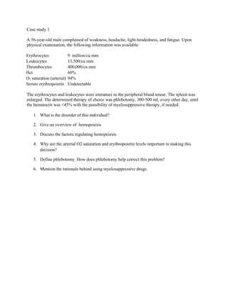
- 1. Case study 1 A 56-year-old male complained of weakness, headache, light-headedness, and fatigue. Upon physical examination, the following information was available: Erythrocytes 9 million/cu mm Leukocytes 13,500/cu mm Thrombocytes 400,000/cu mm Hct 60% O2 saturation (arterial) 94% Serum erythropoietin Undetectable The erythrocytes and leukocytes were immature in the peripheral blood smear. The spleen was enlarged. The determined therapy of choice was phlebotomy, 300-500 mL every other day, until the hematocrit was <45% with the possibility of myelosuppressive therapy, if needed. 1. What is the disorder of this individual? 2. Give an overview of hemopoiesis 3. Discuss the factors regulating hemopoiesis. 4. Why are the arterial O2 saturation and erythropoietin levels important in making this decision? 5. Define phlebotomy. How does phlebotomy help correct this problem? 6. Mention the rationale behind using myelosuppressive drugs.
- 2. Case 2 A 16-year-old girl complained of fatigue and loss of strength. Her appetite was marginal, as she was very conscious of maintaining her body weight at 96 pounds. Her monthly menstrual flow was always heavy and long from its onset at twelve years of age. Relevant laboratory findings included the following: Hematocrit (Hct) 28% Hemoglobin (Hgb) 9 g/dL Iron 16 µg/dL Bone marrow iron Absent Erythrocytes Small and pale Suggested treatment included ferrous sulfate or ferrous gluconate for six months orally between meals, since food may reduce absorption. A well-balanced diet was also suggested. 1. What is the primary disorder of this individual? 2. Explain the stages of erythropoiesis. 3. Discuss the mechanism of Iron absorption 4. What does the ferrous sulfate or ferrous gluconate provide? Why is it necessary? 5. What dietary inclusions would you suggest? 6. Why is bone marrow iron an important clinical indicator in this individual?
- 3. Case Study 3 A 38-year-old male complained of fatigue, anorexia, weight loss, dyspnea on exertion, and a sense of abdominal fullness. An examination of peripheral blood and bone marrow resulted in the following: Complete blood count (CBC): Erythrocytes 2.4 million/cu mm Leukocytes 160,000/cu mm Thrombocytes (platelets) 51,000/cu mm Hematocrit 23% Differential: Lymphocytes = 85% of all leukocytes Bone marrow examination demonstrated a tremendous predominance of small lymphocytes (>90%). The bone marrow was so tightly packed that it aspirated with difficulty. The spleen was moderately enlarged, and there was generalized lymph node enlargement. 1. What is the primary disorder of this person? 2. Define leukocytosis. What is the cause of the leukocytosis? 3. Define thrombocytopenia. What is the cause of the thrombocytopenia? 4. Mention the functions of spleen 5. What is the cause for swollen lymph nodes? 6. Explain the role of lymphocytes in immunity
- 4. Case study 4 Martin and Kim were both twenty-five when they had Michael, their first child. Kim remained very healthy during her pregnancy and went into labor at 9:00 a.m., just 3 days after her due date. Delivery went quite smoothly, and that evening, mother and child rested comfortably. Two days later, Kim and Michael were released from the hospital. That evening at feeding time, Kim noticed that the whites of Michael's eyes seemed just slightly yellow, a condition that worsened noticeably by the next morning. Kim called the pediatrician and made an appointment for that morning. Upon examining Michael, the pediatrician informed Martin and Kim that the infant had neonatal jaundice. The physician told the parents he would like to see Michael every other day in order to monitor blood bilirubin concentration until the bilirubin concentration dropped into the normal range. He recommended that Kim feed Michael frequently and instructed them to place Michael in sunlight whenever possible. 1. Which organs are primarily responsible for removing old or worn red blood cells from circulation? 2. Discuss the patho physiology of neonatal jaundice 3. Explain the normal sequence of steps in the breakdown of hemoglobin. 4. Why would you expect the sclera of the eyes to turn yellow as a result of jaundice? 5. Differentiate between neonatal jaundice and obstructive jaundice.
- 5. Case study 5 Justin Mather, a 23-year-old man, presented to the physician with a long medical history dating back to 1983 when, as a 6-year-old, he was referred to a pediatrician by a dentist. At that time, the dentist was about to administer an anesthetic drug by injection prior to extracting a tooth when he elicited a history of easy bruising in the boy as well as a history of a brother who died at age one from an intracranial hemorrhage after falling from a crib. In addition, Justin has had several instances of severe bruising following minor trauma and progressively worsening arthritis of both knee joints over the past four years. Interestingly, Justin's maternal grandfather died at age 27 of a bleeding complication following an appendectomy. Justin's maternal grandmother remarried, and Justin's mother lost complete touch with her deceased father's family. Justin's mother's three half-brothers are alive and well. Lab findings in Justin at that time (1983) were as follows: -platelet = count 280,000 / mm3 of blood (normal = 150,000 - 350,000 / mm3) -bleeding time = 6 minutes (normal = 3.5 - 10.5 minutes) -prothrombin time (PT) = 11 seconds (normal = 10 - 12 seconds) -partial thromboplastin time (PTT) = 58 seconds (normal = 20 - 30 seconds) -fibrin split-products = 22 µg / ml (normal = 8 - 40 µg / ml) -hematocrit and white blood cell count were both normal Justin was told at that time that he had Hemophilia and would require close medical attention 1. What’s the differences between hemophilia A, B, and C? 2. What’s the difference between Von willebrand's disease and hemophilia . 3. What’s the difference between platelet adhesion and platelet aggregation? 4. Describe the physiologic events that occur following endothelial interruption in the blood vessel. 5. Why is the PTT elevated? Why isn't the PT elevated? In your answer, be sure to explain exactly what is assessed by these tests.
- 6. Case study 5 Justin Mather, a 23-year-old man, presented to the physician with a long medical history dating back to 1983 when, as a 6-year-old, he was referred to a pediatrician by a dentist. At that time, the dentist was about to administer an anesthetic drug by injection prior to extracting a tooth when he elicited a history of easy bruising in the boy as well as a history of a brother who died at age one from an intracranial hemorrhage after falling from a crib. In addition, Justin has had several instances of severe bruising following minor trauma and progressively worsening arthritis of both knee joints over the past four years. Interestingly, Justin's maternal grandfather died at age 27 of a bleeding complication following an appendectomy. Justin's maternal grandmother remarried, and Justin's mother lost complete touch with her deceased father's family. Justin's mother's three half-brothers are alive and well. Lab findings in Justin at that time (1983) were as follows: -platelet = count 280,000 / mm3 of blood (normal = 150,000 - 350,000 / mm3) -bleeding time = 6 minutes (normal = 3.5 - 10.5 minutes) -prothrombin time (PT) = 11 seconds (normal = 10 - 12 seconds) -partial thromboplastin time (PTT) = 58 seconds (normal = 20 - 30 seconds) -fibrin split-products = 22 µg / ml (normal = 8 - 40 µg / ml) -hematocrit and white blood cell count were both normal Justin was told at that time that he had Hemophilia and would require close medical attention 1. What’s the differences between hemophilia A, B, and C? 2. What’s the difference between Von willebrand's disease and hemophilia . 3. What’s the difference between platelet adhesion and platelet aggregation? 4. Describe the physiologic events that occur following endothelial interruption in the blood vessel. 5. Why is the PTT elevated? Why isn't the PT elevated? In your answer, be sure to explain exactly what is assessed by these tests.