GHH - Introduction; Lecture
•
3 gefällt mir•500 views

Student-made hand-out.... General Histology and Histotechnique Lecture; 2012-2013; 1st sem; Midterm handout Credit to the original owner of the pictures used in this pdf document

Empfohlen
Weitere ähnliche Inhalte
Kürzlich hochgeladen
Kürzlich hochgeladen (20)
Empfohlen
Empfohlen (20)
GHH - Introduction; Lecture
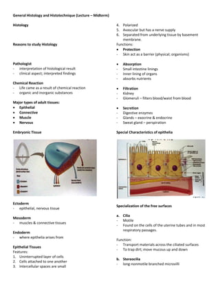
- 1. General Histology and Histotechnique (Lecture – Midterm) Histology 4. Polarized 5. Avascular but has a nerve supply 6. Separated from underlying tissue by basement membrane. Reasons to study Histology Functions: Protection - Skin act as a barrier (physical; organisms) Pathologist Absorption - interpretation of histological result - Small intestine linings - clinical aspect; interpreted findings - Inner lining of organs - absorbs nutrients Chemical Reaction - Life came as a result of chemical reaction Filtration - organic and inorganic substances - Kidney - Glomeruli – filters blood/waist from blood Major types of adult tissues: Epithelial Secretion Connective - Digestive enzymes Muscle - Glands – exocrine & endocrine Nervous - Sweat gland – perspiration Embryonic Tissue Special Characteristics of epithelia Ectoderm Specialization of the free surfaces - epithelial, nervous tissue a. Cilia Mesoderm - Motile - muscles & connective tissues - Found on the cells of the uterine tubes and in most respiratory passages. Endoderm - where epithelia arises from Function: - Transport materials across the ciliated surfaces Epithelial Tissues - To trap dirt; move mucous up and down Features: 1. Uninterrupted layer of cells b. Stereocilia 2. Cells attached to one another - long-nonmotile branched microvilli 3. Intercellular spaces are small
- 2. General Histology and Histotechnique (Lecture – Midterm) - Found on the surface of cells in the epididymis; Ductus deferens Function: for absorption c. Microvilli - Visible as striated brush borders on the epithelium of the small intestine and tubules of kidney Function: for absorption Lateral surface: Cell junctions Cell junctions: b. Adherens junctions (zonula adherence) - made of plaque - Form extensive bands called adhesion belts - Plaque-dense layer of proteins in the inside of plasma membrane - Composed of actin - Help epithelial surfaces to resist separation and adhesion is due to the transmembrane glycoprotein cadherin a. Tight junction - It connects the cells of tissues that line the surfaces of organs and body coverings. - Specifically/specially found in intestine, urinary bladder - contain materials - Known a tights because it prevent/seal lining of organs to prevent leaking of substances towards the blood or tissues - Pose a life threatening situation c. Desmosomes (macula adherens) Location: Most apical part of epithelial - Composed of plaque Zonula – refers to the junction that forms the band. - With tonofilaments Occludens – membrane fusions that closed of - Numerous among cells that make up the epidermis intercellular spaces. and cardiac muscle cells of the heart. - Place a role in dissipating physical forces throughout the cell thru the attachment sites. - Made of intermediate filament (keratin) - Composed of cadherin
- 3. General Histology and Histotechnique (Lecture – Midterm) Basement Membrane - Basal lamina (2 layers) + Reticular lamina Functions of Hemidesmosomes: - cell signaling d. Hemidesmosomes (hemi-half) - Proliferation - Found at the basal surface of some stratified - Apoptosis squamous epithelia Functions: - Connect cells to the basement membrane - Anchor one kind of tissue to another in the body. - Integrin - Presence of keratin Composition of Basal Lamina: 1. Type IV collagen - made of a 3 α chains e. Gap junction - Greatest number of protein found in our body. - The gap is bridged by transmembrane protein channels called connexons. 2. Laminin - Large glycoprotein Functions: - Localize in lamina lucida. - Allow cell and a tissue to communicate - It enables nerve or muscle impulses to spread 3. Proteoglycans rapidly between cells. - With heparan sulfates Function: to give the basal lamina a strong anionic Connexons charge. - Made up of connexins Functions: - Individual unit of gap junctions that forms a manute - For cell attachments fluit-filled tunnels - For selective filter - Composed of connexins 4. Fibronectin Function: Form tunnels w/ different physiological - Present in small amount. properties. - Found in basal lamina - More in the lamina densa - For connecting purpose of underlying membrane.
- 4. General Histology and Histotechnique (Lecture – Midterm) 1. Simple General function of Basal Lamina - Single layer of cells Functions: Diffusion, Filtration, Absorption, secretion. 1. Provide a physical support to the epithelium 2. Stratified 2. Its structural support of type IV collagen gives it - 2 or more layers of cells considerable tensile strength Function: For protection for the underlying tissues 3. It is flexible enough to permit stretching and recoil Ex. Skin to protect hollow organs. 3. Pseudostratified 4. Provides for cell attachment by specific binding sites - False-stratified-single layer in the cell membrane - It looks like many layers. - Which may or may not possess cilia or stereocilia Reasons: - The location of nuclei is at different lavels. - Not all cells reach the apical surface Ex. Trachea - According to cell shape Classifications of Epithelia 1. Squamous – thin flat cells Functions: 2. Cuboidal – cube-shaped Functions: Functions: Secretion & Absorption - Covering and lining 3. Columnar – Tall- cylindrical Covering - Some may have cilia - covers the body surface Function: Secretion / Absorption Ex. Epidermis Lining 4. Transitional - lines the endothelium - Cell changes in shape as the body - outer covering of body cavities (mesothelium) moves/expand/stretched. Ex. Blood vessels Ex. Urinary Bladder, Urethra Glandular Function: for Protection. - secreting portion of glands Ex. Thyroid, adrenal gland Classification according to combined cell layers and shapes Types of epithelial tissue A. Simple Epithelium 1. Simple Squamous - Single layer of flat cells - Nucleus is located at the center - Nucleus – oval/spherical in shape Functions: - Filtration, Diffusion, Osmosis, Secretion, Absorption Location: - lining of lung alveoli, the lining of blood vessels (endothelium), Bowman’s capsule in the kidney and lining of major body cavities (mesothelium)
- 5. General Histology and Histotechnique (Lecture – Midterm) 2. Simple Cuboidal - Single layer cube-shaped cells - Nucleus – centrally located Functions: - Secretion, Absorption, covering Location: - Commonly encountered in glandular ducts, surface of ovary, kidney tubules, capsule of the lens of the eye, surface of thyroid. 3. Simple Columnar - cells appear rectangular with oval nuclei - Nuclei – found at the base or near the base. 2 forms: a. Nonciliated simple columnar - Contains microvilli and goblet cells Microvilli - absorption Goblet cells - are found in the Duodenum of small intestine to secrete mucous.
- 6. General Histology and Histotechnique (Lecture – Midterm) - Lines the GIT from the stomach to the anus, ducts of many glands and gall bladder. Functions: - Absorption, secretion, lubrication, protection. B. Stratified epithelium 1. Stratified squamous Two forms: a. Keratinized stratified squamous - keratinized Keratin – tough fibrous protein deposited in the surface of the cells. Function: Help protect the skin from heat, microorganisms, and other chemicals. - Found in the superficial layer of skin – epidermis - It is dry. b. Ciliated simple columnar Goblet cells present in the upper respiratory tract. Cilia are present for ciliary movement. Ex. Cilia in the uterine tube - Lines a few portion of upper respiratory tract, uterine tubes, uterus, some paranasal sinuses and central canal of spinal cord. b. Non-keratinized stratified squamous - It does not contain keratin - It remains moist - Lines the surface of mouth, esophagus, epiglottis, vagina, & tongue.
- 7. General Histology and Histotechnique (Lecture – Midterm) Location: - Adult sweat glands - Part of male urethra - Developing ovarian follicle 3. Stratified Columnar - Only found at the apical surface Functions: Protection and secretion - Lines part of urethra, large excretory glands, and small areas in the anal mucous membrane and part of the conjunctiva of the eye. 4. Transitional - No definite shape Apical surface - Squamous - stretched - Cuboidal columnar – relax - Lines the urinary bladder and portions of ureters and urethra. Function: To prevent distention – rupture of organs. 2. Stratified Cuboidal - Rare type of epithelial tissue - Cube-shaped cells found in apical surface. Functions: - Most for protection - Limited in secretion and absorption
- 8. General Histology and Histotechnique (Lecture – Midterm) Glandular Epithelium C. Pseudostratified Columnar Epithelium - formed by cells specialized for secretion - Nucleus is found at the base but not at the apical surface. 1. Endocrine - Lines the most upper respiratory tract, line ducts of - Ductless many glands, epididymis, and other part of male - Located in pituitary gland at the base of the brain, urethra. pineal gland in brain, thyroid and parathyroid Functions: glands near larynx, adrenal glands superior to - For secretion kidneys, pancreas near stomach, ovaries in pelvic - Movement of mucous by ciliary action. cavity, testes in scrotum, and thymus gland in thoracic cavity. 2. Exocrine - Duct gland - Located in sweat, oil, earwax and mammary glands of the skin; digestive glands such as salivary glands which secrete into the mouth cavity; and pancreas which secretes into the small intestine – product maybe released at the skin surface or into the lumen (cavity) of the hallow organ.
- 9. General Histology and Histotechnique (Lecture – Midterm) e. Simple branched acinar - Secretory portion is branched & flask-liked Ex. Sebaceous gland Classification: a. Unicellular glands - Common example is the goblet cell - Located in the lining and glands of intestines and 2. Compound gland – branched ducts certain passages of respiratory tract Function: To secrete mucous. a. Compound tubular - Tubular secretory portion b. Multicellular gland Ex. Bulbourethral gland, Copper’s gland - consist of more than one cell Examples: Sweat, Oil and Salivary gland b. Compound Acinar/Alveolar Categorized into two: - Flask-like secretory portion Ex. Mammary gland 1. Branched glands 2. Unbranched glands c. Compound Tubule-acinar/Tubule-alveolar - Tube-like and Flask like secretory portion Multicellular exocrine glands as to structure: Ex. Glands of pancreas 1. Simple gland – single-non-branched duct. a. Simple Tubular - Secretory portion is straight and tubular Ex.: Large intestine gland b. Simple Branched Tubular - Secretory portion is branched and tubular in shape Ex. Gastric gland Functional Classification of exocrine glands c. Simple coiled Tubular *** Base on how the secretions are released: - Secretory portion is coiled and tubular. Ex. Sweat gland a. Merocrine secretion - Produce the secretory products and released it from d. Simple Acinar/Alveolar the cells most exocrine glands. - It has a flask-like secretory portion - Exocytosis Ex. Gland of phenylurethra
- 10. General Histology and Histotechnique (Lecture – Midterm) b. Apocrine secretion - Products is accumulated at the apical surface which will be pinched off then removed apex to released secretions c. Holocrine - Secretory products accumulate at the apical surface which will pinched off. Adenocarcinoma – developed from glandular epithelium.