
Empfohlen
Weitere ähnliche Inhalte
Was ist angesagt?
Was ist angesagt? (20)
Ähnlich wie 4.3._BLOOD_AND_TISSUE_NEMATODES.ppt
Ähnlich wie 4.3._BLOOD_AND_TISSUE_NEMATODES.ppt (20)
Mehr von KnqAutlawzvFb
Mehr von KnqAutlawzvFb (17)
Kürzlich hochgeladen
Kürzlich hochgeladen (20)
4.3._BLOOD_AND_TISSUE_NEMATODES.ppt
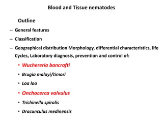
- 1. Blood and Tissue nematodes Outline – General features – Classification – Geographical distribution Morphology, differential characteristics, life Cycles, Laboratory diagnosis, prevention and control of: • Wuchereria bancrofti • Brugia malayi/timori • Loa loa • Onchocerca volvulus • Trichinella spiralis • Dracunculus medinensis
- 2. Learning objective At the end of this unit the student will be able to: – Explain the general features of blood and tissue nematodes – Classify tissue nematodes – Explain the Geographical distribution, Morphology, differential characteristics, life Cycles, prevention and control methods of blood and tissue nematodes – Collect blood samples in appropriate timing – Collect skin snip for the diagnosis of O. volvulus – Detect and identify microfilaria of blood and tissue nematodes
- 3. 2.1.3 Blood and tissue nematodes 2.1.3.1. General features: • Adults which live in the tissues of human (lymphatic system, subcutaneous tissues or muscle) • Long thread - like worms • Requires two host to complete their life cycle. • Females are viviparous, larvae hatch in the uterus • The female produce first stage larvae (L1)
- 4. The immature L1 stage larva is called Microfilariae Small , slender, motile forms L1 require blood sucking insects (IH) to develop to infective form (L3) No reproduction in the vector, rather development
- 5. 2.1.3.2. Classification: • Tissue nematodes can be classified based on: – Habitat in the body – Clinical manifestation – Morphology
- 6. Three families/ groups 1. FAMILY FILARIDAE( Filarial worm) - Common/pathogenic filaria • Wuchereria bancrofti • Brugia malayi • Brugia timori • Loa loa • Onchocerca ovolvulus – Less/non-pathogenic Filaria • Mansonella perstance • Mansonella streptocerca • Mansonella ozardi
- 7. 2. FAMILY TRICHINELOIDAE • Trichinella sps 3. FAMILY DRACUNCULIDAE( guinea worm) • Dracunculus medinensis Animal tissue nematode rarely infect human • Dirofilaria sps • Angiostrongylus cantonensis • Gnathostoma spinigerem
- 8. General features: Filariae live as adults in various human tissues Agents of LF reside in lymphatic vessels and lymph nodes O. Volvulus, Loa loa, M. Ozzardi and M. Streptocerca in subcuraneous tissues M. Streptocerca besides reside in the dermis M. Perstans resides in body cavities and surrounding tissues 8 2.1.3.3. FAMILY FILARIDAE ( Filarial worm)
- 9. Three of these are responsible for most of the morbidity: W. bancrofti and B. malayi cause lymphatic filariasis, and O. volvulus causes onchocerciasis (river blindness). Animal reservoirs play no role of significance in most places, except in sub-periodic B. malayi. 9 Cont ….
- 10. Diagnosis based on Mf findings: • Morphologic features: – Size – Presence or absence of “ sheath” – Appearance i.e. curvature, Kinks, coiling etc – Arrangement of the column of nuclei in the body – Presence of nuclei at the very tip of the tail • Other features: – Periodicity – Source of specimen
- 11. • Factor to be considered when collecting blood –Collect blood at the correct time –Concentration technique recommended –In chronic infection Mf is rarely found in blood –In LF Mf are higher in capillary blood than venous blood –Mf are higher in capillary blood from the ear lob
- 12. FAMILY FILARIDAE( Filarial worm) Morphology • Adult – The adults are long thread like worms. – Measure 2 cm – 120 cm (4 – 10 µ wide) – Live in body cavities, lymphatic, and subcutaneous tissues – Release embryos (microfilaria) which live in blood or dermis (skin) – all require an insect or crustaean vector as intermediate host
- 13. • Microfilaria – The immature first stage larva of filarial worms – Are motile and live in blood or dermis – Measure, 150-350 µ long – Transparent and colorless with rounded or pointed tail in unstained smear – Internal structure can be visualized by the use of fixed stained preparation – Can be sheathed or unsheathed
- 14. Periodicity:- • Microfilaria of pathogenic filarial worms that found in the blood (m.f of filarial worms that causes lymphatic filariasis and Loasis) show periodicity
- 15. Periodicity:- – Mf are found in the blood in greater number in a certain hours of a day or a night – Corresponds to peak biting times of their insect vector • Nocturnal periodicity -mf is high in blood during night hrs • Diurnal periodicity-mf is high in blood during day hrs • Nocturnal or diurnal subperiodicity;- mf can found in blood 24 hrs with slight increase in number during day or night hrs
- 16. 16 Filarial worms (Synonym) Periodicity Main Vector (IH) Reservoir O. volvulus (River blindness) Non Periodic Black fly (Simulium) Human W. ancrofti (LF) Periodic (N) 22 – 04hr (24hr) Culex, Anopheles Human Sub Periodic 20 – 22 (21hr) 14 – 18 (16hr) Aedes Human B. Malayi (LF) Periodic (N) 22 – 04hr (24hr) Anopheles Human Sub Periodic 20 – 22 (21hr) Mansonia Human, Monkey, Cat – Zoonotic B. Timori (LF) Periodic (N) Anopheles Human L. Loa (Eye worm) Periodic (D) Deer fly Man, Monkeys M. streptocerca Non Periodic Midge (Culicoides) M. perstans Sub Periodic Midge (Culicoides) M. ozzardi Non periodic Midge (Culicoides)
- 17. • Flarial worm causes 3 main diseases –lymphatic filariasis (Elephantiasis) –Loasis –Onchocerciasis (river blindness)
- 19. Lymphatic Filariasis • Disease caused by filarial worms living in the human lymphatic system • Causative agents • Wuchereria bancrofti • Brugia malayi • Burigia timori • These worms lodge in the lymphatic system • They live for four to six years, producing millions of minute larvae that circulate in the blood”
- 20. Lymphatic Filariasis • Large numbers are present in the lymphatics of the: Lower extremities (inguinal and obturator groups), Upper extremities (axillary lymph nodes), and, Male genitalia (epididymis, spermatic cord, testicle) - particular for W. bancrofti
- 21. Social consequences It is one of the most debilitating and disfiguring diseases of the world 1. Disfigurement of the limbs and genitals leads to: – Stigma – Anxiety – Ostracization – Psychological trauma – Sexual disability
- 22. Social consequences….. 2. The disease impeds – Mobility – Travel – Educational opportiity – Employment opportunity – Marriage prospect
- 23. Epidemiology of Lymphatic Filariasis • Endemic in 83 countries • 1.2 billion at risk • > 120 million people infected • > 25 million men suffer from genital disease, • > 15 million people suffer from lymphoedema or elephantiasis of the leg • ~ 2/3 of infected people live in India and Africa • Others live in parts of Asia, the Pacific, & in Central and South America.
- 24. Distribution
- 25. Distributin • Wuchereria bancrofti • affects an estimated 119 million individuals and disfigures 40 million. • Wide distribution (Africa, SE Asia, Indonesia, South Pacific Islands) • Brugia malayi • Limited distribution (China, India, SE Asia, Indonesia, Philippines) • Brugia timori • Leser sunda, island of Indonesia
- 26. Wuchereria bancrofti Disease: Bancroftian filariasis, Wuchereriasis, elephantiasis Distribution: tropical and subtropical countries Morphology: 1. Adult: – Thready – Cylinderical oesophagus – Creamy white in color – Male: • About 4cm in lentth • Curved posterior end • 2 unequal spicules and has anal papillae 26
- 27. Wuchereria bancrofti • Female: • About 8 cm in length • 2 sets of genitalia • Vulva opens close to the posterior end • Viviparous 2. Micrfilaria: – 250 x 8 – Body forms graceful curves – Body has a column of nuclei separated by free areas – Rounded anterior and tapered tail ends free of nuclei – Loose sheath (stretched vitelline membrane) closely fits the body but projects beyond the head and tail ends. 27
- 28. Wuchereria bancrofti 3. Infective larvae: 1500 – 2000 x 20 – Cylinderical oesophagus 28
- 29. W. Bancrofti & B. Malayi 29
- 30. Transmission and life cycle
- 31. Cont .... • Requires two host • Human-DH • Mosquitoes-IH • Transmission • Bite female mosquitoes (Genera Culex, Aedes, Anopheles, Mansonia)
- 32. • Infective larvae deposited onto human skin during the mosquito's blood meal • Enter through the mosquito bite puncture wound or local abrasions. • In humans: – Parasites passes to the lymphatic system – Undergo further molts – Become adult male and female worms
- 33. • Adult female worms produce thousands of sheathed microfilariae per day • Mf can be found in blood 9 months after infection (W.bancrofti) and 3 months (Burigia species) • Normally found in peripheral circulation in evening.
- 34. • Microfilariae ingested during blood meal from infected person • Penetrate the mosquito stomach wall • Enter the body cavity (hemocoel) • Migrate to the insect's flight muscles for growth. • After 2 molts, the L3 migrate through the head, • Reach the proboscis of the mosquito.
- 35. Clinical manifestation. • Depends on: –Site occupied by adult –Number of worms, –Length of infection and –Immune response of the host
- 36. Clinical manifestation. 1. Many infections are asymptomatic 2. Circulating Mf probably do not cause pathology 3. The main pathological lesions are: a) Inflammatory manifestations – due to toxic products of living or dead adult worms
- 37. Clinical manifestation. – There may be: • Recurrent attacks of lymphangitis – Funiculits – Epididymitis – Orchitis, etc... • Lymphadenitis • Swelling and redness of affected parts • Fever, chills, headache, vomiting and malais
- 38. b) Obstructive manifestations – • Fibrosis following the inflammatory process • Coiled worms inside lymphatics. • This may result in: Dilatation Rupture of distended lymphatics In the urinary passage – chyluria In the pleura – chylothorax In the peritoneal cavity – chylous ascitis In the testis – chylocoele
- 39. Elephantiasis: Hard and thick, rough and fissured skin Frequently legs & genitalia (scrotum, penis and vulva) Less often arms and breasts.
- 41. Clinical ...... 4. Tropical pulmonary eosinophilia • Pulmonary symptoms: cough, dyspnoea, "asthmatic syndrome". • Chest X-rays: patchy infiltrates • Splenomegaly • High ESR & marked eosinophilia • No microfilariae in the peripheral blood. • Serological tests strongly positive • Responds very well to therapy with DEC 41
- 42. Diagnosis of W. bancrofti 1. BF (taken at night) Thin and thick smears Concentration methods • Lyzed capillary blood technique • Tube centrifugation lyzed blood technique • Microhematocrite tube technique • Membrane filtration technique • Counting chamber technique DEC provocation test 42
- 43. Diagnosis of W. bancrofti Mf in: Aspirates of hydrocele Lymph gland fluid Chylous urine 2. Intradermal test (antigen from Dirofilaria immitis) 3. Serological tests as IHA, IFA 4. Antigen detection: ICT filariasis test 43
- 45. 45
- 46. 46
- 47. Adult female worm of W. bancrofti 47 Adult male worm of W. bancrofti
- 48. Differential diagnosis • Podoconiosis (syn. lymphatic siderosilicosis or lymphoconiosis): – Very slow onset of oedema – Lymphoedema – Elephantiasis (mostly limited to below the knee) • Caused by immune response to certain minerals. • No hydrocoele, eosinophilia, nocturnal microfilaraemia 48
- 49. Treatment of W. bancrofti Diethyl carbamazine (DEC) General measures: Rest, antibiotics, antihistamines, and bandaging Surgical measures for elephantiasis 49
- 50. Prevention and control of W. bancrofti Control of mosquitoes Avoid mosquito bite Treat patients Health education Global LF elimination program strategy: Mass drug administration Care for chronic cases 50
- 51. 2.1.3.5. Loiasis
- 52. Loiasis • Caused by filarial worms living in subcutaneous tissue • Causative agents • Loa loa (Eye worm) • Distributed in Rain Forest areas of West Africa and equatorial Sudan.
- 53. Loa loa (Eye worm) Habitat: Adults live in: Connective tissues under the skin Mesentry Parietal peritoneum Subconjunctival tissue of the eye or thin skinned areas Microfilaria in peripheral blood of man during day time Infective larvae in the gut, mouth parts and muscles of chrysops 53
- 54. Loa loa (Eye worm) Morphology Adult – cylinderical and transparent Microfilariae Has several curves and kinks Sheath which stains best with haematoxylin Body nuclei are not distinct and more dense Nuclei extend to the end of the tail which is rounded 54
- 55. 55
- 56. Transmission • Horse flies (Tabanidae) in genus Chrysops • Day-feeding & forest-dwelling • Also called the “deer fly” or mango fly.
- 57. Life cycle
- 58. Life Cycle • Adult worms continuously migrate through tissue at a rate of about 1 cm per minute. • Found in back, chest, axilla, groin, penis, scalp and eyes.
- 59. Clinical manifastation • Loiasis is often asymptomatic. • Calabar swellings (episodic angioedema) – Itchy, red, and nonpitting swollen areas in the skin – 2-10 cm in diameter, Often painful/may be painless – In any portion of the skin/wrists & ankles most frequent
- 60. Clinical manifastation • Adult worms also migrate in sub-conjunctival tissues. • They can cause inflammation and irritation but not blindness
- 61. Laboratory diagnosis • Mf in stained BF taken during the daytime • Occasionally the Mf can be found in joint fluid • Differentiation from mansonella required (hematoxylin staining) 61
- 63. Loa loa: Sheathed, Relatively dense nuclear column Tail tapers and is frequently coiled, and Nuclei extend to the end of the tail. Mansonella perstans: Smaller than L. loa No sheath Blunt tail with nuclei extending to the end of the tail 63
- 64. 64 Mf of Loa loa Mf of M. pestans
- 66. Onchocerciasis • Is a filarial disease caused by O. Volvulus 66
- 67. ONCHOCERCIASIS • Commonly known as river blindness • The world’s second leading infectious cause of blindness • WHO estimates the global prevalence is 17.7 million, of whom about 270,000 are blind
- 68. DISTRUBUTION MAP Tropical Africa between the 15° north and the 13° south Foci are present in Southern Arabia, Yemen and in S. & C. America
- 69. • Occurs most widely along the courses of fast running rivers in the forests and Savannah areas of west and central Africa • Also occurs in the Yemen, Arab Republic, Central and South America
- 70. Onchocerca volvulus • Habitat: – Adult: • Subcutaneous nodules and in the skin • Adults can live ~ 8 – 10 years in nodules – Microfilariae: • Skin, eye and other organs of the body – Infective larvae in: • Gut, mouth parts and muscles of black fly 70
- 71. Onchocerca volvulus Morphology • Microfilariae: –220 to 360 µm by 5 to 9 µm –No sheath –Head end is slightly enlarged –Anterior nuclei are positioned side by side –No nuclei in the end of the tail –Tail is long and pointed 71
- 72. Onchocerca volvulus • Adult: –Females - 33 to 50 cm in length and 270 to 400 μm –Males - 19 to 42 mm by 130 to 210 μm. 72
- 73. Onchocerca volvulus Transmission: • By the bite of infected vector (simulium species) 73
- 74. Life cycle • During a blood meal, infected blackfly introduces L3 (infective stage) larvae onto the skin of the human • L3 penetrate into the bite wound • In subcutaneous tissues the larvae develop into adult filariae • Adult commonly reside in nodules in subcutaneous connective tissues 74
- 75. Life cycle • The female worms produce Mf for ~ 9 years • Mf have a life span ~ 2 years • Mf found: – Typically in the skin and in the lymphatics of connective tissues – Occasionally in peripheral blood, urine and sputum 75
- 76. • A black fly ingests the Mf during a blood meal • Mf migrate from the blackfly's midgut through the hemocoel to the thoracic muscles • Mf develop into L1 larvae and then to L3 • L3 migrate to the blackfly's proboscis • Infection occurs when the fly takes a blood meal 76
- 77. Life cycle of Onchocerca volvulus 77
- 78. Clinical feature Onchocerciasis • Acute onchocerciasis: – Itchy (pruritic) – Erythematous – Papular rash with thickening of the skin 78
- 79. Clinical feature • Chronic onchocerciasis: – Elephant or lizard skin Hanging groin – Leopard skin River blindness 79
- 80. Clinical feature • Onchocercomata: – Upper part of the body (American onchocerchiasis) – Pelvic region (African form) • Nodules surrounded by concentric bands of fibrous tissue 80
- 81. Laboratory diagnosis • Mf in skin snips • Mf in urine, blood & most body fluids (in heavy infection) – Wet mount preparation Staining 81 Skin biopsy Skin fragment
- 82. 82
- 83. • Mf must be differentiated from Mf of M. Streptocerca and M. Ozzardi. – Mf of O. Volvulus are longer and do not have nuclei to the end of the tail 83
- 84. 84
- 85. 85
- 86. Prevention and control • Destruction of Simulium • Avoiding Simulium bites • Treatment of communities (~APOC) 86
- 87. Treatment • Ivermectin: – Paralysis of worms – Reduces the microfilarial load • Surgical Care: – Nodulectomy – Removes adult worms 87
- 89. Trichinella spiralis A tissue nematode caused by Trichinella spiralis Zoonotic disease Disease in humans: Trichinosis, Trichiniasis, Trichinelliasis, Trichinellosis Distribution: Temperate regions where pork is eaten 1. T. Spiralis spiralis – found in temperate regions 2. T. Spiralis nativa – found in the Arctic 3. T. Spiralis nelsoni – found in Africa and S. Europe 89
- 90. Trichinella spiralis Habitat: Adults in the small intestine of man and animals specially pigs and rats (reservoir hosts) Larvae : encysted in muscles 90
- 91. Trichinella spiralis Morphology; 1. Adults: • Attenuated anterior end • Cellular oesophagus • Anus or cloaca terminal Male: 1.5 mm in length • Posterior end curved ventrally • 2 caudal papillae • One set of genitalia 91
- 92. Morph .... Female: 3.5 mm in length Posterior end bluntly rounded One set of genitalia Vulva opens at the junction of the anterior 5th with the rest of the body Larviparous (viviparous) 92
- 93. Morph .... 2. Encycted larva: in cyst wall formed by tissue reaction Larva (1mm) coiled inside the cyst (0.5 x 0.2 mm) Larva grows from 0.1 to 1mm (~ 2 weeks to become infective) Lies along the longitudinal axis of muscle fibres Cyst usually become calcified 93
- 94. Transmission • Eating flesh of infected pork (raw/undercooked) 94
- 95. Life cycle 95
- 96. Life cycle The same host (animal/man) act as DH & IH After fertilization, males die and are expelled. Females penetrate deeply in the mucosa and lay Female lays ~ 1500 larvae in its life span (~ 2 months) 96
- 97. • Larvae to the circulation • Passes through pulmonary filter • Distributed all over the body (esp. diaphragm, tongue, eye, deltoid, pectoralis, intercostals, etc) Larvae coil and encyst in the long axis of muscles Pigs become infected by eating infected flesh from other pigs or ingestion of infected dead pigs and rats Rats are infected by eating flesh of dead pigs or rats and by canibalism 97
- 98. Life cycle Larvae liberated from the cysts in small intestine and mature to adults Larvae start to be deposited by the female 98
- 99. Pathogenicity Intestinal invasion by adult worms Abdominal pain, nausea, vomiting, diarrhoea and colic. 99
- 100. Migration of larvae 100
- 101. Encystment of larvae Manifestations depend up on organs affected. > 50 – 100 larvae/gm of muscle are symptomatic < 10 larvae are often asymptomatic 101
- 102. Clinical signs & syptoms The main findings are: o Oedema chiefly orbital o Muscle pain and tenderness o Headache, fever, rash, dyspnoea, general weakness o Death occurs in severe cases from exhaustion, heart failure (myocarditis), pneumonia, etc. 102
- 103. 103
- 104. Laboratory diagnosis 1. Immunodiagnosis: a) Intradermal test (Bachman test) b) Serological tests: • Bentonite flocculation (BF) • Latex agglutination (LA) • Counter – current electrophoresis (CEP) • Complement fixation test (CFT) • IFA and IHA 104
- 105. Diagnosis ..... 2. Muscle biopsy: • Direct examination • After digestion in a pepsin hydrochloric acid medium 3. Eosinophilic leucocytosis in the acute stage 105
- 106. 106 Larvae, freed from their cysts Encysted in pressed muscle tissue.
- 107. 107 Adult worm from intestine wall
- 108. Prevention & control Thorough cooking of pork 770c or freezing at – 150c for 20 days – 180c for 24 hours Proper breeding of pigs Sterilizing garbage Antirat campaign Inspection of pork in slaughter houses Trichinoscope. 108
- 109. Treatment Non specific symptomatic treatment: Sedatives Cortisone and ACTH Supportive treatment: Rest, fluids, smooth diet and vitamins Thiabendazole Mebendazole 109
- 110. Dracontiasis
- 111. 2.1.3.8. Dracunculus medinenis “Guinea worm, ”
- 112. Dracunculosis Synonyms: Dracontiasis, Dracunculosis, Dracunculiasis Causative agent Scientific name: Dracunculus medinensis Common name: Medina worm or Guinea worm 112
- 113. Epidemiology • Most common in areas of limited water supply where individuals acquire water by physically entering water sources. –“Walk-in well” –Water holes in parts of Africa
- 114. Distribution of Dracunculus medinensis Global: Nile valley, India and areas where wells are used for water supply Ethiopia:
- 115. Dracunculosis Habitat: Adults in subcutaneous tissues of man/reservoir animals 115
- 116. Morphology I. Adult :thread like, cylinderical oesophagus II. Male: About 3 cm in length Posterior end coiled 2 unequal spicules I. Female: About 30 to 100 cm in length Swollen anterior end Hooked posterior end Inconspicuous vulva near anterior end 116
- 117. D. medinensis 2. Larva (or embryo): 600 x 20 Rhabditiform oesophagus Anterior end rounded Tapering and long tail (1/3 body) 117
- 118. Life Cycle of Dracunculus medinensis • A blister is formed from the female worm's production of embryos released beneath the skin, and due to a burning pain that comes with this, the victims often immerses their legs in water for relief. • With the sudden drop in temperature that follows, the blisters usually rupture, releasing the worms. • These worms may release thousands of infective juveniles at this time, which enter the water.
- 119. Before After The cephalic end of the fertilized female pressing on the skin, produces a papule that becomes a blister and then ruptures forming an ulcer
- 120. Life Cycle of Dracunculus medinensis Infective larvae In water, larvae Must be eaten by Copepod (Crustacean), the IH,
- 121. Life Cycle of Dracunculus medinensis • Once within the copepod, the infective juvenile larvae moves into the hemocoel where they develop into 3rd stage juveniles. • These get consumed when humans drink water with infected copepods.
- 122. Life cycle of D. medinensis Man is infected on drinking water containing cyclops In the small intestine, the cyclops is digested , larvae liberated and penetrate through the duodenal wall and migrate to the subcutaneous tissues probably via lymphatics. • At this point the females are fertilized by the males, and the males die. The females then migrate to the skin, reach sexual maturity, and produce juveniles. They tend to go to parts most likely to come in contact with water as the lower extremities (positive hygrotropism and geotropism) Several months (9 or more) elapse between infection and appearance of the gravid female at the skin surface 122
- 123. Life cycle of D. medinensis Male dies after copulation The cephalic end of the fertilized female pressing on the skin, produces a papule that becomes a blister and then ruptures forming an ulcer When the ulcer contacts water, a loop of the uterus prolapses through a rupture in the anterior end of the worm and larvae are discharged. . They penetrate its intestine and settle in the body cavity to become infective in about 3 weeks 123
- 124. Copepod 124
- 125. Life Cycle of D. medinensis 125
- 126. Pathogencity of D. medinensis Early manifestatiosn are produced when the female worm approaches the skin. It liberates a toxic substance that results in local erythema, tenderness and pain. This is followed by formation of a blister that turns into a cesicle and ultimately ulcerates There may be local or systemic symptoms as urticaria, pruritus, pain, dyspnoea, nausea and vomiting, which subside with rupture of the blister The ulcer may be secondarily infected producing cellulitis and induration Eosinophilia 126
- 127. Adult worm of D. medinensis 127
- 128. Adult worm of D. medinensis 128
- 129. D. medinensis 129 Blister containing the worm Ruptured blister with filamentous worm A B
- 130. D. medinensis 130
- 131. D. medinensis 131 Adult Dracunculus in foot B
- 132. Diagnosis of D. medinensis Laboratory tests to investigate dracunculiasis are limited because the larvae are normally washed into water A diagnosis is usually made when the blister has ruptured and the anterior end of the female worm can be seen 132
- 133. Diagnosis of D. medinensis If required, laboratory confirmation of the diagnosis can be made as follows: 1. Place a few drops of water on the ulcer to encourage discharge of the larvae 1. After a few minutes collect the water in a plastic bulb pipette or pasteur pipette 3. Transfer the water to a slide and examine microscopically using 10x objective – motile larvae will be observed 133
- 135. Prevention & Treatment • People with an open Guinea worm wound should not enter ponds or wells used for drinking water. • Water can be boiled, filtered through tightly woven nylon cloth, or treated with a larvae-killing chemical. • No medication is available to end or prevent infection. – The only treatment is to remove the worm over many weeks by winding it around a small stick and pulling it out a tiny bit at a time. – Sometimes the worm can be pulled out completely within a few days, but the process usually takes weeks or months. – The worm can be surgically removed before the wound begins to swell. – Antihistamines and antibiotics can reduce swelling and ease removal of the worm.
Hinweis der Redaktion
- Intradermal test (Bachman test): antigen prepared from larvae, diluted 1:10000 give an immediate reaction within 30 minutes. It becomes positive about two weeks after infection and remains for several years. Little value in the diagnosis of acute trichinosis (positive reaction after years) & cross – reaction with other nematodes Serological tests: become positive 3 – 4 weeks after infection: Bentonite flocculation (BF), Latex agglutination (LA), Counter – current electrophoresis (CEP), Complement fixation test (CFT), IFA and IHA
- done 3 – 4 weeks after infection. A piece of muscle near the tendon (e.g deltoid) compressed between two slides is examined: Directly or After digestion in a pepsin hydrochloric acid medium (larvae concentrated by centrifugation). NB: wear protective rubber gloves and use forceps to handle the tissue.