SlideShare ist ein Scribd-Unternehmen logo
1 von 1
Downloaden Sie, um offline zu lesen
!!
Development of an In Vitro Model for the Specification of Retinal Neurons from Human Pluripotent Stem Cells
Kirstin Langer1-2, Akshayalakshmi Sridhar2, and Jason S. Meyer2-4
1Department of Natural and Physical Sciences, Olivet College, 2Department of Biology, Indiana University Purdue University Indianapolis, 3Department of Medical and
Molecular Genetics, Indiana University School of Medicine, 4Stark Neurosciences Research Institute, Indiana University School of Medicine, Indianapolis, IN 46202
Neural Differentiation
!!
I would like to thank the members of the Meyer lab who have mentored and
assisted in the completion of this study. I would also like to give thanks to
IUPUI and The National Eye Institute for their generous funding that made
this study possible.
Introduction
Undifferentiated hPSCs
Retinal Progenitor Cells
Retinal Ganglion Cells
Photoreceptors
Modeling FASD with hPSCs
Conclusions
AcknowledgementsFig. 1: Characterization of undifferentiated human pluripotent stem cells
(hPSCs) (A) The phenotypic characterization of hPSCs included numerous
colonies of tightly packed cells enclosed by a clearly defined border. (B) RT-
PCR revealed expression of pluripotent markers and the absence of ectoderm,
mesoderm, and endoderm markers. (C-H) Immunocytochemistry illustrated
expression of transcription factors (red) as well as cell surface markers (green)
commonly associated with a state of pluripotency.
Fig. 2: Characterization of retinal eye field populations. (A) After 10 Days of differentiation,
hPSCs displayed a dense, 3D center and a flattened appearance toward the periphery. (B)
RT-PCR indicated the loss of pluripotent markers from 0-10 days of differentiation, along
with the onset of eye field markers. (C-N) Immunocytochemistry indicated widespread
expression of neural and eye field transcription factors.
Human pluripotent stem cells (hPSCs) have progressed into a vital
tool for in vitro studies of human development as well as studying
the progression of diseases affecting specific cell types. hPSCs
have the ability to self renew and give rise to all cell types of the
body1,2. The ability to derive retinal cells from hPSCs allows for in
vitro analysis of these cells and how they may be adversely
affected in the development of the retina3. Thus, the efforts of this
study focused on examining the ability of hPSCs to give rise to
retinal phenotypes, following a predicted, step-wise differentiation
process that closely mimics what is known about retinogenesis.
The differentiation process followed a step-wise progression, with
cells differentiating through identifiable stages of retinal
development yielding retinal neurons, including photoreceptors
and retinal ganglion cells. Subsequently, the ability to utilize
hPSCs to effectively model fetal alcohol spectrum disorders
(FASD) was initiated, as previous studies have documented retinal
defects in zebrafish following ethanol exposure4. Current efforts
are focused upon examining the effects of ethanol exposure on
retinal development from hPSCs, as retinal development is known
to be adversely affected in FASD. The results of these studies
have the potential to serve as a powerful and novel in vitro model
for retinal development as well as disorders affecting the retina. !
References
1. Borooah S. et al., (2013) Using human induced pluripotent stem cells
to treat retinal disease. Prog Retin Eye Res 37: 163-181.
2. Takahashi K. et al., (2006) Induction of pluripotent stem cells from adult
human fibroblast by define factors. Cell 131: 861-872
3. Meyer J. et al., (2009) Modeling early retinal development with human
embyronic and induced pluripotent stem cells PNAS 106: 16698-16703
4. Muralidharan P. et al., (2014) Zebrafish retinal defects induced by
ethanol exposure are rescued by retinoic acid and folic acid supplement.
Alcohol 49: 149-163.
At Day 20, differentiating cells were separated into three
groups, a control, 50mM EtOH, and 75mM EtOH
•  From Day 20 to Day 30, retinal differentation
medium was changed every other day as well as the
ethanol treatment for each group.
•  At Day 30 ethanol treatment was terminated and the
cells continued to grow in untreated retinal
differentiation medium until Day 60.
Once the cells have differentiated for 60 days, they will be
fixed for cyrostat sectioning and staining
•  Staining of cryostat sections will include retinal
ganglion cells and photoreceptor-specific markers to
determine effects of EtOH exposure (if any) between
the control and ethanol treated groups.
Fig. 3: Characterization of retinal progenitor cells and non-retinal forebrain cells. (A) Retinal
neurospheres, indicated by black arrows, were characterized by a bright ring around the outer
edge of the cell. Non-retinal populations, indicated by the white arrows, displayed a larger,
more uniform, and darker phenotype. (B-C) RT-PCR analysis indicated that retinal
neurospheres express neural and retinal makers but did not express forebrain makers, and
also demonstrated increased expression of retinal progenitor markers from 0 to 30 days. (E-
J) Immunocytochemistry analysis displayed the expression of retinal progenitor markers
Chx10 and Pax6 in retinal neurospheres (H) with the absence of forebrain maker Sox1 (F).
Non-retinal neural populations (D) displayed common forebrain neuron features and also
preserved the expression of forebrain and neural makers (E, G). Pluripotent markers, Oct4
and Nanog, were expressed in populations of cells attached to the retinal neurospheres (I-J).
Fig. 5: Characterization of photoreceptors differentiated from hPSCs. (A) RT-PCR analysis
illustrated the expression of photoreceptor associated transcription factors as well as the
expression of phototransduction proteins. (B-G) Immunocytochemistry displayed
photoreceptor-positive cells by the expression of Otx2, Crx, NeuroD1, and Recoverin in
retinal neurospheres.
Fig. 4: Characterization of retinal ganglion cells differentiated from hPSCs. (A) RT-PCR
analysis revealed the expression of the retinal ganglion makers, BRN3, ISLET1, SNCG, and
RBPMS in retinal neurospheres. (B-G) Immunocytochemistry verified the expression of
BRN3-postive cells within huC/D-expressing RGCs, as well as ISLET1-positive ganglion cells
with MAP2-positive extensions in retinal neurospheres.
•  The differentiation of hPSCs to retinal neurons follows
a step-wise progression of cells differentiating through
identifiable stages of retinal development.
•  Undifferentiated cells expressed pluripotent
transcription factors and cell surface markers.
•  Differentiation after 10 days yielded cells of the eye
field, expressing appropriate markers.
•  Retinal progenitor neurospheres analogous to the
optic vesicle exhibited appropriate retinal progenitor-
associated makers after 30 days.
•  At day 70, hPSCs yielded retinal ganglion cells and
photoreceptor cells, two of the major cells types of the
retina.
C E
D F
Oct4 Nanog
SSEA4 TRA-1-60
Sox2
TRA-1-81
Oct4 Nanog Lin28 Pax6 AFPT
G
H
BA
Day0 Day10Day30
Oct4
Nanog
Otx2
Lhx2
Six3
Six6
Pax6
Chx10
Rax
Pax6FABP7 Rax DLX1 EMX1
A B
C
Chx10/Pax6Sox1/Pax6
BIII Tub/Otx2 Sox1/Pax6
Chx10
Neural Retinal Forebrain
Nanog/Oct4 Chx10/Oct4
D
E
F
G
H
I J
Non-retinal
Non-retinal Non-retinal
Retinal
Retinal Retinal
Retinal
Otx2
Lhx2
Sox1
Pax6
Six6
Rax
Sox2Six3
Otx2
Lhx2
Six6
Six3
Pax6
Rax
Oct4
Nanog
Day 0 Day10
A C D E
F G H
I J
B
K
L M N
Merge
Merge
Merge
Merge
Brn3 huC/D
Islet1 Map2
A
B C D
E
Brn3 Islet1 SNCG RBPMS
Merge
Merge
F G
A
Otx2 Crx
NeuroD1
Crx ND4
Recoverin
Trans. OpsinNRL Arr.
B C D
E F G
Merge
Merge
Overview of Retinal Development
The steps of retinal
d e v e l o p m e n t f r o m
hPSCs yielding retinal
n e u r o n s c a n b e
illustrated at various
s t a g e s i n c l u d i n g
undifferentiated cells
(Day 0), eye field
populations (Day 10),
retinal progenitor cells
(Day 30) and retinal
neurons (Day 70).

Weitere ähnliche Inhalte

Was ist angesagt?

The role of Programmed Cell Death in the drought response of Arabidopsis roots
The role of Programmed Cell Death in the drought response of Arabidopsis rootsThe role of Programmed Cell Death in the drought response of Arabidopsis roots
The role of Programmed Cell Death in the drought response of Arabidopsis rootsMohammed Antar
 
Stem Cells in the Treatment of Alzheimer's Disease
Stem Cells in the Treatment of Alzheimer's DiseaseStem Cells in the Treatment of Alzheimer's Disease
Stem Cells in the Treatment of Alzheimer's DiseaseNatasha Delgado
 
2 2. hyung min chung ppt
2 2. hyung min chung ppt2 2. hyung min chung ppt
2 2. hyung min chung pptJohn Redaelli
 
BIOMIMESYS® Liver, a 3D cell culture model for maintaining and promoting hep...
BIOMIMESYS® Liver, a 3D cell culture model for maintaining and  promoting hep...BIOMIMESYS® Liver, a 3D cell culture model for maintaining and  promoting hep...
BIOMIMESYS® Liver, a 3D cell culture model for maintaining and promoting hep...HCS Pharma
 
ACR2016 Wermuth Exosome poster
ACR2016 Wermuth Exosome posterACR2016 Wermuth Exosome poster
ACR2016 Wermuth Exosome posterKellan Carney
 
Stem cell therapy in neurological disorder
Stem cell therapy in neurological disorderStem cell therapy in neurological disorder
Stem cell therapy in neurological disorderNeurologyKota
 
Webinar_Exosome Isolation and Monitoring- from cell culture to clinically rel...
Webinar_Exosome Isolation and Monitoring- from cell culture to clinically rel...Webinar_Exosome Isolation and Monitoring- from cell culture to clinically rel...
Webinar_Exosome Isolation and Monitoring- from cell culture to clinically rel...Ketil Winther Pedersen
 
A draft single-cell atlas of human glioma
A draft single-cell atlas of human gliomaA draft single-cell atlas of human glioma
A draft single-cell atlas of human gliomaAaron Diaz
 
ISEV2014 - Introduction to EV biogenesis and secretion (C. Thery)
ISEV2014 - Introduction to EV biogenesis and secretion (C. Thery)ISEV2014 - Introduction to EV biogenesis and secretion (C. Thery)
ISEV2014 - Introduction to EV biogenesis and secretion (C. Thery)andyfhill
 
Pluripotent Stem Cells and their applications in disease modelling, drug disc...
Pluripotent Stem Cells and their applications in disease modelling, drug disc...Pluripotent Stem Cells and their applications in disease modelling, drug disc...
Pluripotent Stem Cells and their applications in disease modelling, drug disc...tara singh rawat
 
Dr. Tsang: Stem Cell Treatment Research and Its Possible Applicability to Ach...
Dr. Tsang: Stem Cell Treatment Research and Its Possible Applicability to Ach...Dr. Tsang: Stem Cell Treatment Research and Its Possible Applicability to Ach...
Dr. Tsang: Stem Cell Treatment Research and Its Possible Applicability to Ach...AchromatopsiaConvention
 
Role of Stem Cells in Neurosurgery
Role of Stem Cells in NeurosurgeryRole of Stem Cells in Neurosurgery
Role of Stem Cells in NeurosurgeryAmit Agrawal
 
Stem Cell Therapy of Spinal Cord Injury
Stem Cell Therapy of Spinal Cord InjuryStem Cell Therapy of Spinal Cord Injury
Stem Cell Therapy of Spinal Cord Injurywiseyou
 
Heraud Et Al. S C R
Heraud Et Al.  S C RHeraud Et Al.  S C R
Heraud Et Al. S C Ruvperson
 

Was ist angesagt? (20)

PIIS089662731300319X
PIIS089662731300319XPIIS089662731300319X
PIIS089662731300319X
 
Rowitch, David
Rowitch, DavidRowitch, David
Rowitch, David
 
The role of Programmed Cell Death in the drought response of Arabidopsis roots
The role of Programmed Cell Death in the drought response of Arabidopsis rootsThe role of Programmed Cell Death in the drought response of Arabidopsis roots
The role of Programmed Cell Death in the drought response of Arabidopsis roots
 
JoU
JoUJoU
JoU
 
Stem Cells in the Treatment of Alzheimer's Disease
Stem Cells in the Treatment of Alzheimer's DiseaseStem Cells in the Treatment of Alzheimer's Disease
Stem Cells in the Treatment of Alzheimer's Disease
 
2 2. hyung min chung ppt
2 2. hyung min chung ppt2 2. hyung min chung ppt
2 2. hyung min chung ppt
 
BIOMIMESYS® Liver, a 3D cell culture model for maintaining and promoting hep...
BIOMIMESYS® Liver, a 3D cell culture model for maintaining and  promoting hep...BIOMIMESYS® Liver, a 3D cell culture model for maintaining and  promoting hep...
BIOMIMESYS® Liver, a 3D cell culture model for maintaining and promoting hep...
 
ACR2016 Wermuth Exosome poster
ACR2016 Wermuth Exosome posterACR2016 Wermuth Exosome poster
ACR2016 Wermuth Exosome poster
 
Stem cell therapy in neurological disorder
Stem cell therapy in neurological disorderStem cell therapy in neurological disorder
Stem cell therapy in neurological disorder
 
Medicina
Medicina Medicina
Medicina
 
Webinar_Exosome Isolation and Monitoring- from cell culture to clinically rel...
Webinar_Exosome Isolation and Monitoring- from cell culture to clinically rel...Webinar_Exosome Isolation and Monitoring- from cell culture to clinically rel...
Webinar_Exosome Isolation and Monitoring- from cell culture to clinically rel...
 
A draft single-cell atlas of human glioma
A draft single-cell atlas of human gliomaA draft single-cell atlas of human glioma
A draft single-cell atlas of human glioma
 
ISEV2014 - Introduction to EV biogenesis and secretion (C. Thery)
ISEV2014 - Introduction to EV biogenesis and secretion (C. Thery)ISEV2014 - Introduction to EV biogenesis and secretion (C. Thery)
ISEV2014 - Introduction to EV biogenesis and secretion (C. Thery)
 
Apoptosis - An Introduction
Apoptosis - An IntroductionApoptosis - An Introduction
Apoptosis - An Introduction
 
Pluripotent Stem Cells and their applications in disease modelling, drug disc...
Pluripotent Stem Cells and their applications in disease modelling, drug disc...Pluripotent Stem Cells and their applications in disease modelling, drug disc...
Pluripotent Stem Cells and their applications in disease modelling, drug disc...
 
Dr. Tsang: Stem Cell Treatment Research and Its Possible Applicability to Ach...
Dr. Tsang: Stem Cell Treatment Research and Its Possible Applicability to Ach...Dr. Tsang: Stem Cell Treatment Research and Its Possible Applicability to Ach...
Dr. Tsang: Stem Cell Treatment Research and Its Possible Applicability to Ach...
 
Role of Stem Cells in Neurosurgery
Role of Stem Cells in NeurosurgeryRole of Stem Cells in Neurosurgery
Role of Stem Cells in Neurosurgery
 
Stem Cell Therapy of Spinal Cord Injury
Stem Cell Therapy of Spinal Cord InjuryStem Cell Therapy of Spinal Cord Injury
Stem Cell Therapy of Spinal Cord Injury
 
Apoptosis
ApoptosisApoptosis
Apoptosis
 
Heraud Et Al. S C R
Heraud Et Al.  S C RHeraud Et Al.  S C R
Heraud Et Al. S C R
 

Andere mochten auch

Andere mochten auch (12)

POSTER- 1091210015, 1091210038 & 1091210040
POSTER- 1091210015, 1091210038 & 1091210040POSTER- 1091210015, 1091210038 & 1091210040
POSTER- 1091210015, 1091210038 & 1091210040
 
food colouring
food colouringfood colouring
food colouring
 
Artificial Colors, Flavors And Additives 3 1 1
Artificial Colors, Flavors And Additives 3 1 1Artificial Colors, Flavors And Additives 3 1 1
Artificial Colors, Flavors And Additives 3 1 1
 
Synthetic Food coloring
Synthetic Food coloringSynthetic Food coloring
Synthetic Food coloring
 
Natural dyes
Natural dyesNatural dyes
Natural dyes
 
Natural dyes
Natural dyesNatural dyes
Natural dyes
 
Food colours
Food coloursFood colours
Food colours
 
Natural dye
Natural dyeNatural dye
Natural dye
 
NATURAL DYES
NATURAL DYESNATURAL DYES
NATURAL DYES
 
Food Coloring
Food ColoringFood Coloring
Food Coloring
 
Classification of dyes
Classification of dyesClassification of dyes
Classification of dyes
 
Natural Dyes - a mini presentation
Natural Dyes - a mini presentationNatural Dyes - a mini presentation
Natural Dyes - a mini presentation
 

Ähnlich wie Poster

Differentiation of neural_cells_in_human_embryonic_stem
Differentiation of neural_cells_in_human_embryonic_stemDifferentiation of neural_cells_in_human_embryonic_stem
Differentiation of neural_cells_in_human_embryonic_stemHoney Cheng
 
ChenLab_Rotation_Presentation
ChenLab_Rotation_PresentationChenLab_Rotation_Presentation
ChenLab_Rotation_PresentationAmaris Castanon
 
Poster sot 2015_am_final
Poster sot 2015_am_finalPoster sot 2015_am_final
Poster sot 2015_am_finalPete Shuster
 
Mouse retina count poster draft Final
Mouse retina count poster draft FinalMouse retina count poster draft Final
Mouse retina count poster draft FinalRichard Anderson III
 
koobjexpneuro
koobjexpneurokoobjexpneuro
koobjexpneuroTal Mohn
 
EXTRACTION AND CLASSIFICATION OF BLEBS IN HUMAN EMBRYONIC STEM CELL
EXTRACTION AND CLASSIFICATION OF BLEBS IN HUMAN EMBRYONIC STEM CELLEXTRACTION AND CLASSIFICATION OF BLEBS IN HUMAN EMBRYONIC STEM CELL
EXTRACTION AND CLASSIFICATION OF BLEBS IN HUMAN EMBRYONIC STEM CELLdbpublications
 
Terzic and Maxon et al., 2016
Terzic and Maxon et al., 2016Terzic and Maxon et al., 2016
Terzic and Maxon et al., 2016Jake Maxon
 
Visualizing Human Stem Cell Dynamics by Multicolor, Multiday High-Content Mic...
Visualizing Human Stem Cell Dynamics by Multicolor, Multiday High-Content Mic...Visualizing Human Stem Cell Dynamics by Multicolor, Multiday High-Content Mic...
Visualizing Human Stem Cell Dynamics by Multicolor, Multiday High-Content Mic...InsideScientific
 
Poster - determining the effects of tau on synaptic density in a mouse model ...
Poster - determining the effects of tau on synaptic density in a mouse model ...Poster - determining the effects of tau on synaptic density in a mouse model ...
Poster - determining the effects of tau on synaptic density in a mouse model ...Shaun Croft, MScR
 
Neural Stem Cell Differentiation
Neural  Stem  Cell  DifferentiationNeural  Stem  Cell  Differentiation
Neural Stem Cell Differentiationuvperson
 
Applications of Flow Cytometry | Cell Analysis
Applications of Flow Cytometry | Cell AnalysisApplications of Flow Cytometry | Cell Analysis
Applications of Flow Cytometry | Cell AnalysisUniversity of The Punjab
 
Olivia_Creasey_Science2012_Poster
Olivia_Creasey_Science2012_PosterOlivia_Creasey_Science2012_Poster
Olivia_Creasey_Science2012_PosterOlivia Creasey
 
Determination and comparison rate of expression markers of osteoblast derived...
Determination and comparison rate of expression markers of osteoblast derived...Determination and comparison rate of expression markers of osteoblast derived...
Determination and comparison rate of expression markers of osteoblast derived...IJERD Editor
 
Neuronal and glial differentiation of human pluripotent stem cells
Neuronal and glial differentiation of human pluripotent stem cellsNeuronal and glial differentiation of human pluripotent stem cells
Neuronal and glial differentiation of human pluripotent stem cellsDiana Santos
 

Ähnlich wie Poster (20)

brain chemoarchitecture
brain chemoarchitecturebrain chemoarchitecture
brain chemoarchitecture
 
PNAS-2013-Nikolakopoulou-8714-9
PNAS-2013-Nikolakopoulou-8714-9PNAS-2013-Nikolakopoulou-8714-9
PNAS-2013-Nikolakopoulou-8714-9
 
Differentiation of neural_cells_in_human_embryonic_stem
Differentiation of neural_cells_in_human_embryonic_stemDifferentiation of neural_cells_in_human_embryonic_stem
Differentiation of neural_cells_in_human_embryonic_stem
 
ChenLab_Rotation_Presentation
ChenLab_Rotation_PresentationChenLab_Rotation_Presentation
ChenLab_Rotation_Presentation
 
Poster sot 2015_am_final
Poster sot 2015_am_finalPoster sot 2015_am_final
Poster sot 2015_am_final
 
Mouse retina count poster draft Final
Mouse retina count poster draft FinalMouse retina count poster draft Final
Mouse retina count poster draft Final
 
koobjexpneuro
koobjexpneurokoobjexpneuro
koobjexpneuro
 
EXTRACTION AND CLASSIFICATION OF BLEBS IN HUMAN EMBRYONIC STEM CELL
EXTRACTION AND CLASSIFICATION OF BLEBS IN HUMAN EMBRYONIC STEM CELLEXTRACTION AND CLASSIFICATION OF BLEBS IN HUMAN EMBRYONIC STEM CELL
EXTRACTION AND CLASSIFICATION OF BLEBS IN HUMAN EMBRYONIC STEM CELL
 
Flow cytometry
Flow cytometryFlow cytometry
Flow cytometry
 
Terzic and Maxon et al., 2016
Terzic and Maxon et al., 2016Terzic and Maxon et al., 2016
Terzic and Maxon et al., 2016
 
Visualizing Human Stem Cell Dynamics by Multicolor, Multiday High-Content Mic...
Visualizing Human Stem Cell Dynamics by Multicolor, Multiday High-Content Mic...Visualizing Human Stem Cell Dynamics by Multicolor, Multiday High-Content Mic...
Visualizing Human Stem Cell Dynamics by Multicolor, Multiday High-Content Mic...
 
Poster - determining the effects of tau on synaptic density in a mouse model ...
Poster - determining the effects of tau on synaptic density in a mouse model ...Poster - determining the effects of tau on synaptic density in a mouse model ...
Poster - determining the effects of tau on synaptic density in a mouse model ...
 
Neural Stem Cell Differentiation
Neural  Stem  Cell  DifferentiationNeural  Stem  Cell  Differentiation
Neural Stem Cell Differentiation
 
Applications of Flow Cytometry | Cell Analysis
Applications of Flow Cytometry | Cell AnalysisApplications of Flow Cytometry | Cell Analysis
Applications of Flow Cytometry | Cell Analysis
 
Olivia_Creasey_Science2012_Poster
Olivia_Creasey_Science2012_PosterOlivia_Creasey_Science2012_Poster
Olivia_Creasey_Science2012_Poster
 
Determination and comparison rate of expression markers of osteoblast derived...
Determination and comparison rate of expression markers of osteoblast derived...Determination and comparison rate of expression markers of osteoblast derived...
Determination and comparison rate of expression markers of osteoblast derived...
 
Neuronal and glial differentiation of human pluripotent stem cells
Neuronal and glial differentiation of human pluripotent stem cellsNeuronal and glial differentiation of human pluripotent stem cells
Neuronal and glial differentiation of human pluripotent stem cells
 
Project.2020 ravera
Project.2020 raveraProject.2020 ravera
Project.2020 ravera
 
SBGN comprehensive disease maps at LCSB.
SBGN comprehensive disease maps at LCSB.SBGN comprehensive disease maps at LCSB.
SBGN comprehensive disease maps at LCSB.
 
Power 2
Power 2Power 2
Power 2
 

Poster

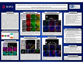
  • 1. !! Development of an In Vitro Model for the Specification of Retinal Neurons from Human Pluripotent Stem Cells Kirstin Langer1-2, Akshayalakshmi Sridhar2, and Jason S. Meyer2-4 1Department of Natural and Physical Sciences, Olivet College, 2Department of Biology, Indiana University Purdue University Indianapolis, 3Department of Medical and Molecular Genetics, Indiana University School of Medicine, 4Stark Neurosciences Research Institute, Indiana University School of Medicine, Indianapolis, IN 46202 Neural Differentiation !! I would like to thank the members of the Meyer lab who have mentored and assisted in the completion of this study. I would also like to give thanks to IUPUI and The National Eye Institute for their generous funding that made this study possible. Introduction Undifferentiated hPSCs Retinal Progenitor Cells Retinal Ganglion Cells Photoreceptors Modeling FASD with hPSCs Conclusions AcknowledgementsFig. 1: Characterization of undifferentiated human pluripotent stem cells (hPSCs) (A) The phenotypic characterization of hPSCs included numerous colonies of tightly packed cells enclosed by a clearly defined border. (B) RT- PCR revealed expression of pluripotent markers and the absence of ectoderm, mesoderm, and endoderm markers. (C-H) Immunocytochemistry illustrated expression of transcription factors (red) as well as cell surface markers (green) commonly associated with a state of pluripotency. Fig. 2: Characterization of retinal eye field populations. (A) After 10 Days of differentiation, hPSCs displayed a dense, 3D center and a flattened appearance toward the periphery. (B) RT-PCR indicated the loss of pluripotent markers from 0-10 days of differentiation, along with the onset of eye field markers. (C-N) Immunocytochemistry indicated widespread expression of neural and eye field transcription factors. Human pluripotent stem cells (hPSCs) have progressed into a vital tool for in vitro studies of human development as well as studying the progression of diseases affecting specific cell types. hPSCs have the ability to self renew and give rise to all cell types of the body1,2. The ability to derive retinal cells from hPSCs allows for in vitro analysis of these cells and how they may be adversely affected in the development of the retina3. Thus, the efforts of this study focused on examining the ability of hPSCs to give rise to retinal phenotypes, following a predicted, step-wise differentiation process that closely mimics what is known about retinogenesis. The differentiation process followed a step-wise progression, with cells differentiating through identifiable stages of retinal development yielding retinal neurons, including photoreceptors and retinal ganglion cells. Subsequently, the ability to utilize hPSCs to effectively model fetal alcohol spectrum disorders (FASD) was initiated, as previous studies have documented retinal defects in zebrafish following ethanol exposure4. Current efforts are focused upon examining the effects of ethanol exposure on retinal development from hPSCs, as retinal development is known to be adversely affected in FASD. The results of these studies have the potential to serve as a powerful and novel in vitro model for retinal development as well as disorders affecting the retina. ! References 1. Borooah S. et al., (2013) Using human induced pluripotent stem cells to treat retinal disease. Prog Retin Eye Res 37: 163-181. 2. Takahashi K. et al., (2006) Induction of pluripotent stem cells from adult human fibroblast by define factors. Cell 131: 861-872 3. Meyer J. et al., (2009) Modeling early retinal development with human embyronic and induced pluripotent stem cells PNAS 106: 16698-16703 4. Muralidharan P. et al., (2014) Zebrafish retinal defects induced by ethanol exposure are rescued by retinoic acid and folic acid supplement. Alcohol 49: 149-163. At Day 20, differentiating cells were separated into three groups, a control, 50mM EtOH, and 75mM EtOH •  From Day 20 to Day 30, retinal differentation medium was changed every other day as well as the ethanol treatment for each group. •  At Day 30 ethanol treatment was terminated and the cells continued to grow in untreated retinal differentiation medium until Day 60. Once the cells have differentiated for 60 days, they will be fixed for cyrostat sectioning and staining •  Staining of cryostat sections will include retinal ganglion cells and photoreceptor-specific markers to determine effects of EtOH exposure (if any) between the control and ethanol treated groups. Fig. 3: Characterization of retinal progenitor cells and non-retinal forebrain cells. (A) Retinal neurospheres, indicated by black arrows, were characterized by a bright ring around the outer edge of the cell. Non-retinal populations, indicated by the white arrows, displayed a larger, more uniform, and darker phenotype. (B-C) RT-PCR analysis indicated that retinal neurospheres express neural and retinal makers but did not express forebrain makers, and also demonstrated increased expression of retinal progenitor markers from 0 to 30 days. (E- J) Immunocytochemistry analysis displayed the expression of retinal progenitor markers Chx10 and Pax6 in retinal neurospheres (H) with the absence of forebrain maker Sox1 (F). Non-retinal neural populations (D) displayed common forebrain neuron features and also preserved the expression of forebrain and neural makers (E, G). Pluripotent markers, Oct4 and Nanog, were expressed in populations of cells attached to the retinal neurospheres (I-J). Fig. 5: Characterization of photoreceptors differentiated from hPSCs. (A) RT-PCR analysis illustrated the expression of photoreceptor associated transcription factors as well as the expression of phototransduction proteins. (B-G) Immunocytochemistry displayed photoreceptor-positive cells by the expression of Otx2, Crx, NeuroD1, and Recoverin in retinal neurospheres. Fig. 4: Characterization of retinal ganglion cells differentiated from hPSCs. (A) RT-PCR analysis revealed the expression of the retinal ganglion makers, BRN3, ISLET1, SNCG, and RBPMS in retinal neurospheres. (B-G) Immunocytochemistry verified the expression of BRN3-postive cells within huC/D-expressing RGCs, as well as ISLET1-positive ganglion cells with MAP2-positive extensions in retinal neurospheres. •  The differentiation of hPSCs to retinal neurons follows a step-wise progression of cells differentiating through identifiable stages of retinal development. •  Undifferentiated cells expressed pluripotent transcription factors and cell surface markers. •  Differentiation after 10 days yielded cells of the eye field, expressing appropriate markers. •  Retinal progenitor neurospheres analogous to the optic vesicle exhibited appropriate retinal progenitor- associated makers after 30 days. •  At day 70, hPSCs yielded retinal ganglion cells and photoreceptor cells, two of the major cells types of the retina. C E D F Oct4 Nanog SSEA4 TRA-1-60 Sox2 TRA-1-81 Oct4 Nanog Lin28 Pax6 AFPT G H BA Day0 Day10Day30 Oct4 Nanog Otx2 Lhx2 Six3 Six6 Pax6 Chx10 Rax Pax6FABP7 Rax DLX1 EMX1 A B C Chx10/Pax6Sox1/Pax6 BIII Tub/Otx2 Sox1/Pax6 Chx10 Neural Retinal Forebrain Nanog/Oct4 Chx10/Oct4 D E F G H I J Non-retinal Non-retinal Non-retinal Retinal Retinal Retinal Retinal Otx2 Lhx2 Sox1 Pax6 Six6 Rax Sox2Six3 Otx2 Lhx2 Six6 Six3 Pax6 Rax Oct4 Nanog Day 0 Day10 A C D E F G H I J B K L M N Merge Merge Merge Merge Brn3 huC/D Islet1 Map2 A B C D E Brn3 Islet1 SNCG RBPMS Merge Merge F G A Otx2 Crx NeuroD1 Crx ND4 Recoverin Trans. OpsinNRL Arr. B C D E F G Merge Merge Overview of Retinal Development The steps of retinal d e v e l o p m e n t f r o m hPSCs yielding retinal n e u r o n s c a n b e illustrated at various s t a g e s i n c l u d i n g undifferentiated cells (Day 0), eye field populations (Day 10), retinal progenitor cells (Day 30) and retinal neurons (Day 70).