1. 12 PRACTICAL GASTROENTEROLOGY • MARCH 2015
FRONTIERS IN ENDOSCOPY, SERIES #16
Douglas G. Adler MD, FACG, AGAF, FASGE, Series Editor
Mirizzi Syndrome: A Rare
but Relevant Biliary Entity
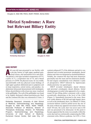
Kimberley Davenport Douglas G. Adler
Kimberley Davenport, University of Utah School
of Medicine, Gastroenterology and Hepatology.
Douglas G. Adler MD, FACG, AGAF, FASGE,
Associate Professor of Medicine, Director of
Therapeutic Endoscopy, Director, GI Fellowship
Program, Gastroenterology and Hepatology,
University of Utah School of Medicine,
Huntsman Cancer Center, Salt Lake City, UT
CASE REPORT
A
64 year-old man presented to our facility with
a one-day history of right upper quadrant pain,
scleral icterus, and intermittent fever and chills.
The patient’s vital signs included a temperature of 97.8,
a pulse of 71, a respiration rate of 14, and a blood
pressure of 124/80. On physical exam, he was mildly
uncomfortable with key physical exam findings of
right upper quadrant abdominal pain to palpation and
on deep inspiration, scleral icterus, and jaundice. An
abdominal ultrasound demonstrated mild intrahepatic
biliary ductal dilation with at least one stone visualized
in the gallbladder. The wall of the gallbladder was found
to be slightly thickened without pericholecystic fluid
and the common bile duct was measured at 11mm. A
contrast-enhanced CT of the abdomen and pelvis was
obtained and revealed prominent intrahepatic ductal
dilation and what was interpreted as choledocholithiasis.
Notably, the common bile duct had more distension
proximally near the stone and less distension distally.
GI and surgery consultations were obtained. A plan
of preoperative ERCP followed by laparoscopic
cholecystectomy was agreed upon.
ERCP revealed intrahepatic ductal dilation
and proximal extrahepatic ductal dilation. Initial
cholangiogram revealed what appeared to be a single,
round 30 mm filling defect consistent with a stone
eccentrically obstructing at the level of the CHD
(Figure 1). Immediately proximal to the stone, there
was visible dilation of the left and right hepatic ducts
as well as the intrahepatic ducts. An inflated 15-18mm
occlusion balloon could be pulled across the site of
the stone without any apparent movement of the stone
itself. Of note, the cystic duct was never seen to fill
and was presumed to be obstructed. At this point it
became apparent that the stone was actually within the
cystic duct and was extrinsically compressing the CBD,
consistent with a Mirizzi syndrome. An 8.5Fr x 12cm
2. FRONTIERS IN ENDOSCOPY, SERIES #16
Mirizzi Syndrome: A Rare but Relevant Biliary Entity
PRACTICAL GASTROENTEROLOGY • MARCH 2015 13
obstructed from mechanical compression and
surrounding inflammation by gallstone impaction
at either the cystic duct or the gallbladder neck.2,5
Compression by the impacted stone results in biliary
obstruction which can then cause obstructive jaundice
and in some patients also lead to cholangitis. 4,6
From
there, this process has the potential to further evolve into
an internal biliary fistula, or even complete obliteration
of the CHD.3
Overall, the incidence of Mirizzi Syndrome is rare,
discovered only in 0.7 – 1.4% of patients undergoing
cholecystectomy.7
Management has historically been
difficult due to the inadequacy of pre-operative diagnosis
as well as the surgical challenge it presents given the
Figure 2. ERCP image of same patient following
placement of a plastic biliary stent to
decompress the biliary tree.
Figure 1. ERCP image of a large stone extrinsically
compressing the CHD. (Arrow)
plastic biliary stent was inserted into the CBD across
the site of extrinsic compression, with the proximal
end of the stent in the CHD. (Figure 2) The patient
improved clinically over the next 24 hours following
biliary decompression. The patient was then referred
for his cholecystectomy.
At surgery, the gallbladder was noted to be inflamed
and adherent to the CBD, with what appeared to be a
cholecystocholedocho fistula between the two at the
level of the gallbladder neck/proximal cystic duct.
Opening of the gallbladder revealed the impacted stone
at the level of the fistula, which was then removed.
Cholecystectomy was then carried out with subsequent
incision of the CHD and removal of the impacted
stone. The CHD was felt to have been damaged too
severely by the stone/fistula combination for simple
T-tube placement. The patient then underwent biliary
reconstruction via a Roux-en-Y hepaticojejunostomy,
although the distal CBD was left in continuity. The
plastic bile duct stent was left in as a guide during
surgery. The patient did well postoperatively and
without complication.
A follow-up ERCP 8 weeks later demonstrated
brisk flow of contrast through the biliary tree as well as
a patent hepaticojejunostomy with free flowing contrast
and bile into the small bowel. No filling defects were
noted, indicating an absence of any residual or recurrent
stones. Overall, the patient was felt to have developed
a Type IV Mirizzi syndrome.
DISCUSSION
In developed countries, the incidence of gallstone
disease has been rising of late with the majority of
cases involving cholesterol stones. In the U.S. alone,
an estimated 20 – 25 million adults suffer from some
form of gallstone disease. Epidemiological studies
have found that gallstone disease correlates highly with
obesity, diabetes and metabolic syndrome.1
Though
gallstones are understood to be multifactorial from an
etiology point of view, one property these risk factors
all share seems to be hypercholesterolemia: a condition
that can allow supersaturation of bile by cholesterol
with subsequent precipitation of gallstones.1
Gallstones
can lead to a variety of conditions including cholangitis,
cholecystisis, gallstone pancreatitis, and even gallstone
ileus.2
Mirizzi Syndrome (MS) is a rare complication
of cholelithiasis.3,4
Classically, the common hepatic
duct (CHD) or the common bile duct (CBD) become
3. Mirizzi Syndrome: A Rare but Relevant Biliary Entity
FRONTIERS IN ENDOSCOPY, SERIES #16
14 PRACTICAL GASTROENTEROLOGY • MARCH 2015
liver enzymes, hyperbilirubinemia, leukocytosis, and
a high CA-19-9.12
Areported 28% of MS types II, III and IV cases also
have also been found to have concurrent gallbladder
cancer.11,12
Notably, longstanding gallstones is one of
the primary risk factors for both MS and gallbladder
cancer.12
Average age of onset for MS is around 61
years.9
Most MS patients have had gallstone disease
for a mean of 29 years.12
Diagnosis
Lack of pathognomonic patterns or reliable clinical
or laboratory indicators have made the diagnosis
of MS difficult to distinguish from common
choledocholithiasis.5,15
Optimal management of MS
and avoidance of iatrogenic bile duct injury during
cholecystectomy depends on accurate preoperative
diagnosis, although this cannot be made in all
circumstances.10
Several modalities are available for
biliary imaging including transabdominal ultrasound,
CT, MRCP, endoscopic ultrasound (EUS) and ERCP.12
Transabdominal ultrasound (US) is typically the initial
imaging test ordered for suspected biliary disease
because it is non-invasive, although it’s ability to detect
choledocholithiasis is limited and the test is highly
operator dependent.2
US has high sensitivity to findings
consistent with MS such as gallstones in the gallbladder
neck, acute cholecystitis, a shrunken gallbladder
and a dilated CHD.10
CT scans have the capacity to
demonstrate dilated intra and extrahepatic ducts with
gallstones in the biliary tree, but in general are a poor
test to detect MS or choledocholithiasis.5
EUS is ideal
for detecting choledocholithiasis but may fail to detect
MS if the stone is located too proximally for the EUS
transducer to visualize.16
MRCP (Magnetic Resonance
Cholangiopancreotography) is noninvasive and has a
high sensitivity for finding pericholicystic inflammation
and cholecystobiliary fistulas.11
However, ERCP
(Endoscopic Retrograde Cholangiopancreotography)
is the gold standard for diagnosis of MS.9
The
sensitivity of ERCP for diagnosis of MS approaches
90%.11,12
ERCP is both diagnostic and therapeutic.11
Diagnostically, ERCP identifies ductal abnormalities
and can distinguish with precision the cause, level and
degree of biliary obstruction.10
Therapeutically, ERCP
is used for stone clearance if the stone is endoscopically
accessible.8
It also facilitates biliary decompression
prior to cholecystectomy in patients with MS via
(continued on page 16)
presence of active inflammation, dense adhesions, and
distorted biliary ductal anatomy.5,8
The term Mirizzi Syndrome refers to a collection of
four subtypes of the disease commonly acknowledged
as the Csendes Classification model.9
Type I is the most
dominant form of MS and is characterized by extrinsic
compression of the CBD due to an impacted stone at the
cystic duct or the gallbladder neck.4,10
Types II, III and
IV all include cholecystocholedochal fistulas.11
Type II
describes a fistula where 1/3 of the CBD circumferential
wall has eroded.4,9
Type III refers to a larger fistula
with erosion of 2/3 of the circumferential wall of the
CBD.4,9
Finally, Type IV denotes a fistula that has
caused circumferential damage to the CBD and thus
requires reconstruction of the CBD.4,9
Pathophysiology
Mirizzi Syndrome begins when a gallstone becomes
impacted at the cystic duct or gallbladder neck and
causes an inflammatory response. The offending
stone then applies external pressure on the bile duct,
eventually leading to erosion of the CBD with possible
fistulization.12
Once this situation is established, episodes
of biliary colic cause fibrosis and gallbladder
atrophy.12,13
Ongoing inflammation and proximity
allow the gallbladder and the CBD to fuse together.12
An impacted gallstone at this location likely causes
a pressure ulcer which then develops necrosis. The
necrotic tissue can enable partial stone migration into
the CBD and ultimately the production of a chronic
fistula.12
Such cholecystocholedocal fistulas are the
hallmark of Type II, II and IV Mirizzi Syndrome, but
they are still exceedingly rare. In fact, most biliary
fistulas are actually cholecystoduodenal, joining the
gallbladder to the small bowel.11
Despite advanced
imaging, most fistulas are found intra-operatively
during cholecystectomy.4,11
Clinical Presentation
Mirizzi Syndrome is difficult to diagnose clinically
because of its non-specific symptom profile.5,6
Because
MS resembles several other conditions, its differential
diagnosis includes choledocholithiasis, cholecystitis,
cholangitis,gallstoneileus,andgallbladdercancer.5,11,12,14
Clinical presentation is non-specific, but obstructive
jaundice is a common symptom – especially for types
II, III and IV.4
RUQ pain is also frequently seen, along
with a fever. Laboratory values often reflect elevated
4. 16 PRACTICAL GASTROENTEROLOGY • MARCH 2015
FRONTIERS IN ENDOSCOPY, SERIES #16
Mirizzi Syndrome: A Rare but Relevant Biliary Entity
stent placement, as was the case in our patient.6,8
If
a preoperative diagnosis of MS is not obtained, most
experienced surgeons would recognize the entity in the
operating room.
Treatment
The goals of treating patients with MS include: removal
of the impacted stone and gallbladder, restoration of
bile drainage and repair of the anatomical defect.6,11
Preoperative ERCP can confirm the presence of MS.9
It also allows for placement of a temporary stent –
providing critical decompression of the obstructed
biliaryduct.12
Suchdecompressionalleviatesprogression
of any fistulas.14
Moreover, the stent will serve as a
landmark in subsequent surgery to help minimize the
risk of ductal injury.4
Preoperative ERCP is also a viable
alternative for patients who are not candidates for open
cholecystectomy (OC).12
Removal of the gallbladder and the impacted
stone along with repair of the malformed anatomy is
performed laparoscopically or by open surgery. OC has
historically been the surgical standard of care for MS.4
However, LC is fast gaining acceptance as a viable
alternative to OC.6,11
The main advantage conferred
by OC is a lower risk of iatrogenic bile duct injury
than LC.14
This is large part due to the feasibility of
a fundus first approach enabled by OC.17
Morbidity
from OC has been reported to be 3% for MS type I.
However, OC performed on types II and III correlate
with a higher morbidity rate (15 – 26%) due to more
complicated anatomical deformities.4
The biggest risk
associated with OC is iatrogenic injury to the CBD.5
Such risk stems from the technical challenge presented
by anatomical distortions, cholecystocholedocal
fistulas, and adhesions in Calot’s triangle encountered
in MS patients undergoing OC.4,5
Disadvantages of OC
include a larger incision which provides a higher risk
of infection and wound dehiscence.8
Additionally, OC
entails a longer recovery.6
Laparoscopic Cholecystectomy (LC) for treatment
of MS has been somewhat controversial.10
Indeed,
some surgeons still consider MS as a contraindication
to laparoscopic cholecystectomy.4,6,9
Nonetheless, LC
has become a viable alternative to OC as experience and
enhanced instruments continue to improve outcomes
with this procedure.6
LC involves removal of the
gallbladder, the impacted stone and suturing the biliary
defect.8
Its advantages are that it is minimally invasive
(continued from page 14) and by extension entails a lower risk of infection,
produces less blood loss, and offers a shorter hospital
stay.8
Some disadvantages are that it is technically
demanding, requires a high degree of expert skill and
thus a longer duration for the procedure.8
In general, LC is indicated for the less complicated
MS as in Type I. Open surgery is indicated for higher
risk cases of MS, difficult dissections (due to anatomical
malformations or acute inflammation), and patients who
have failed LC.8
In some cases, higher grades of MS:
Type II, III and IV, may require open biliary-enteric
bypass to repair extensive destruction of the bile ducts
or strictures.11
Such complete biliary reconstruction is
termed a Roux-en-Y hepatico-jejunostomy and involves
anastomosis of the jejunum with the fistula to close
the eroded duct.18
Lastly, postoperative ERCP after
complete healing of the site removes the temporary
stent.8
The incidence of iatrogenic complications is
reported to range between 0 – 14%.3
Such adverse
events can be severe and include cholangitis, bile
leakage from perforation of the CBD or perforation of
the gallbladder, bile duct strictures from fistula repair,
post-ERCP pancreatitis, and sepsis.3
Regardless of
the intervention, a high risk of morbidity exists with
treatment of MS.9
CONCLUSION
Mirizzi Syndrome is a rare clinical entity that presents
a formidable challenge when encountered. The
constellation of a contracted gallbladder, obstructive
jaundice, gallstones, pain and cholecystitis should
arouse suspicion of MS. A preoperative diagnosis is
critical to optimal management and is best confirmed by
cholangiography during ERCP, although not all patients
have the diagnosis made prior to interventions. Once a
definitive diagnosis of MS is made, a multidisciplinary
team approach including a skilled endoscopist and
surgeon offers the best outcome. Treatment for each
patient must be individualized, as specific MS types
necessitate distinct surgical approaches. Preoperative
ERCP with stenting can restore bile flow and mitigate
cholangitis, thus enhancing safety of subsequent
surgery.
References
1. Shaffer EA. Gallstone disease: Epidemiology of gall-
bladder stone disease. Best Pract Res Clin Gastroenterol.
2006;20(6):981-96. Review. PubMed PMID: 17127183.