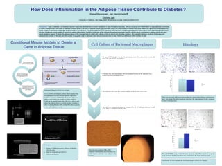
This document summarizes research into how inflammation in adipose tissue contributes to diabetes. Mouse models were used where certain inflammatory signaling genes were conditionally knocked out in adipose tissue. Histological analysis found that obese knockout mice had similar numbers of fat cells as obese wildtype mice, but a higher level of apoptotic cells in fat tissue. The results suggest the knocked out gene affects cell viability and survival in fat tissue. Conditional gene knockout was achieved using the Cre-Lox system to selectively delete genes in adipose tissue.
How Does Inflammation in the Adipose Tissue Contribute to Diabetes
1. How Does Inflammation in the Adipose Tissue Contribute to Diabetes?
Kiana Khosravian, Jan Heinrichsdorff
Olefsky Lab
University of California, San Diego, 9500 Gilman Drive, La Jolla, California 92093-0378
Introduction: Type 2 Diabetes is a metabolic disorder due to the development of insulin resistance in cells throughout the body. We are studying how inflammation in adipose tissue contributes
to insulin resistance and the onset of diabetes. Excess lipids in the cells of adipose tissue (lipid overload) cause the cells to leak the excess fatty acids leading to higher DAG (diacylglycerols)
levels, a type of secondary messenger to accumulate in muscle cells. The accumulation of DAG interferes with the insulin receptor signaling and inhibits insulin from regulating glucose levels1.
We use conditional mouse models to knock out certain inflammatory signaling molecules in the adipose tissue and investigate how this affects insulin resistance in adipose tissue and other
insulin sensitive organs. To study the adipose tissue of the mice we have to collect cell cultures which we turned into histology sections. We looked at histology sections of fat tissue and
assessed the number of fat cells and the number of apoptotic cells in wild type mice versus knockout mice to see if there is a difference in the number of cells.
Conditional Mouse Models to Delete a
Gene in Adipose Tissue
The Cre-Lox System was
used to knock out a certain
gene in the mouse’s genome.
DNA from mouse tails was
isolated and used for PCR
(Polymerase Chain Reaction)
genotyping.
Gel Electrophoresis was used
to separate the DNA and to
determines which mice were
wildtype and which have the
gene deleted.
Mouse # 767 is Cre+
which means that Cre
is expressed and will
delete the targeted
gene
+
-
770
768
767
763
786
Ladder
L 756 763 767 768 770 778 785 798 813 814 815 816 861 862
864 865 866 867 868 869 870 - +
778 785 798 813 814 815 816 861 862 864 784 865 866 867 W+
868 865 870 756 763 767 768 770 W+
Cell Culture of Peritoneal Macrophages
1
• We injected 3% thioglycolate into the peritoneal cavity of the mice which irritates the
mice cells and attracts macrophages.
2
• Two days later, the macrophages that accumulated because of the injection were
flushed out of the peritoneal cavity.
3
• The collected cells were then washed and the red blood cells were lysed.
4
• The cells were counted and plated at a density of 2.5 X 10⁵ cells per well in a 24 well
plate. Medium DEMEM 10% FCS P/S
Histology
KO #51 WT #56
0
50
100
150
200
250
300
350
400
450
500
1
numberoffatcellsperfield
WT
TAK1 F-KO
There was not much difference between the cell counts of the wildtype and knockout
mice. This means The obese knockout mice have the same amount of cells compared
to obese wild type mice.
KO #53 WT #74
0
100
200
300
400
500
600
700
800
900
1
ApoptoticCellsPerField
WT
TAK1 F-KO
We used TUNNEL assay to label the dead, apoptotic cells. There was more apoptosis
in the fat tissue of obese knockout mice compared to the obese wild type mice.
References:
1. Taubes, G (2009) Prosperity’s Plague. SCIENCE
325, 256-260.
2. http://en.wikipedia.org/wiki/Cre-
Lox_recombination
Mice with a band of DNA amplified in our
PCR similar to 867 show that their gene has
been floxed which means that a particular piece
of DNA has been inserted with a section of
loxP on either side of it. The larger the DNA
the slower it migrates on the gel so this
indicated that there is a larger piece of DNA
which contains the loxP site.
Here are some pictures of the cells I
cultured. Unfortunately after three days the
wells without antibiotics were infected with
bacteria.
Conclusion: We can conclude that the knockout gene affects cell viability.
Schematic Diagram of Cre-Lox System2.
Cre is a DNA recombinase protein which catalyzes the
recombination of DNA between specific target sites.
LoxP is a sequence of DNA that flanks two sides of a
DNA section which you would recombine with Cre.
LoxP are the specific target sites. The Cre is able to only
affect the adipose tissue in a mouse because we place it
after a certain promoter (AP2) in the DNA that is specific
to the cells in adipose tissue.
N=6
N=6
Apoptotic Cell
*