1. BIOCHEMISTRY AND MOLECULAR BIOLOGY PROGRAM
Characterization of Cell Lines Generated by Conditional
Reprogramming of Cells (CRC) Technology
Kholood Ahmad1, Bassem R. Haddad2
1
Department of Biochemistry and Molecular Biology, Georgetown University School of Medicine, 2
Lombardi
Comprehensive Cancer Center, Georgetown University Medical Center, 3900 Reservoir Rd, Washington, DC 20057-145
Introduction:
Introduction
Methods
Conditional reprogramming of cells (CRC), a technique developed at Georgetown University, has shown to be successful in diagnostic and therapeutic studies
as well as in regenerative medicine studies. It was developed to keep human primary somatic cells viable without transduction by oncogenes or other
exogenous materials that change the nature of the original cell1
. Regenerated cells were found to be identical to the primary cells and had no change in the
karyotype. However, the efficiency of this technique for the growth of malignant cells as a representative model of the primary cells in cancer research is still
under investigation. In this study, we used FISH (fluorescent in situ hybridization) to determine if the breast cancer cells cultured with the CRC technique are
representative of the primary malignant tumor cells. We tested four individual breast cancer cell lines derived from four patients with HER2 positive invasive
breast cancer. Using the FISH technique, we found that the resultant cells cultured by the CRC technique do not demonstrate amplification of the HER2 gene
and have a normal number of chromosomes. There is a possibility that the four primary tumor samples did not have abundance of the malignant cells, so this
technique may favor the growth of the normal cells more than the malignant cells. On the other hand, there is a possibility that this culture technique may switch
off amplification of the HER2 gene or affect the growth of malignant cells in vitro.
Finding a method to maintain the viability and
propagation of human somatic cells for biomedical
research is a big challenge. The CRC method,
developed at Georgetown University, uses irradiated
fibroblast feeder cells and a Rho kinase inhibitor (Y-
27632) to grow human epithelial cells that are taken
from needle or excision biopsies to generate large
numbers of cells without transduction with a virus or
other oncogenic cellular particles which may affect
the pathological nature of the original cells1
.
Many studies have shown that the CRC technique is
an effective way to increase the cloning efficiency of
keratinocytes2
. These cells were used as a model to
study many diseases that have epithelial cell origin,
and were also used in organotypic culture for
regenerative medicine studies, like in epidermal auto
graft2
. Additionally, the CRC technique has been
used to study respiratory papillomatosis in a patient,
by allowing the investigators to detect a mutation in
the virus that caused the disease (HPV-11 virus) and
also to find an effective treatment for the advanced
stage of the disease3
. This technique has a
promising future in cancer research as well as in
personalized medicine1
.
In many studies, the cells cultured with the CRC
technique were representative of the original normal
cells with a normal karyotype, growth, and
differentiation potential4
. Even in the respiratory
papillomatosis study, the tumor cells are benign, so
the cells have a normal cell character. The validity of
this technique depends on the identity of the growing
cells in culture relative to the primary tumor cells. In
this study, we wanted to determine if the breast
cancer cells grown with the CRC technique are
representative of the original malignant tumor cells
that are strongly positive for HER2 gene, as
described in the pathology reports.
Abstract
Results
CRC Culture:
Culture and irradiation of feeder cells
The conditional F medium preparation
Primary cell culture in conditional F
medium containing Y-27632 compound
Passaging the primary cells in direct
culture with the feeder fibroblast cells.
(Figure obtained from Liu et al.1
)
Harvesting Cell lines: After harvesting the cells, we prepared 3
slides from each breast cancer cell line. We selected the best
slide (that has more metaphases and interphases) for each
cell line for FISH.
FISH Technique:
Nick Translation: Probes for the HER-2 gene and centromere
region of chromosome 17 were prepared by this procedure.
FISH: Pretreated slides and prepared probes containing control
chromosome 17 centromeric probe (green) and a HER2 probe
(red), were denatured and incubated overnight. Stringency
washes were performed with 50% formamide, and 1X SSC the
next day. Following the stringency washes, the slides were
incubated with a blocking solution, followed by detection
solutions. FISH signal was detected using a fluorescence
microscope.
In order for the CRC technique to be a useful technology for cancer research, the cell lines generated
should be representative of the primary cancer cells originally obtained from biopsy material. In this
study, we found that the growing cells did not show HER2 amplification similar to the primary breast
cancer cells, as indicated in the original pathology reports.
The possibility that there are no cancer cells in the four primary samples should be excluded first. The
CRC Lab has to be sure that the primary samples that are given to them have malignant tumor cells
before culture.
The CRC technique can be a promising tool in cancer diagnostic and therapeutic research, and in
personalized medicine, perhaps used in drug screening, However, more studies should be done to
support the validity of this technique to grow and maintain the malignant cells in vitro.
Conclusions
Acknowledgments
I would like to express my thanks and appreciation to Dr. Bassem R. Haddad, MD and Janice D. Rone, PHD for
the opportunity to work in their laboratory and helpful advice throughout my internship. I would also sincerely like
to thank the members of the CRC Lab for providing us with the CRC cell lines. My thanks also to Dr. Cynthia
Rosenthal, PHD, Program Director, for her advice and help.
References:
1
Liu, Xuefeng, et al. "ROCK inhibitor and feeder cells induce the conditional reprogramming of epithelial cells." The American journal of pathology 180.2
(2012): 599-607.
2
Chapman, Sandra, et al. "Human keratinocytes are efficiently immortalized by a Rho kinase inhibitor." The Journal of clinical investigation 120.7 (2010):
2619.
3
Yuan, Hang, et al. "Use of reprogrammed cells to identify therapy for respiratory papillomatosis." New England Journal of Medicine 367.13 (2012):
1220-1227.
4
Suprynowicz, Frank A., et al. "Conditionally reprogrammed cells represent a stem-like state of adult epithelial cells." Proceedings of the National
Academy of Sciences 109.49 (2012): 20035-20040.
Spring 2015
Georgetown University
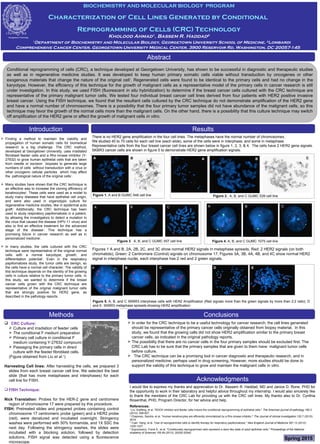
There is no HER2 gene amplification in the four cell lines. The metaphases have the normal number of chromosomes.
We studied 40 to 70 cells for each cell line (each slide), some of the cells were in interphase, and some in metaphase.
Representative cells from the four breast cancer cell lines are shown below in figure 1, 2, 3, & 4. The cells have 2 HER2 gene signals.
SKBR3 cancer cells are shown in figure 5 to demonstrate HER2 gene amplification signals.
Figure 4. A, B, and C GUMC 1075 cell line
Figure 5. A, B, and C SKBR3 interphase cells with HER2 Amplification (Red signals more than the green signals by more than 2:2 ratio). D
and E: SKBR3 metaphase spreads showing HER2 amplification
A
Figure 1. A and B GUMC 548 cell line Figure 2. A, B, and C GUMC 539 cell line
Figure 3. A, B, and C GUMC 557 cell line
Figures 1 A and B, 2A, 2B, 2C, and 3C show normal HER2 signals in metaphase spreads. Red: 2 HER2 signals (on both
chromatids); Green: 2 Centromere (Control) signals on chromosome 17. Figures 3A, 3B, 4A, 4B, and 4C show normal HER2
signal in interphase nuclei, each interphase has 2 red and 2 green signals.
A B
A AB C B C
A B CA B C D E
A B A B C