
Empfohlen
Weitere ähnliche Inhalte
Was ist angesagt?
Was ist angesagt? (20)
Ähnlich wie Dop phys (1)
Ähnlich wie Dop phys (1) (20)
Mehr von KamalEldirawi
Mehr von KamalEldirawi (20)
Kürzlich hochgeladen
Kürzlich hochgeladen (20)
Dop phys (1)
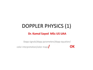
- 1. DOPPLER PHYSICS (1) Dr. Kamal Sayed MSc US UAA Dopp signals/dopp parameters/dopp equation/ color interpretation/color maps/ OK
- 2. What is the principle of ultrasound? An electric current passes through a cable to the transducer and is applied to the crystals, causing them to deform and vibrate. This vibration produces the ultrasound beam. The frequency of the ultrasound waves produced is predetermined by the crystals in the transducer. • • Correct interpretation of flow images requires knowledge of physical and technical factors that influence the Doppler signal. • Artefacts caused by physical limitations of the modality or inappropriate equipment settings may result in displayed flow conditions that may differ considerably from the actual physiological situation. • As a consequence, artefacts in Doppler imaging may be confusing and lead to misinterpretation of flow information. •
- 3. • DOPPLER SIGNAL • The Doppler effect is a change in wavelength (frequency) of sound resulting from motion of a source, receiver or reflector. As the US TXR is a stationary source and receiver, the Doppler effect arises from reflectors in motion—for all practical purposes these are the erythrocytes. • When a pulse is reflected from erythrocytes, the frequency of the wave received differs from that, which is transmitted. • This difference is known as the Doppler shift, named after the Austrian physicist and mathematician Chr. Andreas Doppler, who first described the phenomenon for light in 1843.5
- 4. • motion—for all practical purposes these are the erythrocytes. When a pulse is reflected from erythrocytes, the frequency of the wave received differs from that, which is transmitted. This difference is known as the Doppler shift, named after the Austrian physicist and mathematician Chr. Andreas Doppler, who first described the phenomenon for light in 1843.5
- 5. • There are two successive Doppler shifts involved: • First, the sound from the stationary transmitting TXR is received by the moving erythrocytes. • Second, the erythrocytes act as moving sources as they • re-eradiate the US back toward the transducer, which is now the stationary receiver. • These two Doppler shifts account for factor 2 in the • Doppler equation: • fd = ft – fr = 2 ft v cos^(ceta) / C
- 6. Doppler Equation : • fd = ft – fr = 2 ft v cos^(ceta) / C • where: fD is the Doppler shift, ft is the transmitted frequency, fr is the received frequency, v is the blood velocity, θ (ceta) is the insonation angle (the angle between the US beam and the blood flow), and c is the speed of sound. • The Doppler shift is thus directly proportional to the velocity of the flow, v, cosine to the insonation angle, θ, and the transmitted frequency of the US, ft.6 •
- 7. • • Pulsed doppler • The Doppler circuitry determines the change in frequency indirectly. With a series of pulses the phase of the returning signals are compared with the phase of the emitted signal. • A change in phase translates to a change in frequency; eg, when the returning signal is compared with the emitted one, wave tops will not meet wave tops because the distance between wave tops has changed. The number of these pulses per second is called the pulse repetition frequency (PRF).
- 8. • Insonation angle, (Doppler angle) • This is the angle between the path of the Doppler pulses and the direction of flow in the vessel. • When this angle is 90°, there will be no frequency shift as can be seen from the equation above: cos(90°) = 0. • The maximum frequency shift of a given vessel is obtained when the direction of flow matches the direction of the Doppler pulses (Doppler angle = 0, flow directly towards or away from the transducer).
- 9. • Blood velocity versus Doppler shift • The Doppler circuitry determines the change in frequency and, this may only be translated into a blood velocity if the insonation angle is recorded and is included in the calculation. Nevertheless, all newer equipment report blood velocities (both in spectral Doppler and on the colour bar) assuming that the Doppler angle is zero. •
- 10. • This is, however, more often wrong than right and we are in fact dealing with frequency information unless angle correction has been performed (the process of informing the machine of the insonation angle). • Angle correction is only possible in spectral Doppler and is not an issue in a rheumatological setting.
- 11. • COLOUR DOPPLER, VELOCITY DOPPLER AND COLOUR FLOW MAPPING • In CFD, real-time presentation of flow information in colour is superimposed on the grey-scale morphological image. • In colour Doppler, analysis is performed in the colour box, which is divided into cells. • Each cell behaves like an independent Doppler gate with its own Doppler analysis.
- 12. • The mean frequency shift for each cell is computed and displayed as a colour. • The colours that arise from the detected Doppler shifts primarily indicate qualitative direction of flow. • Generally, red is used to indicate a flow towards the transducer and blue away from the transducer. • Different hues of red (or blue) indicate different velocities (in reality different frequency shifts). • Lighter hues are used to indicate higher frequency shifts.
- 13. • Interpreting the colours • The Doppler circuitry merely detects movements up and down in the image plane. • A dark red spot may therefore be blood moving slowly, directly towards the transducer or blood moving quickly at an angle close to 90°. • As we generally do not know the insonation angle to the vessels we cannot compare velocities between vessels.
- 14. • POWER DOPPLER (ENERGY DOPPLER) • PD displays power of the Doppler shift in each cell instead of the mean frequency shift. This gives PD a theoretical advantage over colour Doppler with regard to sensitivity. Disregarding direction of flow (negative or positive frequency shift) and disregarding velocity (high or low frequency shift) the power (energy) of the many different frequency shifts inside a cell are added to form the power signal. • The power mode does not measure velocity or direction and is very sensitive to flow. Therefore, it is almost angle independent and without aliasing.7
- 15. • The wall filter in ultrasound is a way of filtering out low or high frequency Doppler signals. ... • In clinical ultrasound, it is usually used to filter out very low frequencies that may add noise to a spectral Doppler waveform. • A typical use is removing the low frequency reverberation of an arterial wall. •
- 16. • DOPPLER PARAMETERS • The most important adjustable parameters are Doppler frequency, Doppler gain, PRF, colour priority, filter, focus, persistence, colour box position and size. • Patient positioning and scanning technique further influence the quality of the Doppler examination.
- 17. • Doppler frequency • A lower Doppler frequency will allow more penetration but also a more grainy Doppler image (larger colour pixels). Thus, higher Doppler frequency gives a more detailed image of the vessels but at the expense of penetration. • The trade-off between penetration and sensitivity is somewhat unpredictable and resolution is in this context really not an issue.
- 18. • The ability to depict slow flow in a small vessel (with a weak Doppler reflection) is enhanced by a lower frequency (because the weak reflection has more penetration) but is also enhanced by a higher frequency because the Doppler shift is higher (if the reflection is powerful enough to penetrate). • The optimum frequency must be found in practice and not in theory.
- 19. • Colour box • The numerous Doppler analyses inside the colour box are quite demanding on computing power of the US unit. • The frame rate goes down when colour is added and in order to obtain as live an image as possible (high frame rate) it is therefore generally recommended to make the box as small as possible. • We do, however, recommend always letting the box go to the top of the image in order to be aware of reverberation artefacts (see reverberations).
- 20. • Scale and pulse repetition frequency • PRF is the Doppler sampling frequency of the transducer and is reported in Hz. • The maximum Doppler shift frequency that can be sampled without aliasing is PRF/2, which is called the Nyquist limit.8 • The Nyquist limit may be presented on-screen as a blood velocity (the maximum measurable velocity of blood moving directly towards or away from the transducer) or in Hz (maximum measurable Doppler shift).
- 21. • If the blood velocity is above the Nyquist limit, the machine will misinterpret the velocity and aliasing will occur. This is not an issue with PD. • The sensitivity of both colour Doppler and PD is affected by PRF adjustments. When a high PRF is chosen it is assumed that the investigator is interested in high velocities, and therefore filters that remove low flow to remove noise are applied (so-called linked controls). • Selecting a high PRF therefore makes the system insensitive to lower velocities because of the linked controls.
- 22. • Colour priority (threshold) • When colour information is obtained, grey-scale information will often also be present and the machine has to decide whether to show one or the other. • Colour priority is a function that makes this decision for the machine. This function allows valid grey-scale information to override false Doppler information, eg, it helps suppress motion artefacts in the relatively hyperechoic tissue surrounding a pulsating artery (above a certain grey level, grey overrides colour).
- 23. • This function also allows supposedly valid Doppler information to override false grey-scale information, eg, inside vessels colour overrides the relatively weak grey-scale reverberation artefacts (below a certain grey level colour overrides grey). This function explains why some Doppler artefacts apparently prefer to appear in dark regions of the image. It also explains why grey-scale gain may influence the amount of colour in the image (increasing grey-scale gain may result in more grey information being above the threshold where colour is suppressed).
- 24. • Filters • Every Doppler instrument has high-pass filters, which eliminate the lowest Doppler shifts from the display. • The Doppler shifts originate from motion of the vessel wall and solid tissue. • These unwanted shifts are referred to as clutter or motion artefacts. •
- 25. • The filters—also called wall filters—may, however, eliminate signals from low velocity flow as the filters separate by frequency alone.9 10 • The PRF and wall filter are linked controls—the lowest possible wall filter is lower for a low PRF than a high. • The filters should be kept at their lowest setting for use in rheumatology.
- 26. • Gain • The Doppler gain is independent of grey-scale gain. • The gain setting determines the sensitivity of the system to flow. • By lowering the gain, noise and motion artefacts may be prevented but weak flow signals will go undetected. • 11 A gain setting that is too high results in random noise.12 • It is appropriate to set the Doppler gain by turning it up until random noise is encountered and then lowering it until the noise disappears.11
- 27. • Persistence • Persistence is a function that averages colour information over a number of frames. • Most brochure images are made with maximum persistence because this results in all colour information over time being displayed in one image. • All vessels are then filled with colour. The dynamic nature of flow is, however, lost. • With low or no persistence the high or low resistance nature of arterial flow may be seen as blinking colour foci or colour foci with pulsating intensity, respectively. • In a rheumatological setting there is no advantage with high persistence
- 28. • Patient positioning • The patient must be positioned comfortably, and the area under investigation must be completely relaxed; otherwise, tension in the muscles and tendons will produce slight tremor resulting in movement artefacts. When scanning the hand and elbow regions, the arm should not rest on the abdomen or thorax because of respiratory movement. Some patients should also keep quiet during the Doppler examination because the voice itself may produce movement artefacts and because some patients cannot talk without involuntary movement of the hands.
- 29. • Scanning technique • Most important in Doppler examinations is that very little pressure should be applied by the transducer. • The pressure will affect the haemodynamics with resulting decreased flow. • The use of generous amounts of scanning gel with visible gel between the transducer and skin will ensure light pressure.
- 30. • Focusing • The Doppler uses the same focus point as the grey-scale image. In the focal zone, the pulse is most narrow and the pulse therefore has higher spatial peak energy. • As a consequence, the echoes generated in the focal zone have higher amplitudes. • It is therefore not surprising that the Doppler is very dependent upon focus positioning .
- 31. • Some machines help us to some extent. When Doppler is activated, these machines move the focus point into the colour box (if it was outside) and then to a predetermined position inside the box. • If multiple focus points were in use, the machine changes that to single focus point and then inside the colour box. Still, within the colour box focus positioning affects flow detection • . Substantial differences may be falsely generated or falsely overlooked in longitudinal studies if the focal point is not consistently in the area under investigation.
- 32. • Pressure • False findings of absence of flow may occur if the examiner presses too hard on the tissue with the transducer, thereby blocking the flow. • When scanning a concave or convex surface it may be tempting to press the surface flat with the transducer. Instead, a generous amount of scanning gel should be used, which obviates the need for pressure to obtain good acoustic contact.
- 33. • Color Maps • Doppler shifts yield information regarding velocity • Color Doppler uses a “dictionary” or look-up table to convert • measured velocities into colors. • You can choose the dictionary: • » velocity mode or variance mode • Velocity Mode : varies up & down • Variance mode : varies up & down • and side-to-side • Image slide ( 34)
- 34. Color maps