Research Abstract Submitted to Biomedical Engineering Society
1. Measuring andModeling PlasmonicHeating byGold Nanoelectrodes for Stimulation of Neurons
Daniel Corral1
, Parveen Bazard1,3
, Kalen Hall1
, Robert D. Frisina1,2,3
, Joseph P. Walton2,1,3
, Venkat R. Bhethanabotla1,3
1
Dept.of Chemical & Biomedical Engineering, 2
Dept.ofCommunication Sciences & Disorders and 3
Global Center for Hearing &
Speech Research
University of South Florida, Tampa, FL 33620, USA
Introduction: In recent years, there has been considerable interest in use of metal nanoparticles (NPs) for
biomedical applications. This is because at the nanoscale, these metals possess useful optical properties that can
alter biological function. More specifically, with light interactions, metal NPs,like gold, generate heat surrounding
the particle, a phenomena known as plasmonic heating. We are investigating the possibility of using plasmonic
heating for stimulation of neurons and cardiomyocytes which has the potential to give better spatial resolution than
the existing electrical stimulation approaches. Hence there is a potential significant impact on the field of neural
prosthesis and cardiac stimulation. For this, we have fabricated nanoelectrodes (glass micropipettes coated with
gold NPs),and developed an alternative method to measure temperature change at the nanoelectrode surface. We
are also modeling arrays of NPs as occurs with nanoelectrodes. The aim of the present study is to understand and
quantify mechanisms of plasmonic stimulation.
Materials and Methods: The experimental design involves several steps. The first is the synthesis of gold NPs
using a standard citrate method; reduction of chloroauric acid solution using a sodium citrate solution. Next, these
gold NPs were coated onto glass micropipettes to fabricate the gold nanoelectrodes, and the nanoelectrodes were
characterized using scanning electron microscopy. We then used a pipette resistance method to indirectly measure
local temperature changes using a patch clamp system. Changing resistances in the presence of light allowed us to
calculate temperatures using our resistance vs. temperature calibration curve. We are also modeling temperature
distributions of NPs using a finite elements software package: COMSOL Multiphysics 4.4.
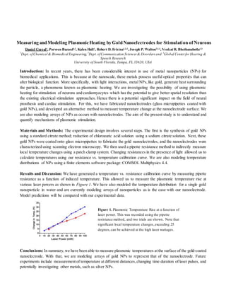
Results and Discussion: We have generated a temperature vs. resistance calibration curve by measuring pipette
resistance as a function of induced temperature. This allowed us to measure the plasmonic temperature rise at
various laser powers as shown in Figure 1. We have also modeled the temperature distribution for a single gold
nanoparticle in water and are currently modeling arrays of nanoparticles as is the case with our nanoelectrode.
Model predictions will be compared with our experimental data.
Conclusions: In summary,we have been able to measure plasmonic temperatures at the surface of the gold-coated
nanoelectrode. With that, we are modeling arrays of gold NPs to represent that of the nanoelectrode. Future
experiments include measurement of temperature at different distances,changing time duration of laser pulses, and
potentially investigating other metals, such as silver NPs.
Figure 1. Plasmonic Temperature Rise at a function of
laser power. This was recorded using the pipette
resistance method, and two trials are shown. Note that
significant local temperature changes,exceeding 25
degrees,can be achieved at the high laser wattages.