
Empfohlen
Weitere ähnliche Inhalte
Was ist angesagt?
Was ist angesagt? (20)
Ähnlich wie anaesthesia for Lung resection surgeries
Ähnlich wie anaesthesia for Lung resection surgeries (20)
Kürzlich hochgeladen
Kürzlich hochgeladen (20)
anaesthesia for Lung resection surgeries
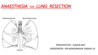
- 1. ANAESTHESIA FOR LUNG RESECTION PRESENTED BY---JUNAID ARIF MODERATOR---DR MANORANJAN BANSAL Sir
- 2. THORACIC SURGERIES MAY INVOLVE • INFECTIONS---Lung abscess,empyema,bronchiectasis • MALAGNANCIES----Pulmonary, esophageal and mediastinal • Lung Transplantation • Lung Volume Reduction Surgeries-lobectomy,thoracotomy,VATS, pneumonectomy,sleeve resection segmental/wedge resection
- 3. Fundamental ToAnaesthetic Management For The Majority Of Thoracic Procedures Are TwoTechniques 1. Lung isolation to facilitate surgical access within the thorax 2. Management of one lung anaesthesia
- 4. Pre-anesthetic evaluation • A detailed medical history for any coexisting disease.optimal treatment and control of associated medical condition should be achieved • The patient’s functional capacity should be assessed. • Since patients with lung cancer are usually smokers,a history of smoking and of symptoms suggestive of COPD should be elicited. • The patient should be evaluated for ischaemic heart disease since they tend to be chronic smoker,which predispose them to atherosclerosis. • Airway evaluation should be preformed keeping in mind that these patients are candidates for lung isolation intraoperatively. • Patients may receive chemotherapy preoperatively and should be evaluated for chemotherapy related toxicity. • Investigations should include CBC-this may show polycythemia due to COPD or leucocytosis which indicate active pulmonary infection SPUTUM culture and sensitivity –to guide appropriate antibiotic therapy LIVER and RENAL function test –should be assessed in view of age CHEST X RAY-should be evaluated for tracheal deviation or obstruction to predict difficulty with intubation or ventilation ECG-should be evaluated for signs of left or rt heart dysfunction. ECHO- to rule out pulmonary hypertension. • Pulmonary function tests should be carried out to diagnose obstructive or restrictive abnormalities.
- 5. ASSESSMENT OF RESPIRATORY FUNCTION 1. The best assessment of resp functions comes from a detailed history of the pts quality of life 2. All pts undergoing a pulmonary resection should have a baseline simple spirometry done. 3. Resp function can be divided into three • respiratory mechanics • gas exchange • cardiopulmonary interaction
- 6. The number of subsegments of each lobe is used to calculate the predicted postoperative (ppo) pulmonary function (e.g., after a right lower lobectomy, a patient with a preoperative FEV1 [or DLco] 70% of normal would be expected to have a ppoFEV1 of 70% × ( 1 − 29/100) = 50%. Ppo FEV1 >40% -Lowrisk 30-40%- mod risk < 30% - highrisk ppoFEV1% = preop FEV1% ×(1- %Functional lung tissue removed/100) RESPIRATORY MECHANICS • Most valid single test for post thoracotomy respiratory complications is the predictive postoperative FEV1 {ppo FEV1%} which is calculated as
- 7. LUNG PARENCHYMAL FUNCTION • Traditionally paO2 < 60 and paCO2 > 45 have been used as a cutoff values for pulmonary resection------useful as warning indicators for increased risk • The most useful test of the gas exchange of the lung is the diffusion capacity of carbon monoxide DLC0 • DLC0 correlates with the total functioning surface area of alveolar capillary interface • Can be calculated by same formula as for FEV1 • A ppo DLC0 < 40% correlates with both increased respiratory and cardiac complications and is usually independent of FEV1 The National Emphysema Treatment Trial has shown that patients with a preoperative FEV1 or DLco less than 20% had an unacceptably frequent perioperative mortality rate.These can be considered as the absolute minimal values compatible with a successful outcome
- 8. CARDIOPULMONARY INTERACTION • Formal laboratory exercise is currently the Gold standard for assessment of cardiopulmonary function and the minimal oxygen consumption ( VO2max ) is the most useful predictor of the post thoracotomy outcome • The risk of morbidity and mortality is HIGH preoperative VO2max< 15ml/kg/min VERY HIGH preoperativeVO2max <10ml/kg/min • Few patients with a VO2max >20 mL/kg/min have respiratory complications
- 9. SIX MINUTE WALK TEST The distance that a copd pt can can walk during a 6 min test shows an excellent correlation with VO2max . VO2max can be estimated from the 6-minute walk test distance in meters divided by 30 (i.e., 6-minute walk text of 450m: estimated VO2max = 450/30 = 15 mL/kg/min) SHUTTLE WALKING TEST Pt walks at a fixed and gradually increased rate between 2 marks 10 meters apart.A distance of <250meters correlates with a VO2max <10mL/kg/min. EXERCISE-OXIMERTRY Patients with a decrease of oxygen saturation (SpO2) greater than 4% during exercise are also at increased risk. STAIR CLIMBING The ability to climb fiVe flights of stairs correlates with a VO2max > 20 mL/kg/min, and climbing two flghts corresponds to a VO2max of 12 mL/kg/min
- 10. • Most valid test :ppoFEV1 • Threshold for increased risk:<30%-40% Respiratory mechanics • Most valid test:ppoDLCO • Threshold for increased risk:<30%-40% Caediopulmonary reserve • Most valid test: VO2max • Threshold for increased risk :<15ml/kg/min Lung parenchymal function The “THREE –LEGGED” stool of prethoracotomy Respiratory Assessment
- 11. Functional capacity >2 METS SPIROMETRY: ppoFEV1 and ppoDLCO BOTH > 60% EITHER 60%-30 EITHER <30% Simple exercise testing 6MWT>400M 6MWT<400M Proceed with scheduled Pulmonary Resection Cardiopulmonary Exercise testing VO2max >10ml/kg/min VO2max <10ml/kg/min Increased risk for scheduled pulmonary resection Functional capacity < 2MET Defer for medical consultation and optimisation High Risk Consider alternative therapies Algorithm for preop respiratory evaluation for pulmonary resection
- 12. VENTILLATION PERFUSION SCINTIGRAPHY • If the lung region to be resected is non functioning or minimally functioning, the prediction of postoperative function can be modified accordingly. This is particularly useful in pneumonectomy patients and V/˙ Q˙ scanning should be considered for any pneumonectomy patient who has a preoperative FEV1 and/or DLco less than 80%. However, V/˙ Q˙ scanning is of limited usefulness to predict postlobectomy function
- 13. CONCOMITANT MEDICAL CONDITIONS CARDIAC DISEASES Cardiac complications are the second most cause of perioperative mortality/morbidity in the thoracic surgical population ISCHAEMIA 1. Elective pulmonary resection surgery is an intermediate risk procedure in terms of perioperative cardiac ischemia ( incidence 5% and peaks on days 2 to 3 postoperatively) 2. Non invasive testing is indicated in pts with major ( unstable angina, remote infarction , previous ccf , diabetes) clinical predictors of myocardial risk. 3. Limiting the delay to 4-6 weeks in a medically stable and fully investigated and optimized pt seems acceptable after M.I 4. The appropriate delay after coronary stenting is conventionally 4 to 6 weeks after bare metal stents and 12 months after drug-eluting stents.
- 14. Recommendations for prevention of postoperative atrial fibrillation (AF) in thoracic surg. All patients -Continue β blocker if taken Preoperatively -Magnesium if serum level is low Or suspected total body stores depleted High risk for AF -Includes:anterior mediastinal mass,lobectomy,Pneumonectomy,esophagectomy -Diltiazem.,if preserved cardiac function and not taking β blocker -Consider amiodarone -Continue statins ARRYTHMIA 1. INCIDENCE-30% TO 50% of pts in the first week postoperatively 2. Of these arrythmias 60% to 70% are atrial fibrillation
- 15. PULMONARY HYPERTENSION 1. Pts with pulmonary hypertension are at increased risk of respiratory complications and the need for prolonged intubation after noncardiac surgery 2. Prevalence in severe chronic lung disease ranges from 40% to 50% Management principles for pulmonary hypertension secondary to lung disease 1.Avoid hypotension and vasodilatory anesthetic agents when ever possible 2.Ketamine does not exacerbate pulmonary hypertension 3.Support mean systolic pressure with vassopressors:norepinephrine,phenylephrine,vasopressin. 4.Use inhaled pulmonary vasodilators(nitric oxide,prostacyclin) in preference to IV vasodilators when necessary 5.Use thoracic epidural local anesthetics cautiously and with inotropes when necessary 6.Monitor cardiac output.
- 16. RENAL DYSFUNCTION 1. Recently post thoracotomy renal dysfunction as assessed by significant increase in serum creatinine levels has been found to be associated with prolonged length of stay but not increased mortality . 2. Predictive factors for renal dysfunction include:- -preoperative hypertension, -angiotensin II receptor blockers , -use of hydroxyethyl starch -open thoracotomies.
- 17. AGE RELATED FACTORS 1. There is no maximum age that is a cutoff for pulmonary resection 2. Rate of respiratory complications (40%) was double and the rate of cardiac complications (40%) was nearly triple than what would be seen in younger patients 3. In older pts,thoracotomy is considered a high –risk procedure for cardiac complications and cardiopulmonary function is the most important part of the preop assessment
- 18. Algorithm for the preop cardiac assesment of older pts for thoracic sx OLDER PATIENT (age >70yr) Transthoracic Echocardiography Rule out pulmonary hypertension Excellent exercise tolerance and no history of coronary artery disease or diabetes or congestive failure Lung resection surgery Moderate/poor exercise tolerance Or history of coronary artery disease or diabetes or congestive failure Myocardial perfusion imagign: dobutamine-stress echo or persantine-thallium scan Increased risk Coronary angiography Cardiac surgery not indicated Low risk Candidate for surgical Revascularization
- 19. COPD PATIENTS • Most common concurrent illness in the thoracic surgical population • Increase in PaCO2 should be anticipated and monitored • The only therapy to improve long term survival and decrease rt heart strain is increase in concentration of O2 • COPD pt who have resting PaO2<55mmhg and <44mmhg with exercise should receive supplemental O2 @ home • GOAL:To maintain PaO2 60-65mmhg • tage II and stage III copd pts need an ABG analysis • Auto PEEP has been found to develop in most COPD pts during OLV • Four treatable complications of COPD that must be sought and therapy begun at the time of initial prethoracotomy
- 20. ASSESSMENT OF THE PATIENTWITH LUNG CANCER • Mass effects---Obstructive pneumonia, lung abscess, SVC syndrome, tracheobronchial distortion, Pancoast syndrome, recurrent laryngeal nerve or phrenic nerve paresis, chest wall or mediastinal extension • Metabolic Effects----Lambert-Eaton syndrome, hypercalcemia, hyponatremia, Cushing syndrome • Metastasis----Particularly to brain, bone, liver, adrenal • Medications-----chemotherapy agents , pulmonary toxicity ( bleomycin, mitomycin), cardiac toxicity (doxorubicin),renal toxicity (cisplatin) THE ‘4M’
- 21. DIFFICULT ENDOBRONCHIAL INTUBATION • Plain Chest Radiograph-most useful predictor • CT SCAN • Saber Sheath Trachea-side to side compression of distal trachea can cause obstruction of the tracheal lumen of a left sided DLT during ventilation of the dependent lung for a left thoracotomy. • Extrinsic compression or intraluminal obstruction of a mainstem bronchus that can interfere with endobronchial tube placement may only be evident on CT Scan
- 22. Preoperative preparation 1. CESSATION OF SMOKING:term cessation(>8 weeks) leads to improvement in mucociliary function with increased sputum clearance and also reduced airway reactivity and sputum production 2. A trial of BRONCHODILATOR THERAPY and measurement of there effects on pulmonary function should be performed in patients showing evidance of airflow obstruction. 3. STEROIDS:inhaled steroids improve symptoms,lung function. They decrease mucosal edema & prevent release of bronchoconstricting agents 4. HYDRATION and MUCOLYTIC THERAPY will help in loosening of secreations and along with chest physiotherapy will help in clearing the chest. 5. Correction of hypovolaemia and electrolyte imbalance should be done before surgery. 6. A comprehensive programe of pulmonary rehabilitation involving physiotherapy , nutrition ,exercise,education can improve functional capacity of for pts with severe COPD.This should be arranged at time of initial preop assessment. 7. At time of pre anesthetic assessment the risk and benefit of various forms of post thoracotomy analgesia to be explained.
- 23. INTRAOPERATIVE MONITORING • OXYGENATION---significant desaturation (spo2< 90%) during OLV can occur in 1% to 10% of the surgical patients despite a high FiO2.So apart from pulse oximeter measurement of arterial PaO2 via ABG maybe required. • CAPNOMETRY----the tidal CO2 (Pet co2 ) is a less reliable indicator of the PaCO2 during OLV than during TLV and PaCO2 ---PetCO2 gradient tends to increase during OLV • ARTERAIL LINE---Transient severe hypotension from surgical compression of heart or great vessels can occur during intrathoracic procedures.For this reason ,it is useful to have beat- to-beat assessment of systemic blood pressure. • CVP---Not reliable in the lateral position with the chest open.Maybe useful postoperatively for fluid management , as a vascular access or vasopressor/inotropes infusions
- 24. • PULMONARY ARTERY CATHETERS-- Not reliable in the lateral position with the chest open. It is possible that thermodilution CO data maybe unreliable if there are significant transient unilateral differences in perfusion between the lungs as can occur in OLV. • FIBEROPTIC BRONCHOSCOPY---Placement of DLTs or blockers should be performed with fiberoptic bronchoscope guidance and should be reconfirmed after placing the pt in surgical position • CONTINOUS SPIROMETRY---Particularly useful during pulmonary resection. The breath by-breath monitoring of inspired and expired tidal volumes gives early warning of accidental changes in the intraoperative position of a DLT,with loss of lung isolation if the expired volume suddenly decreases. The development of a persistent end-expiratory flow during OLV, which correlates with the development of auto-PEEP, can be seen on the flow- volume loop.Also accurately measures the differences in inspiratory and expiratory TVs is useful in assessing and managing air leaks.
- 25. • TRANSESOPHAGEAL ECHOCARDIOGRAPHY---Useful for continuous real time monitor of myocardial function and preload. • Potential indications for intraoperative TEE that apply to thoracic surgery include -hemodynamic instability, -pericardial effusions, -cardiac involvement by tumour , -air emboli, -pulmonary thromboendarterectomy, -thoracic trauma, -lung transplantation -pleuropulmonary disease. • Also can detect right to left interarterial shunt during or after thoracic surgery.
- 26. TEMPERATURE MONITORING--- Maintenance of body temperature can be a problem during thoracic surgery because of heat loss from the open hemithorax. This is particularly a problem at the extremes of the age spectrum. Most of the body’s physiologic functions, including HPV, are inhibited during hypothermia. Increasing the ambient room temperature, fluid warmers and the use of lower and/or upper body forced-air patient warmers are the best methods to prevent inadvertent intraoperative hypothermia.
- 27. PREMEDICATION • Patients with moderate to severe respiratory compromise should receive little or no sedative premedication. Although anticholinergics can theoretically inspissate secretions and increase dead space , clinically they are very useful in reducing copious secretions and helpful during repeated laryngoscopies and fibrooptic bronchoscope. • Mild sedation such as an IV short acting BZD • Antisialagogue e.g. glycopyrrolate • Antibacterial Px such as cephalosporins
- 28. LUNG ISOLATION TECHNIQUES Three techniques can be employed 1. placement of a double-lumen bronchial tube 2. use of a single-lumen tracheal tube in conjunction with a bronchial blocker 3. insertion of a conventional endotracheal tube into a mainstem bronchus.
- 29. Patient-related • Confine infection to onelung • Confine bleeding to onelung • Separate ventilation to each lung ----Bronchopleural fistula, Tracheobronchial disruption,Large lung cyst or bulla • Severe hypoxemia due to unilateral lungdisease Procedure-related • Repair of thoracic aorticaneurysm • Lung resection---Pneumonectomy,Lobectomy,Segmental resection • Thoracoscopy • Esophageal surgery • Single-lung transplantation • Anterior approach to the thoracicspine • Bronchoalveolar lavage INDICATIONS FOR ONE LUNG VENTILLATION
- 30. SINGLE LUMEN TUBE • This technique is rarely used today in adult practice (except in some cases of difficult airways ,carinal resection, or after a pneumonectomy ), owing to the limited access to the nonventillated lung and the difficulty in positioning a standard SLT in the bronchus. • It is still used when needed in infants and small children: an uncuffed uncut paediatric ETT is advanced into the main stem bronchus under guidance of infant bronchoscope.
- 31. DOUBLE LUMEN TUBES Carlens tube Robertshaw tube White Advantages Quickest to place successfully Repositioning rarely required Bronchoscopy to isolated lung Suction to isolated lung CPAP easily added Can alternate OLV to either lung easily Placement still possible if bronchoscopy not available Disadvantages Size selection more difficult Difficult to place in patients with difficult airways or abnormal tracheas Not optimal for postoperative ventilation Potential laryngeal trauma Potential bronchial trauma
- 33. carlens DLT
- 35. Fibre-optic view of tracheal and bronchial carina with left sided double lumen tube in situ
- 36. BRONCHIAL BLOCKERS • Bronchial blockers are inflatable devices that are passed alongside or through a single-lumen tracheal tube to selectively occlude a bronchial orifice. • A single-lumen tracheal tube with a built-in side channel for a retractable bronchial blocker is available • The tube is placed with the blocker fully retracted; its natural curve is such that turning the tube with the curve concave toward the right preferentially directs the bronchial blocker toward the right bronchus. Turning the tube with the curve concave toward the left usually directs the blocker toward the left bronchus • The bronchial blocker must be advanced, positioned, and inflated under direct visualization via a flexible bronchoscope
- 37. Wire-Guided Endobronchial Blocker (Arndt Blocker) The Cohen Bronchial Blocker EZ BLOCKER FUJI UNIBLOCKER
- 38. CHARACTERISTICS OF THE COHEN, ARNDT, FUJI, AND EZ BRONCHIAL BLOCKERS Cohen Blocker Arndt Blocker Fuji Uniblocker EZ Blocker
- 39. UNIVENT TUBE... • Developed by Dr. Inoue • Movable blocker shaft in external lumen of a single-lumen ET tube • Easier to insert and properly position than DLT (diff airway, Cervico-spinal injury, pediatric or critical patients) • No need to change the tube for post-op ventilation • Selective blockade of some lobes of the lung • Suction and delivery CPAP to the blocked lung DISADVANTAGES-Same as for BBs ETT portion has higher air flow resistance than regular ETT ETT portion has larger diameter than regular ETT -
- 40. Fogarty Catheter • Single-lumen balloon tipped catheter with a removable stylet • In the parallel fashion, the Fogarty catheter is inserted prior to intubation • In the co-axial fashion, the Fogarty catheter is placed through the endotracheal tube • Both techniques require fiberoptic bronchoscopy to direct the Fogarty catheter into the correct pulmonary segment • Once the catheter is in place, the balloon is inflated, sealing the airway
- 41. ANESTHETIC MANAGEMENT POSITIONING----The majority of thoracic procedures are performed in the lateral position, most often the lateral decubitus position, but depending on the surgical technique, a supine, semisupine, or semiprone lateral position may be used. monitors will be placed and anesthesia will usually be induced in the supine position and the anesthetized patient will then be repositioned for surgery. All lines and monitors will have to be secured during positioning changes and their function reassessed after repositioning Endobronchial tube/blocker position and the adequacy of ventilation must be rechecked by auscultation and fiberoptic bronchoscopy after patient repositioning
- 42. Neurovascular Injuries Specific to the Lateral Position: Routine “Head-to-Toe Survey • Dependent eye • Dependent ear pinna • Cervical spine in line with thoracic spine • Dependent arm: (a) brachial plexus, (b) circulation • Nondependent arm: (a) brachial plexus, (b) circulation* • Dependent and nondependent suprascapular nerves • Nondependent leg sciatic nerve • Dependent leg: (a) peroneal nerve, (b) circulation
- 43. Fluid Management for Pulmonary Resection Surgery Total positive fluid balance in the fist 24-hour perioperative period should not exceed 20 mL/kg. For an average adult patient, crystalloid administration should be limited to less than 3 L in the fist 24 hours. No fluid administration for third-space fluid losses during pulmonary resection Urine output greater than 0.5 mL/kg/h is unnecessary. If increased tissue perfusion is needed postoperatively, it is preferable to use invasive monitoring and inotropes ratherthan to cause fluid overload. • Because of hydrostatic effects, excessive administration of intravenous fluids can cause increased shunting and subsequently lead to pulmonary edema of the dependent lung, particularly during prolonged surgery.
- 44. CHOICE OF ANESTHETIC • All of the volatile anesthetics inhibit HPV in a dose dependent fashion with halothane > enflurane > isoflurane • In doses of less than or equal to 1 MAC, the modern volatile anesthetics (isoflurane, sevoflurane, and desflurane) are weak, and equipotent, inhibitors of HPV. • Nitrous oxide/oxygen (N2O/O2) mixtures is associated with a higher incidence of postthoracotomy radiographic atelectasis and also it tends to increase pulmonary artery pressures in patients who have pulmonary hypertension, and N2O inhibits HPV. For these reasons, N2O is usually avoided during thoracic anesthesia. • Because of the frequent incidence of coexisting reactive airwaysdisease in the thoracic surgical population, an anesthetic technique that decreases bronchial irritability should bechosen. • The principles of anesthetic management are the same as they are for any asthmatic patient avoid manipulation of the airway in a lightly anesthetized patient, use bronchodilating anesthetics, and avoid drugs that release histamine. For intravenous induction of anesthesia, either propofol or ketamine can be expected to diminish bronchospasm. • For maintenance of anesthesia, propofol and/or any of the volatile anesthetics will diminish bronchial reactivity. Sevoflurane may be the most potent bronchodilator of the volatile anesthetics.
- 45. VENTILLATION STRATEGIES DURING ONE LUNG VENTILLATION PARAMETER SUGGESTED GUIDELINES/EXCEPTION Tidal volume 5-6 ml/kg Maintain Peak airway pressure <35cmH2o Plateau airway pressure< 25cmH2o PEEP 5-10 cm H20 Patients with COPD, no added PEEP RESPIRATORY RATE 12 breaths/min Maintain normal PaC02, Pa-etCO2 will usually increase 1-3mm Hg during OLV Mode Volume or Pressure controlled Pressure control for pts at risk of lung injury (e.g., bullae,pneumonectomy,post lung transplantation)
- 46. PREDICTION OF DESATURATION DURING OLV Factors That Correlate with an Increased Risk of Desaturation During One-Lung Ventilation 1.High percentage of ventilation or perfusion to the operative lung on preoperative V/Q scan 2.Poor PaO2 during two-lung ventilation,particularly in the lateral position 3.Right-sided thoracotomy 4.Normal preoperative spirometry (FEV1 or FVC) or restrictive lung disease 5.Supine position during one-lung ventilation
- 48. MANAGEMENT OF ONE LUNGVENTILLATION • During OLV, the anesthesiologist has the unique and often conflicting goals of trying to maximize atelectasis in the nonventilated lung to improve surgical access while trying to avoid atelectasis in the ventilated lung (usually the dependent lung) to optimize gas exchange. • It is important to thoroughly de-nitrogenate the operative lung, by ventilating with oxygen, immediately before it is allowed to collapse. • During the period of two-lung anesthesia before the start of OLV, atelectasis will develop in the dependent lung. It is useful to perform a recruitment maneuver to the dependent lung (similar to a Valsalva maneuver, holding the lung at an end- inspiratory pressure of 20 cmH2O for 15 to 20 seconds) immediately after the start of OLV to decrease this atelectasis. Recruitment is important to maintain PaO2 levels during subsequent OLV.
- 49. HYPOXEMIA There is no universally acceptable figure for the safest lower limit of oxygen saturation during OLV.A saturation greater than or equal to 90% (PaO2 >60 mm Hg) is commonly accepted, and for brief periods a saturation in the high 80s may be acceptable in patients without significant comorbidity. Previously, hypoxemia occurred frequently during OLV.Current reports describe an incidence of less than 5%. The anesthesiologist’s aim, to optimize pulmonary blood flow redistribution during OLV,is to maintain the ventilated lung as close as possible to its FRC while facilitating collapse of the nonventilated lung to increase its PVR.
- 50. HYPOXIC PULMONARY VASOCONSTRICTION HPV is thought to be able to decrease the blood flow to the nonventilated lung by 50%. The stimulus for HPV is primarily the alveolar oxygen tension (PAO2), which stimulates precapillary vasoconstriction, redistributing pulmonary blood flow away from hypoxemic lung regions via a pathway involving nitric oxide and/or cyclooxygenase synthesis inhibition. The mixed venous PO2 (PVo2) is also a stimulus, although considerably weaker than PAO2 HPV has a biphasic temporal response to alveolar hypoxia. The rapid- onset phase begins immediately and reaches a plateau by 20 to 30 minutes. The second (delayed) phase begins after 40 minutes and plateaus after 2 hours .The offset of HPV is also biphasic, and pulmonary vascular resistance may not return to baseline for several hours after a prolonged period of OLV.This may contribute to increased desaturation during the collapse of the second lung during bilateral thoracic procedures
- 51. Factors known to inhibit HPV (increasing venous admixture),and thus worsen the right-to-left shunting (1) very high or very low pulmonary artery pressures (2) hypocapnia (3) high or very low mixed venous Po2 (4)vasodilators such as nitroglycerin, nitroprusside, phosophodiesterase inhibitors (milrinone and inamrinone), β- adrenergic agonists, calcium channel blockers (5) pulmonary infection (6) inhalation anesthetics.
- 52. Factors that decrease blood flow to the ventilated lung canbe equally detrimental; they counteract the effect of HPV by indirectly increasing blood flow to the collapsed lung. Such factors include:- (1)high mean airway pressures in the ventilated lung due to high positive end- expiratory pressure (PEEP), hyperventilation, or high peak inspiratory pressures; (2)a low Fio2 which produces hypoxic pulmonary vasoconstriction in the ventilated lung (3)vasoconstrictors that may have a greater effect on normoxic vessels than hypoxic ones and (4) intrinsic PEEP that develops due to inadequate expiratory times
- 53. INTERMITTENT REINFLATION OF NONVENTILATED LUNG HPV becomes more effective during repeated hypoxic exposure. Often after reinflation, the oxygen saturation will be more acceptable during a second period of lung collapse. Reexpansion can be performed by regular reexpansion of the operative lung via an attached CPAP circuit.
- 54. PARTIAL VENTILLATION METHOD---- These techniques are useful in patients who are particularly at risk of desaturation, such as those who have had previous pulmonary resections of the contralateral lung. These alternatives include: 1. Intermittent positive airway pressure to the nonventilated Lung: This can be performed by a variety of methods
- 55. 2. Selective insufflation of oxygen to recruit lung segments on the side of surgery but remote from the site of surgery A useful technique in minimally invasive surgery is intermittent insuffltion of oxygen using a fiberoptic bronchoscope. A 5-L oxygen flow is attached to the suction port of a fiberoptic bronchoscope that is passed under direct vision into a segment of the ung remote from the site of surgery, which is then reinflated by triggering the suction on the fiberoptic bronchoscope. The surgeon aids this technique by observing the lung inflation with the videoscope to avoid over distention of the recruited segment(s). 3.Selective lobar collapse of only the operative lobe in the open hemithorax. This is accomplished by placement of a blocker in the appropriate lobar bronchus of the ipsilateral operative lung.
- 56. EXTUBATION Post thoracotomy anesthetic management: Predicted postoperative FEV1 (ppoFEV1 %) >40% 30%–40% <30% Extubate in operating Consider extubation Staged weaning room if: based on: from mech. ventilation patient AWaC Exercise tolerance Consider extubation if (alert, warm, and DLCO >20% plus: comfortable) V/Q scan Thoracic epidural Associated analgesia diseases
- 57. POSTOPERATIVE MANAGEMENT • Patients are observed in the PACU, and, in most instances, at least overnight or longer in an ICU or intermediate care unit. • Postoperative hypoxemia and respiratory acidosis are common. These effects are largely caused by atelectasis and “shallow breathing (‘splinting’)”due to incisional pain. Gravity-dependent transudation of fluid into the intraoperative dependent lung may also be contributory. Re-expansion edema of the collapsed nondependent lung can also occur. • Postoperative haemorrhage complicates about 3% of thoracotomies and may be associated with up to 20% mortality. Signs of hemorrhage include increased chest tube drainage (>200 mL/h), hypotension, tachycardia, and a falling hematocrit. • Routine postoperative care should include maintenance of a semiupright (>30°) position, supplemental oxygen (40% to 50%), incentive spirometry, electrocardiographic and hemodynamic monitoring, a postoperative chest radiograph (to confirm proper position of all thoracostomy tube drains and central lines and to confirm expansion of both lung fields), and adequate pain relief.
- 58. POSTOPERATIVE ANALGESIA • There are multiple sensory afferents that transmit nociceptive stimuli after thoracotomy . These include The incision (intercostal nerves T4-T6), Chest drains (intercostal nerves T7-T8), Mediastinal pleura(vagus nerve, CN X), Central diaphragmatic pleura (phrenic nerve, C3-C5), Ipsilateral shoulder (brachial plexus). There is no one analgesic technique that can block all of these various pain afferents, so analgesia should be multimodal.
- 59. OPOIDS • Systemic opioids alone are effective in controlling background pain, but the acute pain component associated with cough or movement requires plasma levels that produce sedation and hypoventilation in most patients. Even when administered by patient-controlled devices, pain control is generally poor and patients have interrupted sleep patterns when serum opioid levels fall below the therapeutic range. NSAIDs • NSAIDs can reduce opioid consumption more than 30% after thoracotomy and are particularly useful in treating the ipsilateral shoulder pain that is often present postoperatively and is poorly controlled with epidural analgesia. Acetaminophen is an antipyretic/analgesic with weak COX inhibition and can be administered orally or rectally in doses of up to 4 g/day. It is effective against shoulder pain and has a low toxicity compared with more potent COX-inhibiting NSAIDs.
- 60. KETAMINE • Low-dose intramuscular ketamine (1 mg/kg) is equivalent to the same dose of meperidine and causes less respiratory depression. Ketamine can also be administered as a low dose intravenous infusion and may be useful in patients who are refractory to other therapies or if there is a contraindication to more common techniques.The possibility of psychomimetic effects with ketamine is always a concern but is rarely seen with analgesic, subanesthetic doses. DEXMEDETOMIDINE– is a useful adjunct for postthoracotomy analgesia and can significantly decrease the requirement for opioids when used in combination with epidural local anesthetics.
- 61. INTERCOSTAL NERVE BLOCKS • These can be done percutaneously or under direct vision when the chest is open. • Indwelling intercostal catheters are an option but they can be difficult to position reliably percutaneously. • useful supplements for the pain associated with the multiple small-port incisions and chest drains after VATS. EPIDURAL ANALGESIA • A meta-analysis of respiratory complications after various types of surgery has shown that epidural techniques reduce the incidence of respiratory complications. • The use of a paramedian approach to the epidural space in the midthoracic levels has improved the success rate for many clinicians.
- 62. PARAVERTEBRAL BLOCK • A catheter can be placed in the thoracic paravertebral space either percutaneously or by approaching the space anteriorly and directly when the chest is open intraoperatively. • Paravertebral local anesthetics provide a reliable multilevel intercostal blockade that tends to be unilateral, with a low tendency to spread to the epidural space. Clinically the analgesia is comparable with that from epidural local anesthetics. It has not yet been demonstrated whether paravertebral analgesia can contribute to a decrease in respiratory morbidity in high-risk cases, which has been shown for thoracic epidural analgesia.